Summary

質量分析による3つの DNA 病変の定量化と、周囲の微細粒子状物質に曝露したマウスの組織におけるそのレベルの評価

Published: May 29, 2019
doi:

Summary

ここでは、病変の高感度かつ正確な定量のための方法 8-オキソ-7, 8-ジヒドロ-2′-デオキシグアノシン (8-oxodGuo), 1,n6-etheno-2′-デオキシアデノシン (1,n6-茶道) および 1,n2-etheno-2′-デオキシグアノシン (1,N2-dGuo) DNA で.この方法は、曝露された A/J マウスの組織 (肺、肝臓および腎臓) における周囲微細粒子状物質 (PM2.5) の影響の評価に適用した。

Abstract

Dna 付加物および酸化 DNA 塩基は、electrophilic された物質の毒性評価のために有用なバイオマーカーである DNA 病変の例であり、生体内変換時に反応性毒物を発生させ、または酸化ストレスを誘導する。酸化核酸塩基のうち、最も研究されたものは 8-オキソ-7、8-dihydroguanine (8-oxoGua) または 8-オキソ-7, 8-ジヒドロ-2′-デオキシグアノシン (8-oxodGuo) であり、DNA における酸化的誘発塩基損傷のバイオマーカーである。脂質過酸化プロセスに起因するアルデヒドおよび epoxyaldehydes は、etheno 付加体1、n2-etheno-2′ のような変異原性 exocyclic DNA 付加体を形成することができる electrophilic 分子である (1,n2-εdGuo) および 1,n6-etheno-2′-デオキシアデノシン (1,n6-εdAdo) は、炎症の病態生理学における潜在的なバイオマーカーとして示唆されている。DNA における選択的で高感度な方法は、細胞の突然変異率や慢性疾患 (例えば、がん、神経変性疾患) の発症を遅らせる予防戦略の開発に必要である。その検出のために利用可能な高感度の方法の中でも (電気化学的またはタンデム質量分析検出器に結合された高性能液体クロマトグラフィー、彗星アッセイ、イムノアッセイ、 32P-postlabeling)、最も選択的なものであるタンデム質量分析 (HPLC ESI-MS/MS) に結合された高性能液体クロマトグラフィー。複雑な生体試料や、DNA、尿、血漿、唾液などの生物学的マトリックス中の修飾ヌクレオシドを定量化するためのゴールドスタンダードとして進化した HPLC-MS/MS は、選択性が重要な利点です。同位体ラベルの内部標準を使用することにより、DNA 加水分解および検体濃縮ステップにおける分子損失の補正、およびサンプル間の分析種イオン化の違いによる利点が加わります。また、複数のピークが存在する場合に、正しいクロマトグラフィーピークの識別にも役立ちます。

ここでは、A/J マウスの肺、肝臓および腎臓 DNA における 8-oxodGuo、1、n6-茶道および 1,2-dGuo の定量化に首尾よく適用された、高感度、正確かつ正確な HPLC − ms/ms 法を提示する。アンビエント PM2.5暴露の影響の評価。

Introduction

一部の活性酸素種 (ROS) は、DNA 塩基の炭素二重結合とデオキシリボース部分の炭素を酸化させ、酸化された塩基を生成し、DNA 鎖が1を壊すことがある。窒素と酸素原子が豊富な負の荷電分子として、DNA は、nucleophilic 部位 (窒素と酸素) と共有的に反応する electrophilic グループの標的でもあり、DNA 付加物2と呼ばれる製品を与えている。そこで、DNA 付加物および酸化 DNA 塩基は、electrophilic された物質の毒性評価のために有用なバイオマーカーである DNA 病変の例であり、生体内変換に対して反応性毒物を発生させ、または酸化ストレス1を誘導し、 2修飾された DNA 塩基は塩基切除修復 (BER または NER) によって DNA から除去されるが、dna 病変の生成と除去との間の不均衡の誘導は、DNA 残業におけるそれらのレベルの正味増加をもたらす。/c5 >。結果は、DNA 突然変異率の増加、遺伝子発現の減少、およびタンパク質活性の低下 (24567) と密接に関連している効果病気の開発。DNA の変異は、細胞のシグナル伝達、細胞周期、ゲノムの完全性、テロメアの安定性、エピゲノム、クロマチン構造、RNA のスプライシング、タンパク質の恒常性、代謝、アポトーシス、細胞分化などの多様な細胞機能に影響を与える可能性があります8 9。細胞突然変異率および慢性疾患の発症を遅らせるための戦略 (例えば、癌、神経変性疾患) は、変異源の知識を通過し、それらのうち、DNA 病変およびそれらの原因に関する。

過剰に内因的に発生した ROS は、汚染物質の曝露に起因して、持続性の炎症、疾患の病態生理学 (例えば、糖尿病) など、DNA および脂質損傷1を含む生体分子損傷の重要な原因である。一例として、遷移金属イオン (Fe2o3+、Cu+) による h2o-2 から形成される高反応性ヒドロキシルラジカル (OH) は、dna 塩基の酸化、dna 糖部分および多価不飽和脂肪酸を拡散制御レート10.80はすでに酸化核酸塩基3を特徴付けられているが、最も研究したものは 8-オキソ-7, 8-dihydroguanine (8-oxoGua) または 8-オキソ-7, 8-ジヒドロ-2′-デオキシグアノシン (8-OxodGuo,図 1), GT transversions を誘導することができる病変である哺乳類細胞1011。グアニンのモノラル電子酸化によって、または DNA1におけるグアニンのヒドロキシルラジカルまたは一重項酸素攻撃によって形成される。多価不飽和脂肪酸は、高反応性酸化剤の他の重要な標的であるOH、脂質過酸化のプロセスを開始する1,12。それは、マロンジアルデヒド、4-ヒドロキシ-2-ノネナール、2、4-decadienal、4、5-エポキシ-(2E)-decenal、ヘキセナール、アクロレイン、crotonaldehyde などの electrophilic アルデヒドおよび epoxyaldehydes に分解することができる脂肪酸ヒドロペルオキシドを生じさせるマロンジアルデヒド-、propano、または etheno 付加体11213などの変異原性 exocyclic DNA 付加を形成することができる。Etheno 付加体 1,n2-etheno-2′-デオキシグアノシン (1,n2-εdGuo,図 1) および 1,n6-etheno-2′-デオキシアデノシン (1,n6-εdAdo,図1) は、炎症14,15の病態生理学における潜在的なバイオマーカーとして示唆されている。

Figure 1
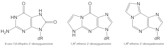
図 1.DNA 病変の化学構造は、本研究において定量化した。dR = 2 ́-デオキシリボース。この図は、オリベイラ et al.34から変更されています。この図の大規模なバージョンを表示するには、ここをクリックしてください。

1980年代初頭に実施された研究は、電気化学的検出 (HPLC ECD) に結合した高性能液体クロマトグラフィーによる 8-oxodGuo の高感度検出を可能にした。いくつかの生物学的システムにおける HPLC − oxodGuo の定量化は酸化条件に供され、DNA1,16における酸化的誘発塩基損傷のバイオマーカーとしての8− oxodGuo の認識につながった。堅牢で、低 fmol 範囲17で 8-oxodGuo の定量化を可能にするが、HPLC ECD 測定は検体同定のための検体保持時間の精度と、干渉を避けるためのクロマトグラフィー分解能に依存します。他のサンプル成分。電気化学検出では、移動相において塩 (例えば、リン酸カリウム、酢酸ナトリウム) を使用する必要があるため、適切な分析条件の維持には、ルーチンカラムおよび機器の洗浄時間が必要です。

あるいは、formamidopyrimidine DNA glycosylase (FPG) およびその後、ヒト 8-oxoguanine glycosylase 1 (hOGG1) の細菌 DNA 修復酵素の使用は、DNA からの 8-oxoGua の検出および除去のための、DNA アルカリ不安定の誘導のための方法として浮上したサイト。アルカリ不安定部位は、DNA 鎖切断に変換され、アルカリ単細胞ゲル電気泳動 (「彗星アッセイ」) による 8-oxoGua の非常に高感度な間接定量を可能にします。細胞 DNA の抽出の必要性のない高い感受性そして分析の達成はこのタイプの試金の主な利点である。これは、DNA において 8-oxoGua の最低定常状態レベルを与え、典型的には、HPLC に基づくシステム法によって得られたレベルよりも低い7-10 倍である。しかし、それは 8-oxoGua の間接測定であり、いくつかの欠点は、1,16,18使用される修復酵素の特異性または未知の効率の欠如である。

免疫アッセイは、1、n6-茶道および1、n2-dGuo12のような 8-oxoGua1および exocyclic DNA 付加物の検出のために使用される方法の他のセットである。感受性にもかかわらず、dna 病変の検出のための抗体の使用の欠点は、正常な dna 塩基1,12を含む生物学的試料の他の成分に対する交差反応性による特異性の欠如である。Exocyclic DNA 付加体としては、1、n6-茶道および 1,n2-dGuo を含む、高感度の32P − postlabeling アッセイ12によって検出および定量することもできる。32P-postlabeling の高感度は、1010通常塩基あたり約1付加体の検出のための非常に少量の DNA (例えば、10μ g) の使用を可能にします。しかし、ラジオ-化学物質の使用は、化学的特異性および低精度の欠如は、いくつかの欠点19,20である。

上記で引用した方法の共有制限は、所望の分子の検出のための低い選択性または特異性である。このシナリオで、エレクトロスプレーイオン化タンデム質量分析法 (HPLC-ESI-MS/MS および HPLC-ms3) に結合された hplc は、DNA、尿、血漿および唾液のような生物学的マトリクスにおける修飾ヌクレオシドの定量化のための金本位として進化した1,19,20. HPLC − MS/ms 法の利点は、感度 (典型的には低 fmol 範囲) と i によって提供される特異性が高いことである) クロマトグラフ分離、ii) 質量内部の分子断片化の特徴と既知のパターン分光器衝突チャンバ、および iii) は、多重反応監視モード1,19において選択された質量を電荷比 (m/z) で正確に測定する。同位体ラベルの内部標準を使用することにより、DNA 加水分解および検体濃縮ステップにおける分子損失の補正、およびサンプル間の分析種イオン化の違いによる利点が加わります。また、1121920の複数のピークが存在する場合に、正しいクロマトグラフィーピークの同定にも役立ちます。

さまざまな生物学的サンプルから抽出された DNA において、oxodGuo、1、N6-茶道および 1,n2-dGuo の定量には、HPLC − ms/ms に基づくいくつかの方法が用いられる12,15,20 212223242526272829 日.微粒子 (PM2.5) は、多環芳香族炭化水素 (PAHs)、ニトロ-PAHs、アルデヒド、ケトン、カルボン酸、キノリン、金属、水溶性イオンなどの有機および無機化学物質を運び、炎症を誘発する可能性があり酸化ストレスは、生体分子損傷および疾患の発生を優先する条件30313233である。ここでは、oxodGuo、1、n6-茶道および 1,2-dGuo の肺、肝臓および腎臓 DNA における定量化のために首尾よく適用された検証済みの HPLC ESI/ms 法を提示します。アンビエント PM2.5暴露34の影響

Protocol

4週齢の雄 A/J マウスは、特定の病原体を遊離し、Fundação オズワルドクルス (FIOCRUZ)、リオデジャネイロ、ブラジルの実験動物の育種センターから得られ、そしてそれに応じて医学部の倫理委員会に扱われたサンパウロ (プロトコル no 1310/09) の。 1. マウスの組織のコレクション キシラジンとケタミンで動物を麻酔。体重が 30 g のマウスの場合、2.63 を含む?…

Representative Results

マウスの肝臓から得られた平均 DNA 濃度 (± SD)、0.2 肺 (~ 1g の組織) と腎臓 (~ 0.4 g の組織) は、それぞれ5068± 2615, 4369 ± 1021, 3223 ±723μ g/mL の最終的な容量を200μ l とした。精製 DNA の HPLC −パパによって得られた代表的なクロマトグラムを図 3に示す。4つの 2 ‘-deoxynucleosides の存在は、対応する 2 ‘-deoxynucleosides の直前に溶出する RNA ribonucleosides から?…

Discussion

HPLC 法による 8-oxodGuo 解析で見られる主要な問題は、dna 抽出の workup 手順、dna 加水分解、および dna 加水分解物22,38の濃度におけるその形成の可能な誘導である。8-oxodGuo artifactual 形成の問題を最小限に抑えるために、すべての DNA 抽出、貯蔵および加水分解溶液へのデフェロキサミンの添加、DNA 抽出におけるヨウ化ナトリウムカオトロピック法の使?…

Divulgations

The authors have nothing to disclose.

Acknowledgements

FAPESP (Fundação ・デ・アンパロà Pesquisa はメヒコ・デ・サンパウロ、2012/22190-3 および 2012/08616-8)、CNPq (Proc. 454214/2014-6 および 429184/2016-6)、ケープ、PRPUSP (Pró・デ・ Reitoria ・ダ・ Pesquisa ・デ・サンパウロ)、セアラ INCT (MCT/INAIRA/CNPQ/岬/FAPEMIG/FAPERJ/FAPESP;Proc. 573813/2008-6)、INCT Redoxoma (FAPESP/CNPq/岬;573530/2008-4)、NAP Redoxoma (PRPUSP;2011.1.9352.1.8) と CEPID Redoxoma (FAPESP;Proc. 2013/07937-8)。F. オリベイラは、FAPESP (Proc. 2012/21636-8, 2011/09891-0, 2012/08617-4) とケープ (Coordenação ・デ・ Aperfeiçoamento ・デ・ Pessoal ・デ・ Nível ・スーペリア) から奨学金を受けました。M. h. g. メデイロス、p. ディ Mascio、p. h. Saldiva、および a. p. m. Loureiro は CNPq からのフェローシップを受け取りました。

この作品に含まれるいくつかの図および表は、ブラジルのサンパウロ市からの濃縮された周囲微細粒子状物質 (PM2.5) に曝露したマウスにおけるオリベイラ A.A.F. et Al. 遺伝毒性および epigenotoxic 効果において最初に出版した。粒子および繊維毒性学。15、40 (2018)。

Materials

[15N5]-2’-deoxyadenosine Cambridge Isotope Laboratories NLM-3895-25
[15N5]-2’-deoxyguanosine Cambridge Isotope Laboratories NLM-3899-CA-10
acetonitrile Carlo Erba Reagents 412413000
alkaline phosphatase from bovine intestinal mucosa Sigma P5521
ammonium acetate Merck 101116
calf thymus DNA Sigma D1501
cell lysis solution QIAGEN 158908
chloroform Carlo Erba Reagents 412653
deferoxamine Sigma D9533
deoxyribonuclease I (DNase I) Bio Basic Inc DD0649
ethanol Carlo Erba Reagents 414542
formic acid Sigma-Aldrich F0507
HPLC-ESI-MS/MS system HPLC: Agilent 1200 series            ESI-MS/MS: Applied Biosystems/MDS Sciex Instruments HPLC: binary pump (G1312B), isocratic pump (G1310A), column oven with a column switching valve (G1316B), diode array detector (G1315C), auto sampler (G1367C).                                                            ESI-MS/MS: Linear Quadrupole Ion Trap mass spectrometer, Model 4000 QTRAP.
HPLC/DAD system Shimadzu Two pumps (LC-20AT), photo diode array detector (DAD-20AV), auto-injector (Proeminence SIL-20AC), column oven (CTO-10AS/VP)
HPLC column (50 x 2.0 mm i.d., 2.5 µm, C18) Phenomenex 00B-4446-B0
HPLC column (150 x 2.0 mm i.d., 3.0 µm, C18) Phenomenex 00F-4251-B0
HPLC column (250 x 4.6 mm i.d., 5.0 µm, C18) Phenomenex 00G-4252-E0
HPLC C18 security guard cartridge (4.0 x 3.0 mm i.d.) Phenomenex AJO-4287
isoamyl alcohol Sigma-Aldrich M32658
isopropyl alcohol (isopropanol) Carlo Erba Reagents A412790010
ketamine Ceva Commercial name: Dopalen
magnesium chloride Carlo Erba Reagents 349377
magnesium chloride Sigma M2393
methanol Carlo Erba Reagents L022909K7
phosphodiesterase I from Crotalus atrox Sigma P4506
protein precipitation solution QIAGEN 158912
proteinase K Sigma-Aldrich P2308
ribonuclease A Sigma R5000
sodium chloride Sigma-Aldrich S9625
SPE-C18 (Strata-X) Phenomenex 8B-S100-TAK
tris(hydroxymethyl)-aminomethane Carlo Erba Reagents 489983
xylazine Syntec do Brasil Commercial name: Xilazin

References

  1. Cadet, J., Davies, K. J. A., Medeiros, M. H. G., Di Mascio, P., Wagner, J. R. Formation and repair of oxidatively generated damage in cellular DNA. Free Radical Biology and Medicine. 107, 13-34 (2017).
  2. Barnes, J. L., Zubair, M., John, K., Poirier, M. C., Martin, F. L. Carcinogens and DNA damage. Biochemical Society Transactions. 46, 1213-1224 (2018).
  3. Cadet, J., Davies, K. J. A. Oxidative DNA damage & repair: An introduction. Free Radical Biology and Medicine. 107, 2-12 (2017).
  4. Cao, H., Jiang, Y., Wang, Y. Stereospecific synthesis and characterization of oligodeoxyribonucleotides containing an N2-(1-carboxyethyl)-2′-deoxyguanosine. Journal of the American Chemical Society. 129, 12123-12130 (2007).
  5. Breyer, V., et al. Analysis and biological relevance of advanced glycation end-products of DNA in eukaryotic cells. The FEBS Journal. 275, 914-925 (2008).
  6. Tamae, D., Lim, P., Wuenschell, G. E., Termini, J. Mutagenesis and repair induced by the DNA advanced glycation end product N2-1-(carboxyethyl)-2′-deoxyguanosine in human cells. Biochimie. 50, 2321-2329 (2011).
  7. Hecht, S. S. Lung carcinogenesis by tobacco smoke. International Journal of Cancer. 131, 2724-2732 (2012).
  8. Garraway, L. A., Lander, E. S. Lessons from the cancer genome. Cell. 153, 17-37 (2013).
  9. Ong, T. P., Loureiro, A. P. M. Nutritional interventions in age-related genetic and epigenetic instability and cancer. Anti-ageing nutrients: Evidence-based prevention of age-associated diseases. , (2015).
  10. Evans, M. D., Dizdaroglu, M., Cooke, M. S. Oxidative DNA damage and disease: induction, repair and significance. Mutation Research. 567, 1-61 (2004).
  11. Moriya, M. Single-stranded shuttle phagemid for mutagenesis studies in mammalian cells: 8-oxoguanine in DNA induces targeted GC → TA transversions in simian kidney cells. Proceedings of the National Academy of Sciences of the United States of America. 90, 1122-1126 (1993).
  12. Medeiros, M. H. G. Exocyclic DNA adducts as biomarkers of lipid oxidation and predictors of disease. Challenges in developing sensitive and specific methods for clinical studies. Chemical Research in Toxicology. 22, 419-425 (2009).
  13. Guéraud, F. 4-Hydroxynonenal metabolites and adducts in pre-carcinogenic conditions and cancer. Free Radical Biology and Medicine. 111, 196-208 (2017).
  14. Nair, U., Bartsch, H., Nair, J. Lipid peroxidation-induced DNA damage in cancer-prone inflammatory diseases: A review of published adduct types and levels in humans. Free Radical Biology and Medicine. 43, 1109-1120 (2007).
  15. Pang, B., et al. Lipid peroxidation dominates the chemistry of DNA adduct formation in a mouse model of inflammation. Carcinogenesis. 28, 1807-1813 (2007).
  16. Møller, P., et al. Harmonising measurements of 8-oxo-7,8-dihydro-2′-deoxyguanosine in cellular DNA and urine. Free Radical Research. 46, 541-553 (2012).
  17. Hofer, T., Moller, L. Optimization of the workup procedure for the analysis of 8-oxo-7,8-dihydro-2′-deoxyguanosine with electrochemicaldetection. Chemical Research in Toxicology. 15, 426-432 (2002).
  18. Collins, A., El Yamani, N., Dusinska, M. Sensitive detection of DNA oxidation damage induced by nanomaterials. Free Radical Biology and Medicine. , 69-76 (2017).
  19. Zubel, T., Buerkle, A., Mangerich, A. Mass spectrometric analysis of sulfur mustard-induced biomolecular adducts: Are DNA adducts suitable biomarkers of exposure?. Toxicology Letters. 293, 21-30 (2018).
  20. Tretyakova, N., Goggin, M., Sangaraju, D., Janis, G. Quantitation of DNA adducts by stable isotope dilution mass spectrometry. Chemical Research in Toxicology. 25, 2007-2035 (2012).
  21. Churchwell, M. I., Beland, F. A., Doerge, D. R. Quantification of multiple DNA adducts formed through oxidative stress using liquid chromatography and electrospray tandem mass spectrometry. Chemical Research in Toxicology. 15, 1295-1301 (2002).
  22. Chao, M. R., Yen, C. C., Hu, C. W. Prevention of artifactual oxidation in determination of cellular 8-oxo-7,8-dihydro-2′-deoxyguanosine by isotope-dilution LC-MS/MS with automated solid-phase extraction. Free Radical Biology and Medicine. 44, 464-473 (2008).
  23. Danielsen, P. H., et al. Oxidative stress, inflammation, and DNA damage in rats after intratracheal instillation or oral exposure to ambient air and wood smoke particulate matter. Toxicological Sciences. 118, 574-585 (2010).
  24. Danielsen, P. H., et al. Oxidative stress, DNA damage, and inflammation induced by ambient air and wood smoke particulate matter in human A549 and THP-1 cell lines. Chemical Research in Toxicology. 24, 168-184 (2011).
  25. Garcia, C. C. M., et al. [13C2]-Acetaldehyde promotes unequivocal formation of 1,N2-propano-2′-deoxyguanosine in human cells. Journal of the American Chemical Society. 133, 9140-9143 (2011).
  26. Angeli, J. P. F., et al. Lipid hydroperoxide-induced and hemoglobin-enhanced oxidative damage to colon cancer cells. Free Radical Biology and Medicine. 51, 503-515 (2011).
  27. Yu, Y., et al. Comprehensive assessment of oxidatively induced modifications of DNA in a rat model of human Wilson’s disease. Molecular and Cellular Proteomics. 15, 810-817 (2016).
  28. Torres-Cuevas, I., Aupi, M., Asensi, M. A., Vento, M., Ortega, &. #. 1. 9. 3. ;., Escobar, J. 7,8-Hydroxy-2′-deoxyguanosine/2′-deoxiguanosine ratio determined in hydrolysates of brain DNA by ultrachromatrography coupled to tandem mass spectrometry. Talanta. 170, 97-102 (2017).
  29. Wu, D., et al. Detection of 8-hydroxydeoxyguanosine (8-OHdG) as a biomarker of oxidative damage in peripheral leukocyte DNA by UHPLC-MS/MS. Journal of Chromatography B. 1064, 1-6 (2017).
  30. IARC. . Monographs on the Evaluation of Carcinogenic Risks to Humans: Outdoor Air Pollution. 109, (2016).
  31. De Martinis, B. S., Kado, N. Y., Carvalho, L. R. F., Okamoto, R. A., Gundel, L. A. Genotoxicity of fractionated organic material in airborne particles from São. Mutation Research. 446, 83-94 (1999).
  32. Karlsson, H. L., Nygren, J., Möller, L. Genotoxicity of airborne particulate matter: The role of cell-particle interaction and of substances with adduct-forming and oxidizing capacity. Mutation Research. 565, 1-10 (2004).
  33. Bell, M. L., Dominici, F., Ebisu, K., Zeger, S. L., Samet, J. M. Spatial and temporal variation in PM2.5 chemical composition in the United States for health effects studies. Environmental Health Perspectives. 115, 989-995 (2007).
  34. Oliveira, A. A. F., et al. Genotoxic and epigenotoxic effects in mice exposed to concentrated ambient fine particulate matter (PM2.5) from São Paulo city, Brazil. Particle and Fibre Toxicology. 15, 40 (2018).
  35. Loureiro, A. P. M., Zhang, W., Kassie, F., Zhang, S., Villalta, P. W., Wang, M., Hecht, S. S. Mass spectrometric analysis of a cyclic 7,8-butanoguanine adduct of N-nitrosopyrrolidine: comparison to other N-nitrosopyrrolidine adducts in rat hepatic DNA. Chemical Research in Toxicology. 22, 1728-1735 (2009).
  36. Loureiro, A. P. M., Marques, S. A., Garcia, C. C. M., Di Mascio, P., Medeiros, M. H. G. Development of an on-line liquid chromatography-electrospray tandem mass spectrometry assay to quantitatively determine 1,N2-etheno-2′-deoxyguanosine in DNA. Chemical Research in Toxicology. 15, 1302-1308 (2002).
  37. Mangal, D., et al. Analysis of 7,8-dihydro-8-oxo-2′-deoxyguanosine in cellular DNA during oxidative stress. Chemical Research in Toxicology. 22, 788-797 (2009).
  38. ESCODD (European Standards Committee on Oxidative DNA Damage). Comparative analysis of baseline 8-oxo-7,8-dihydroguanine in mammalian cell DNA, by different methods in different laboratories: an approach to consensus. Carcinogenesis. 23, 2129-2133 (2002).
  39. Helbock, H. J., et al. DNA oxidation matters: The HPLC-electrochemical detection assay of 8-oxo-deoxyguanosine and 8-oxo-guanine. Proceedings of the National Academy of Sciences of the United States of America. 95, 288-293 (1998).
  40. Risom, L., et al. Oxidative DNA damage and defence gene expression in the mouse lung after short-term exposure to diesel exhaust particles by inhalation. Carcinogenesis. 24, 1847-1852 (2003).
  41. Risom, L., et al. Repeated inhalations of diesel exhaust particles and oxidatively damaged DNA in young oxoguanine DNA glycosylase (OGG1) deficient mice. Free Radical Research. 41, 172-181 (2007).
  42. Tsurudome, Y., et al. Changes in levels of 8-hydroxyguanine in DNA, its repair and OGG1 mRNA in rat lungs after intratracheal administration of diesel exhaust particles. Carcinogenesis. 20, 1573-1576 (1999).
  43. Marie-Desvergne, C., Maître, A., Bouchard, M., Ravanat, J. L., Viau, C. Evaluation of DNA adducts, DNA and RNA oxidative lesions, and 3-hydroxybenzo(a)pyrene as biomarkers of DNA damage in lung following intravenous injection of the parent compound in rats. Chemical Research in Toxicology. 23, 1207-1214 (2010).
  44. Iwai, K., et al. Early oxidative DNA damages and late development of lung cancer in diesel exhaust-exposed rats. Environmental Research. 84, 255-264 (2000).
  45. Ichinose, T., et al. Lung carcinogenesis and formation of 8-hydroxy-deoxyguanosine in mice by diesel exhaust particles. Carcinogenesis. 18, 185-192 (1997).
  46. Schmerold, I., Niedermu, H. Levels of 8-hydroxy-2′-deoxyguanosine in cellular DNA from 12 tissues of young and old Sprague Dawley rats. Experimental Gerontology. 36, 1375-1386 (2001).
  47. Garcia, C. C. M., Freitas, F. P., Di Mascio, P., Medeiros, M. H. G. Ultrasensitive simultaneous quantification of 1,N2-etheno-2′-deoxyguanosine and 1,N2-propano-2′-deoxyguanosine in DNA by an online liquid chromatography-electrospray tandem mass spectrometry assay. Chemical Research in Toxicology. 23, 1245-1255 (2010).
  48. Godshalk, R., et al. Comparison of multiple DNA adduct types in tumor adjacent human lung tissue: effect of cigarette smoking. Carcinogenesis. 23, 2081-2086 (2002).
  49. Dechakhamphu, S., et al. Lipid peroxidation and etheno DNA adducts in white blood cells of liver fluke-infected patients: protection by plasma alpha-tocopherol and praziquantel. Cancer Epidemiology Biomarkers and Prevention. 19, 310-318 (2010).
  50. Arab, K., et al. Typical signature of DNA damage in white blood cells: a pilot study on etheno adducts in Danish mother-newborn child pairs. Carcinogenesis. 30, 282-285 (2009).
  51. Nair, J., et al. High dietary omega-6 polyunsaturated fatty acids drastically increase the formation of etheno-DNA base adducts in white blood cells of female subjects. Cancer Epidemiology Biomarkers and Prevention. 6, 597-601 (1997).
check_url/fr/59734?article_type=t

Play Video

Citer Cet Article
Franco de Oliveira, T., Falcão de Oliveira, A. A., Lemos, M., Veras, M., Saldiva, P. H. N., Gennari de Medeiros, M. H., Di Mascio, P., de Melo Loureiro, A. P. Quantification of three DNA Lesions by Mass Spectrometry and Assessment of Their Levels in Tissues of Mice Exposed to Ambient Fine Particulate Matter. J. Vis. Exp. (147), e59734, doi:10.3791/59734 (2019).

View Video