Here we describe a new method of detecting successful establishment of shared blood circulation of two parabionts through a caudal vein injection of glucose, which causes minimal damage and is not fatal to the parabionts.
Parabiosis is an experimental method for surgically combining two parallel animals along the longitudinal axis of the body. We present a protocol for detecting the successful establishment of blood chimerism in parabionts by a caudal vein injection of glucose. Parabiotic mice were constructed. Glucose was injected into the donor mouse through the tail vein, and the fluctuation of blood glucose level was measured in both mice using a blood glucometer at different time points. Our results showed that after glucose injection, the blood glucose level in donor mice increased sharply after 1 min and decreased slowly thereafter. Meanwhile, the blood glucose level of the recipient mice peaked 15 min after injection. Similar results were obtained with Evans blue, used as a positive control for glucose. The synchronous fluctuation of blood glucose levels indicates that blood flow between the two mice was established successfully.
Parabiosis is a modeling method in which two living organisms are joined together surgically and develop as a single physiological system with a shared circulatory system1. Such models have been widely used for studying physiology owing to the advantage that the substances produced by a single individual can act on both animals at the same time via the shared circulatory system. Since the mid-1800s when parabiotic experiments were pioneered by Paul Bert2, the methods for constructing parabiotic models have become standardized. However, a straightforward and convenient method for verifying the successful establishment of blood chimerism has been missing. It has been reported that cross-circulation can be successfully assessed by intraperitoneally injecting 0.5% Evans blue dye in one of the parabionts followed by measurement of the absorbance of Evans blue in the blood of both parabionts with a microplate reader3. Another method requires a specific mouse breed that contains CD45.1+– and CD45.2+-labeled monocytes in each parabiont. Cell cytometry is then used to determine blood chimerism by measuring the frequency of the two markers in monocytes from spleen or blood4. However, these methods are often lethal or cumbersome to the animals, and a safe and simple method for quick and reliable verification of parabiotic models is highly desirable. In this study, we established a new method for this purpose, which was validated in a mouse model of parabiosis. Glucose concentration in blood samples drawn from a tail vein is measured using a glucometer, and the pattern of changes of glucose level in donor and recipient mice is considered an indication of circulation chimerism. We named this method the "glucose fluctuation method". The application of this validation method is not limited to mice but could be extended to diverse pathological models except for those with serious dysregulation of glucose metabolism. The procedure is simple, timesaving, and safe.
All procedures involving animals and their care were approved by the Institutional Animal Care and Use Committee of Harbin Medical University.
NOTE: The tools and equipment required for the method are listed in the Table of Materials.
1. Preparation of materials and animals
2. Parabiosis
3. Validation of circulation chimerism
In six donor mice, blood glucose levels sharply increased to 26.5 µmol/L (173% increase) at an average of 1 min after the injection of 100 µL of glucose (1.2 g/kg) through the tail vein and then gradually decreased to 13.3 µmol/L at 60 min. In recipient mice, blood glucose slowly increased after injection and reached the first peak level at 15 min (47% increase, 12.2 µmol/L). Based on the above results, the standard for circulation chimerism was set as follows: 1) a sharp increase in the blood glucose level (a minimum 100% increase or >20 µmol/L) in donor mice within 1 min after glucose injection, and 2) a significant increase in the blood glucose level in recipient mice 15 min after injection (a minimum 37% increase) (Figure 1).
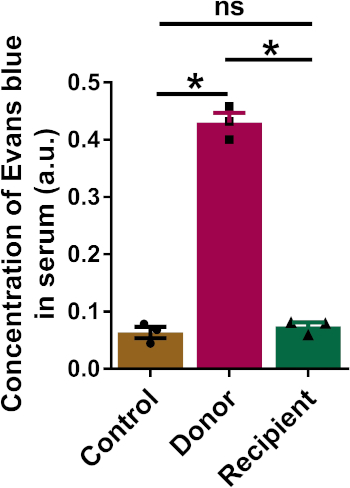
The concentration of Evans blue dye in the serum of parabionts also indicated the successful construction of circulation chimerism (Figure 2). We euthanized the parabionts after blood glucose measurement using 5 mL of 2,2,2-tribromoethanol (20 g/L). The subcutaneous vascular junctions between the parabionts were clearly observed (Figure 3).
Supplementary Figure 1 shows that the donor mice had a significant blood glucose level increase 1 min after glucose injection, while the blood glucose level of the recipient mice was not elevated, which demonstrated that the circulation chimerism in parabionts was not successfully established 1 day after parabiosis surgery. Similarly, the blood OD level in recipient mice was not as elevated as that of donor mice (Supplementary Figure 2).
Based on the results from the glucose fluctuation method, we found that two pairs of parabionts did not establish blood chimerism 15 days after parabiosis surgery. As shown in Supplementary Table 1, the two recipient mice did not have an increased blood glucose level within 60 min after glucose injection into the donor mice. The blood concentration of Evans blue in the two recipients was also not elevated (Supplementary Table 2), which demonstrated that the glucose fluctuation method was as sensitive as the Evans blue method.
Moreover, to evaluate the influence of injected glucose on insulin metabolism, we detected the blood insulin level 1 h and 3 h after injection of 100 µL of glucose (1.2 g/kg) in mice (Supplementary Figure 3). The blood insulin level was remarkably decreased 1 h after glucose injection because of quickly increased glucose and recovered to normal levels at 3 h. These results demonstrated that the effects of glucose we injected on insulin metabolism were restorable.
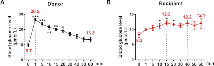
Figure 1: Changes of blood glucose level in parabionts after injection of glucose through the caudal vein. (A) Blood glucose level of donor mice. (B) Blood glucose level of recipient mice (n = 6). The data are presented as the mean ± SEM. *p < 0.05, **p < 0.01, ***p < 0.001 vs. 0 min. Please click here to view a larger version of this figure.
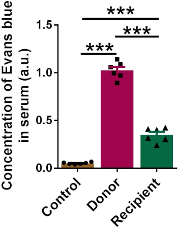
Figure 2: The concentration of Evans blue in serum samples of parabionts measured by a microplate reader. The data are presented as the mean ± SEM. ***p < 0.001 (n = 6). Please click here to view a larger version of this figure.
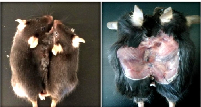
Figure 3: Generation of subcutaneous vasoganglions in connected skin between the parabionts. Left: Representative image of the parabiosis mice. Right: A representative image of the subcutaneous vasoganglion between the parabionts. Please click here to view a larger version of this figure.
Supplementary Figure 1: The blood glucose level of parabionts was tested 1 day after parabiosis surgery. Please click here to view a larger version of this figure.
Supplementary Figure 2: The concentration of Evans blue measured by microplate reader in the serum of the parabionts 1 day after parabiosis surgery. Please click here to view a larger version of this figure.
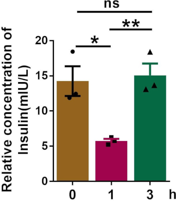
Supplementary Figure 3: The concentration of insulin in the serum of the mice after glucose injection. Please click here to view a larger version of this figure.
0 min | 5 min | 15 min | 20 min | 40 min | 60 min | |
Donor-1 | 5.7 | 26.2 | 21.2 | 17.6 | 16.9 | 15.4 |
Recipient-1 | 6.7 | 5.8 | 5.9 | 6.2 | 5.2 | 5.4 |
Donor-2 | 8.4 | 25.5 | 21.1 | 20.5 | 17.4 | 13.8 |
Recipient-2 | 6.7 | 5.8 | 5.9 | 6.2 | 5.2 | 5.4 |
Supplementary Table 1: Blood glucose level (µmol/L) in parabionts 15 days after parabiosis surgery.
Control-1 | Donor-1 | Recipient-1 | Control-2 | Donor-2 | Recipient-2 | |
OD value | 0.059 | 0.935 | 0.062 | 0.068 | 0.862 | 0.073 |
Supplementary Table 2: Blood concentration of Evans blue (OD value) in parabionts 15 days after parabiosis surgery.
Parabiosis refers to the surgical technique of connecting two living animals to establish a common vascular system by experimental means6,7,8. The advantage of this model is that the substances produced by a single individual can act on both animals at the same time. Thus, the parabiosis model can be used to explore the role of a substance or factor in a related disease, producing many meaningful and innovative conclusions. In view of its great application value, the model has brought about a better understanding of cardiovascular system diseases8,9,10,11,12,13, nervous system disorders14,15, organ transplantation16, and diabetes17,18.
However, verification of the successful establishment of circulation chimerism is the first and determining step for studies with parabiosis. Current studies have reported some methods for detecting circulation chimerism. Loffredo et al.4 reported that blood chimerism was confirmed in parabiotic pairs by measuring the mixed frequency of monocytes in the spleen with different labeled markers in the donor (CD45.1+) and recipient (CD45.2+) mice. In this method, specific mice with CD45.1+– or CD45.2+-labeled monocytes for each parabiont were used for parabiosis. Cell cytometry was needed to determine blood chimerism by measuring the integration of the marked blood cells. In addition, Marta et al.3 assessed cross-circulation by intraperitoneally injecting 200 µL of 0.5% Evans blue dye in one of the parabionts. Blood from both parabionts was collected 2 h later by cardiac puncture. Blood chimerism was determined by an increased Evans blue concentration in the recipient mice, which was tested by a microplate reader. Although these strategies allow us to determine blood chimerism, there are still many limitations that cannot be ignored. First, for lethal methods, the blood chimerism can only be confirmed at execution. However, in our method, the establishment of blood chimerism can be tested any time after parabiosis surgery. Parabionts with unsuccessful established circulation chimerism can be excluded for further study in advance to reduce unnecessary workload. Indeed, using our glucose fluctuation method we were able to pick out the mice with unsuccessful construction of blood chimerism in certain parabionts, which might be due to insufficient time of parabiosis, unstable surgery manipulation, delayed wound healing, or disconnected body tissue caused by animal struggling. In such circumstances, the failure of blood chimerism was also confirmed by using the Evans blue method. These results indicate that the glucose fluctuation method was as effective as the Evans blue method (Supplementary Figure 1 and Supplementary Figure 2, Supplementary Table 1 and Supplementary Table 2). Second, the currently used methods of validation are excessively time-consuming and difficult, as opposed to this new method, which is simple and effective.
In the present study, we successfully tested for circulation chimerism in parabiotic mice by the glucose fluctuation method. The mice were anesthetized for the glucose injection and blood glucose level measurements, which made their manipulation easier. We injected 100 µL of glucose (1.2 g/kg) through the tail vein of the mice within 10 s. Under these conditions, a regular fluctuation of blood glucose levels in donor and recipient mice was observed. Importantly, the amount of glucose we used tended to stably elevate the blood glucose level within a certain range. Moreover, the results showed that the blood insulin level recovered to the normal range 3 h after glucose injection (Supplementary Figure 3), suggesting that the amount of glucose injected into the mice had minimal effects on insulin metabolism. In addition, the dosage of glucose we gave to the donor was lower than that used for the glucose tolerance test of mice (for the GTT, 2 g/kg glucose is intraperitoneally injected to the mice, equaling approximately 1.6 g/kg through caudal vein injection19,20,21), which means that the dosage of glucose for the glucose fluctuation method would not cause hyperglycemia and other damaging alterations. In conclusion, the method we used is effective, harmless, and timesaving. However, it might be limited to diabetic mice in parabiosis, which still need further experiments for confirmation.
The authors have nothing to disclose.
This work was supported by the National Nature Science Foundation of China (81570399 and 81773735), the National Key Research and Development Program of China – Traditional Chinese Medicine Modernization Research project (2017YFC1702003), and Hei Long Jiang Outstanding Youth Science Fund (JC2017020).
curved forceps | JZ surgical Instruments, China | J31340 | |
fine scissors | JZ surgical Instruments, China | WA1030 | |
needle forcep | JZ surgical Instruments, China | 60017961 | |
2.5 mL syringes | Agilent, USA | 5182-9642 | |
Tribromoethano | Sigma-Aldrich, USA | T48402-5G | |
penicillin | Solarbio, China | IP0150 | |
tramadol | Yijishiye, China | YJT712520 | |
glucometer | Roche Diabetes Care, Indiana | Accu-Chek Active test strips | |
Evan's blue Counterstain | Solarbio, China | G1810 | |
depilatory cream | Nair, USA | LL9161 | |
Warming Blanket (Heating pad) | Kent Scientific Corp, USA | TP-22G | |
Electrical shaver | Codos, China | CP-5000 | |
3-0,5-0 surgical suture |
Shanghai Medical Suture Needle Factory, China | SYZ 3-0#, SYZ 5-0# |