Described is a protocol to establish a Doxorubicin-induced dilated cardiomyopathy (DCM) model in mice via long-term intraperitoneal injection of Doxorubicin.
Dilated cardiomyopathy (DCM) refers to a spectrum of heterogeneous myocardial disorders characterized by ventricular dilation and depressed cardiac performance in the absence of hypertension, valvular, congenital, or ischemic heart diseases, and which may be related to infection, autoimmune or metabolic abnormalities, or family inheritance. It can progress into congestive heart failure with a poor prognosis. Doxorubicin (Dox) is widely employed as a chemotherapeutic drug, but its use is limited because it causes DCM-like changes of the myocardium. Its myocardial toxicity is attributed to oxidative stress, chronic inflammation, and cardiomyocyte apoptosis. A model of DCM exploiting these Dox-induced DCM symptoms has not been established.
One of the most common causes of heart failure, DCM is characterized by ventricular dilatation and decreased cardiac function and is the most common reason for heart transplantation worldwide1. In order to further investigate its pathogenesis and find effective treatments, access to mature animal models is especially important. The purpose of the described experiments is to establish a stable mouse model of DCM that resembles human DCM.
Due to the complex pathogenesis of DCM, there are many different methods to make corresponding animal models. Spontaneous DCM models2 are relatively stable, but they are expensive and not easily available. Genetically modified animal models3 are not well-established and require more experimental use. DCM animal models induced by viral infection4 or autoimmune defects5 are easy to obtain, but they are not wholly representative of DCM. Models associated with myocardial toxicity include alcohol-induced DCM models and Dox-induced DCM animal models.
The Dox-induced cardiomyopathy model is obtained by intraperitoneal injection of Dox6. The model exploits the most severe chronic side effect of Dox: After Dox exposure, patients develop late onset DCM symptoms with clinical uniformity7. Dox-induced oxidative stress8 and mitochondrial damage9, which lead to cardiomyocyte apoptosis, are symptoms in the pathogenesis of DCM. There are acute and chronic Dox treatment models: a single high dose of Dox (15 mg/kg) induces a short-term model for cardiomyopathy10, while repetitive low-dose Dox injections (six weekly, 3 mg/kg) induce a long-term model for cardiomyopathy11. Based on the presented study, wild type mice intraperitoneally injected once a week for a month at a dose of 5 mg/kg display morphology and histology of the heart consistent with the characteristics of DCM by the end of the treatment, providing an ideal way to establish a DCM model.
Animal experiments were approved by Institutional Animal Care and Use Committee (IACUC) of Nanjing Drum Tower Hospital.
1. Preparation of the reagents and animals
2. Establishment of DCM animal model
3. Echocardiographic examination
4. Histological staining
Cardiac function
Dilated cardiomyopathy is characterized by progressive ventricular dilatation and contractile dysfunction. Figure 2 shows representative echocardiographic images of the two groups. Dox-treated mice showed markedly reduced left ventricular ejection fraction and left ventricular fractional shortening (Figure 3A,B). The LV diameter also increased in both the diastolic and systolic phases (Figure 3C,D). This revealed that Dox-treated mice had an impaired cardiac function.
Histological staining
Pathological staining was performed to observe the pathological changes of saline-treated mice and Dox-treated mice. Myocardial fibers of the mice in the control group were neatly arranged, without infiltrating leukocytes. In the Dox group, myocardial myofibers were disordered and broken, and cardiomyocytes were smaller and thinner (Figure 4A). Masson staining showed that Dox-treated mice had more interstitial fibrosis (Figure 4B).
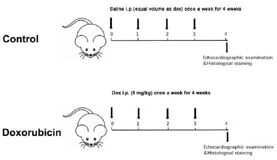
Figure 1: Schematic diagram of a Dox-induced dilated cardiomyopathy. Please click here to view a larger version of this figure.
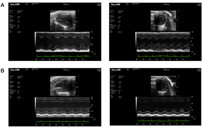
Figure 2: The echocardiography of the two groups. (A) Control group; (B) Dox group. Left is the long-axis section and right is the short-axis section. Please click here to view a larger version of this figure.
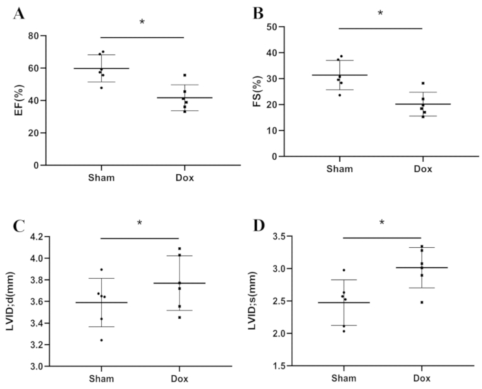
Figure 3: The cardiac function variables between groups. (A) EF; (B) FS; (C) LVEDd; (D) LVEDs. *P < 0.05, Student’s t-test. Please click here to view a larger version of this figure.
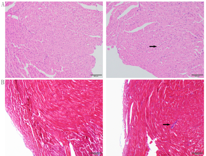
Figure 4: Histological staining. (A) H&E staining; (B) Masson staining. Left is the control group and right is the Dox group. The upper arrow shows that Dox-treated cardiomyocytes were smaller and thinner and the myofibers were disordered and broken. The lower arrow denotes the presence of interstitial fibrosis. Please click here to view a larger version of this figure.
5
Dox is a nonspecific periodic antitumor chemotherapy drug commonly used in clinical practice12. Its main side effect is cardiotoxicity, characterized by cardiomyopathy and subsequent heart failure13. The underlying mechanism includes damage of myocardial lipid peroxidation, inhibition of myocardial sarcoplasmic reticulum Ca2+-ATPase activity, and activation of the myocardial local renin-angiotensin system, resulting in increased AT II production and intracellular calcium overload14. Due to the lethal effect of a large dose of intraperitoneal injection of Dox, an alternative way to establish a reliable model was used in this protocol.
The DCM model established by this method is very similar to the pathogenesis of human DCM. In addition to heart failure, the most common complication of Dox, ascites were observed in DCM mice. Therefore, during model development, attention should be paid to sterile operation and timely replacement of syringes. In addition, for the Dox dose, two methods of administration were compared: a long course of treatment using a small dose, and short-term treatment with a high dose. The results show that continuous injection of Dox at 5 mg/kg for four weeks can result in an accurate model of DCM, lower mice mortality, and a shorter time for model establishment.
In summary, a stable, reliable, and economical mouse DCM animal model can be successfully established by intraperitoneal injection of Dox. This research provides an ideal model for studying DCM. It will contribute to reveal the pathogenesis of dilated cardiomyopathy or chronic heart failure and explore new drugs or treatments for future clinical applications.
The authors have nothing to disclose.
This work was supported by Key Project Medical Science and Technology Development Foundation, Nanjing Department of Health (No.YKK16098).
4% paraformaldehyde | servicebio | CAS30525-89-4 | |
C57BL/6 mice | Model Animal Research Center of Nanjing University | ||
Doxorubicin hydrochloride | Pfizer | CAS25316-40-9 | |
echocardiography | Visualsonics | ||
Hematoxylin and Eosin staining kit | Solarbio | G1120 | |
Masson staining kit | Solarbio | G1343 | |
phosphate buffer solution | Sigma | P5368 | |
potassium chloride | Sigma | CAS7447-40-7 | |
sterilized syringe | Millipore | SLGP033RB |