We present a microfluidic system for high throughput studies on complex life machinery, which consists of 1500 culture units, an array of enhanced peristaltic pumps and an on-site mixing modulus. The microfluidic chip allows for the analysis of the highly complex and dynamic micro-environmental conditions in vivo.
Mimicking in vivo environmental conditions is crucial for in vitro studies on complex life machinery. However, current techniques targeting live cells and organs are either highly expensive, like robotics, or lack nanoliter volume and millisecond time accuracy in liquid manipulation. We herein present the design and fabrication of a microfluidic system, which consists of 1,500 culture units, an array of enhanced peristaltic pumps and an on-site mixing modulus. To demonstrate the capacities of the microfluidic device, neural stem cell (NSC) spheres are maintained in the proposed system. We observed that when the NSC sphere is exposed to CXCL in day 1 and EGF in day 2, the round-shaped conformation is well maintained. Variation in the input order of 6 drugs causes morphological changes to the NSC sphere and the expression level representative marker for NSC stemness (i.e., Hes5 and Dcx). These results indicate that dynamic and complex environmental conditions have great effects on NSC differentiation and self-renewal, and the proposed microfluidic device is a suitable platform for high throughput studies on the complex life machinery.
High throughput techniques are crucial for biomedical and clinical studies. By parallelly conducting millions of chemical, genetic, or live cell and organoid tests, researchers can rapidly identify genes that modulate a bio-molecular pathway, and customize sequential drug input to one’s specific needs. Robotics1 and microfluidic chips in combination with a device control program allow complex experimental procedures to be automated, covering cell/tissue manipulation, liquid handling, imaging, and data processing/control2,3. Therefore, hundreds and thousands of experimental conditions can be maintained on a single chip, according to the desired throughput4,5.
In this protocol, we described the design and fabrication procedure of a microfluidic device, which consists of 1500 culture units, an array of enhanced peristaltic pumps and on-site mixing modulus. The 2-level cell culture chamber prevents unnecessary shear during medium exchange, which ensures an undisturbed culture environment for long-term live cell imaging. The studies demonstrate that the proposed microfluidic device is a suitable platform for high throughput studies on the complex life machinery. Moreover, the advanced features of the microfluidic chip allow automated reconstitution of highly complex and dynamic microenvironmental conditions in vivo, like the everchanging cytokines and ligands compositions6,7, the completion of which takes months for conventional platforms like 96-well plate.
1. Microfluidic chips design
2. Chip fabrication and operation
3. Generation of dynamic inputs in cellular microenvironments
The conventional on-chip peristaltic pump was firstly described by Stephen Quake in 2000, using which the peristalsis was actuated by the pattern 101, 100, 110, 010, 011, 001 8,10. The number 0 and 1 indicate "open" and "close" of the 3 horizontal control lines. Studies using more than 3 valves (e.g., five) have also been reported11. Even though the peristaltic pump composed by 3 control lines and 3 flow lines provides nanoliter accuracy, the transportation rate is too slow to feed 1,500 culture chambers. To solve the problem, we include more flow lines (i.e., the 10 connected channels) to increase the liquid volume driven by per pumping cycle (i.e., 101, 100, 110, 010, 011, 001). Thus, the nutrients and drugs can be effectively delivered to designated chambers. The peristaltic pump transported liquid volume can increase by 16x to ~ 50 nanoliters per pumping cycle. As the array of peristaltic pumps is controlled by 3 connected control channels (Figure 1b), the solutions from each inlet are delivered simultaneously to the chip and allow instantaneously mixing. Combinatorial and sequential inputs can, therefore, be generated by timely on-off of the inlets connected to different solutions. Using the same methodology, dynamic varying cytokine and ligand concentrations can also be generated. For example, selected combinations of 1, 0.9, 0.2, 0.05, 0.01 g/L and 4 blank culture media can generate a sine wave fluctuation (between 0 to 0.5 g/L) in concentration with step sizes ranging from 0.0005 to 0.01 g/L.
For the culture of primary cells, it is crucial to maintain a stable microenvironment. Shear stress and exhaustion of conditioned medium during medium exchange will affect cellular behavior and cell fate12. To overcome these problems, we design a buffer layer to prevent unwanted shear stress on cells during medium exchange (Figure 1c). Unlike the conventional culture units, the main advantages of the device (i.e., valve-controlled) are their capabilities of on-site mixing, delivery and maintenance of independent conditions. In Figure 2d, we demonstrated that a complex Chinese word can be created on chip via precise delivery of liquids to designated positions and maintenance of an unaffected condition in individual culture chambers. The capabilities of the active fluidic device in on-site mixing are illustrated with a video clip, showing the dynamically changing FITC concentrations in a culture chamber (Figure 3c and Video 7:47-7:50), which is accomplished by controlling the pumping rate of 2 independent peristaltic pumps connected to 2 inlets12. Numerical simulation suggests that when the medium is directed top-down through the culture chambers, shear flow quickly reaches the bottom of the culture well (Video 4:07-4:25). The shear forces can be effectively prevented when the solution is directed from left to right. Even at an input flow rate of 10 mm/s, cell or micrometer-sized tissue remains undisturbed at the bottom of the culture unit.
As is shown in Figure 3b(1), each inlet is control by a valve other than the lower peristaltic pump. When one drug is selected to be delivered into designated chambers, the inlet connected to the drug is open and the operation of the array of the peristaltic pump transports only this drug. For 2 or multiple drugs, we simply open the connected inlets to generate a mixture of drugs at equal volume. We also integrate one independent peristatic pump independent from the array (Figure 2g), which could operate at a different pumping rate from the array and therefore generate different dilutions. To mimic the in vivo NSC dynamic environmental conditions, sequential and combinatorial condition of 6 ligands (i.e., Jagged, DLL, EGF, PACAP, CXCL, PDGF), which consists of 720 and 56 different conditions, were generated using the array of peristaltic pumps and delivered to the designated chambers. In detail, sequential conditions can be represented as Sij = {ligand i is added on day j} and a combinatorial condition as Ci = {ligand i is present}, where ligand i = Jagged, DLL, EGF, PACAP, CXCL, PDGF and j=1,2,3,4,5,6. Responses of the NSC cells and spheres were recorded every 2 hours during culture and stimulation.
NSC spheres are maintained in the 400 µm by 400 µm cell culture chamber (Figure 2e and Figure 4). Differentiation and stemness (i.e., self-renewal) of the stem cells are represented by the expression level of Dcx and Hes5, respectively. We observed that when the NSC sphere is exposed to CXCL in day 1 and EGF in day 2, the round-shaped conformation is well maintained and there is obvious increase in the sphere size. NSCs with high-Dcx expression level die on the 3rd day when EGF is replaced by PACAP, suggesting effects on the differentiated cells13,14,15,16. Cells start to attach to untreated PDMS surface upon stimulation by DLL on the 4th day, and dissociate into individual cells with the addition of PDGF on the 5th day and Jagged on the 6th day. Changes in the input order of these 6 ligands bring distinctive NSC status (Figure 5). For example, the evolvement of NSCs varies dramatically when CXCL switches position with PDGF along the sequence (Figure 5a and 5b). These results demonstrate that the dynamic varying environmental conditions have great effects on NSCs differentiation and self-renewal, which paves the way for developing brain-on-chip platforms for biomedical studies as well as clinical applications.
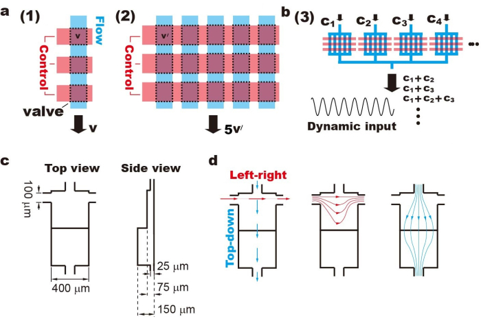
Figure 1: Design of the high-throughput microfluidic chip. a. The enhanced peristaltic pump consists of multiple flow channels and widened control channels, which leads to increase in the transferred liquid volume per pumping cycle. b. The array of peristaltic pumps is controlled by 3 connected control lines. Therefore, the inclusion of different inlets at programmed time points can generate combinatorial, sequential and dynamic varying input signals. c. To prevent unwanted shear flow, we designed a 2-level culture unit. Cells are maintained at the bottom of the lower level, which is 150 µm in depth. d. Each culture chamber is connected to 4 channels, allowing solution to be directed through top-down and left-right directions. Please click here to view a larger version of this figure.
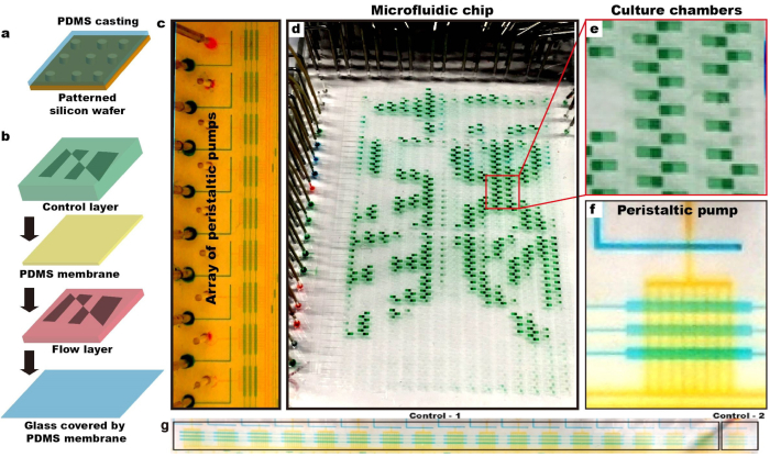
Figure 2: Fabrication of the high-throughput microfluidic chip. a. Microstructures of control and flow layers were firstly patterned on silicon wafer, on which PDMS is casted for replication. b. The control, membrane and flow layers are aligned and bonded together using plasma etching and thermal bonding. c-f. The advanced features of the chip are demonstrated using green, blue and yellow food dyes. We show that by timely on-off of the valves, solutions of nanoliter accuracy can be delivered to the designated chambers. g. The array of peristaltic pumps is controlled by 3 connected control lines (i.e., Control-1), and one independent peristaltic pump controlled by other 3 control lines (i.e., Control-2). Please click here to view a larger version of this figure.
Figure 3: A schematic showing the active fluidic device. A schematic showing that (a) cells flow through the bypass channel; (b) valves control the inlet and the peristaltic pump; and (c) cells flow into the culture chambers. Please click here to view a larger version of this figure.
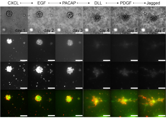
Figure 4: Representative images of the NSC sphere when being stimulated by sequential drug input. Bright field (top row), Hes5-GFP (second row), Dcx-Desred (third row) images show that upon sequential stimulation, substantial cell death occurs among both Dcx-high and Hes5-high cells. The emergence of the dark area suggests the death of stem cells, which localize mostly at the core region. Scale bar: 100 µm. Please click here to view a larger version of this figure.
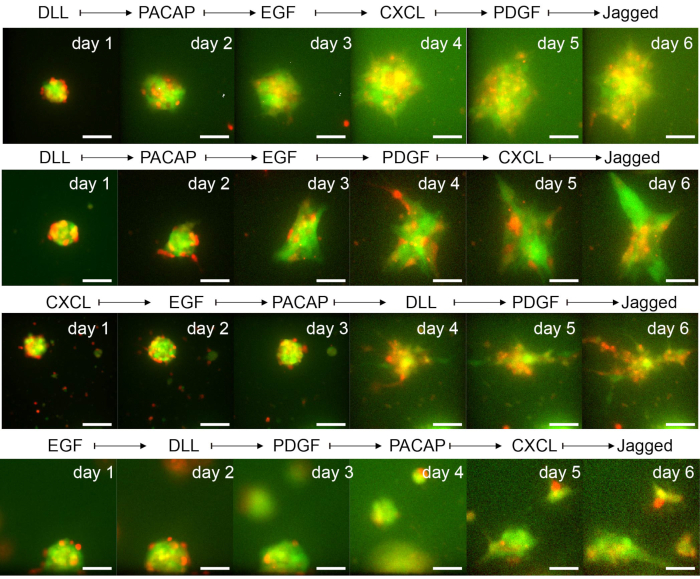
Figure 5: Variations in NSC sphere conformation, Dcx and Hes5 expression level, when being stimulated by different sequential inputs. It is demonstrated that variation in the input order of 6 drugs causes changes in tissue, morphological, and expression level of signaling molecules, indicating the sensitivities of NSC sphere to the dynamic environmental conditions. The input sequences: (a) DLL>> PACAP>> EGF>> CXCL>> PDGF>> Jagged, (b) DLL>> PACAP >> EGF >> PDGF >> CXCL >> Jagged, (c) DLL >> PACAP >> PDGF >> CXCL >> EGF >> Jagged, (d) PDGF >> CXCL >> PACAP >> EGF >> DLL >> Jagged. Scale bar: 100 µm. Please click here to view a larger version of this figure.
Various microfluidic devices have been developed to perform multiplexed and complex experiments17,18,19,20. For example, microwells made of an array of topological recesses can trap individual cells without the use of external force, showing advantageous characters including small sample size, parallelization, lower material cost, faster response, high sensitivity21,22,23,24. Droplet and surface tension confined droplet eliminate the need for auxiliary hardware such as a micropump, making the transportation of droplets more low-cost and eco-friendlier25,26. Drops of liquids can be readily deposited on the surface by micropipettes or sprayer nozzles, and after the experiments, the systems may be facilely refreshed and reusable27,28. The slipchip technique allows multiplexed reactions without the involvement of integrated pumps or valves, and therefore user- and producer-friendly29,30. However, these systems face a number of technical hurdles, such as storing nanoliter solutions in micro wells, controlling humidity, and limitations of parallel low volume liquid-dispensing technologies.
In this paper, we described a protocol for high throughput biomedical experiments using live cell imaging system and a customized microfluidic device. The procedures of chip fabrication including UV and soft lithography, and setup parameters for automatic fluorescent imaging are demonstrated in detail. The results show that the advanced functional features (i.e., the 2-level culture chamber and enhanced peristaltic pump) of the proposed microfluidic chip outperforms previously reported devices and meet the needs to conduct thousands of parallel experiments. We also described the crucial parameters (e.g., the maintenance of conditioned medium) that one should carefully control to maintain healthy environments for the culture of primary and stem cells.
Cell-based biomedical experiments, which include complex protocols and programmed addition of chemicals, are often deemed time-consuming and laborious. For example, besides the cell manipulation procedures, combinatorial inputs of 6 drugs with hourly refilling requires at least 250 pipetting steps per hour, and repetition till the end of the experiment. Furthermore, modeling dynamic environmental conditions in vivo requires timely addition of one or multiple cytokines, ligands and drugs at the accuracy of seconds, which is impossible for manual operations. We herein demonstrate that by connecting the inlets of the chip to multiple drugs, or different concentrations of single drug, combinatorial, sequential and dynamically varying input signals can be generated and maintained in the shear-free cellular environments. The morphological and chemical responses of cells and tissues, which are maintained in the parallelly running culture chambers, can then be monitored in real-time using the commercial microscope software.
With increasing throughput of microfluidic devices and automated data collection during bioimaging, the live cell imaging system can generate thousands of images every hour. For example, continuous running of the 1500-unit chip for one week generates 252,000 images. It may take months to manually track the evolution of tissues and cell populations (e.g., the expression level of the fluorescently tagged biomolecules) at the single cell level. Using a customized MATLAB program, the massive data can be automatically sorted, formatted and analyzed. Traces showing the mobility, morphological features, and expression level of signaling molecules can be retrieved (shown in the video), which avoids human error and saves considerable amount of time.
Therefore, the methodology we demonstrate here show suitable protocols for performing high throughput biomedical experiments with automated cell and tissue manipulation, fluorescent imaging and data analysis. The on-off switching of membrane valves provides optimal liquid volume, time, and spatial resolution to reconstruct the ever-changing and complex environmental conditions for in vitro like an organ-on-chip. Even though the protocol is considerably more complex compared to the transitional biomedical approaches (e.g., manual procedures using 96-well plates), the presented protocol and platform are not restricted to studies on cell and tissue functions. The screening of combinatorial and sequential drug input may allow us to develop therapies for clinical applications.
The authors have nothing to disclose.
Authors acknowledge the technical support from Zhifeng Cheng of Chansn Instrument (China) LTD. This work was supported by grants (National Natural Science Foundation of China,51927804).
2713 Loker Avenue West | Torrey pines scientific | ||
AZ-50X | AZ Electronic Materials, Luxembourg | ||
Chlorotrimethylsilane(TMCS) 92360-25mL | Sigma | ||
CO2 Incubator HP151 | Heal Force | ||
Desktop Hole Puncher for PDMS chips WH-CF-14 | Suzhou Wenhao Microfluidic Technology Co., Ltd. | ||
DMEM(L-glutamine, High Glucose, henol Red) | Invitrogen | ||
Electronic Balance UTP-313 Max:600g, e:0.1g, d:0.01g | Shanghai Hochoice Apparatus Manufacturer Co.,LTD. | ||
FBS | Sigma | ||
Fibronection 0.25 mg/mL | Millipore, Austria | ||
Glutamax 100x | Gibco | ||
Heating Incubator BGG-9240A | Shanghai bluepard instruments Co.,Ltd. | ||
Nikon Model Eclipse Ti2-E | Nikon | ||
Pen/Strep 10 Units/mL Penicillin 10 ug/mL Streptomycin | Invitrogen | ||
Plasma cleaner PDC-002 | Harrick Plasma | ||
polydimethylsiloxane(PDMS) | Momentive | ||
polylysine 0.01% | Sigma | ||
Spin coater ARE-310 | Awatori Rentaro | ||
Spin coater TDZ5-WS | Cence | ||
Spin coater WH-SC-01 | Suzhou Wenhao Microfluidic Technology Co., Ltd. | ||
SU-8 3025 | MicroChem, Westborough, MA, USA | ||
SU-8 3075 | MicroChem, Westborough, MA, USA |