Summary

세포외 소포의 정량화 및 크기 측정을 위한 나노 입자 추적 분석

Published: March 28, 2021
doi:

Summary

우리는 마우스 perigonadal 지방 조직 및 인간 혈장에서 분리된 세포외 소포의 크기 분포 및 총 입자 농도를 추정하기 위하여 새로운 나노 입자 추적 분석 계기를 사용하는 방법을 보여줍니다.

Abstract

세포 외 소포 (EV)의 생리학적 및 병리학적 역할은 점점 더 인식되고 있으며, EV 분야는 빠르게 진화하는 연구 영역으로 만듭니다. EV 격리를 위한 다양한 방법이 있으며, 각각 은하류 수율과 EV순도에 영향을 미치는 고유한 장점과 단점이 있습니다. 따라서 선택한 방법에 의해 지정된 소스로부터 분리된 EV 준비의 특성화는 다운스트림 결과를 해석하고 실험실 전체의 결과를 비교하는 데 중요합니다. 질병 상태에 의해 또는 외부 조건에 응하여 변경될 수 있는 전기 의 크기와 양을 결정하기 위한 각종 방법이 존재합니다. 나노 입자 추적 분석 (NTA)은 개별 EV의 높은 처리량 분석에 사용되는 저명한 기술 중 하나입니다. 여기서, 우리는 현장에서 주요 발전을 나타내는 NTA에 대한 획기적인 기술을 사용하여 마우스 페리고나달 지방 조직 및 인간 플라즈마로부터 분리 된 전기 의 정량화 및 크기 측정을위한 상세한 프로토콜을 제시합니다. 결과는 이 방법이 전송 전자 현미경검사법에 의해 확인된 바와 같이 다른 방법을 사용하여 다른 소스로부터 분리된 전기에 대한 재현 가능하고 유효한 총 입자 농도 및 크기 분포 데이터를 제공할 수 있음을 보여준다. NTA에 대한이 악기의 적응은 EV 연구에서 엄격과 재현성을 높이기 위해 NTA 방법의 표준화의 필요성을 해결합니다.

Introduction

세포외 소포(Ev)는 거의 모든 세포 유형 1에 의해 분비되는 작은 (0.03-2 μm) 막 결합소포이다. 그들은 종종 방출 및 크기2의그들의 기계장치에 따라 “엑소좀”, “microvesicles” 또는 “apoptotic 바디”로 불립니다. 처음에는 전기자동차가 항상성을 유지하기 위해 세포에서 폐기물을 제거하는 수단이라고생각되었지만,우리는 이제 DNA, RNA(mRNA, microRNA), 지질 및 단백질4,5를 포함한 분자 물질의 전송을 통해 세포간 통신에 참여할 수 있다는 것을 알고 있으며, 정상 생리학뿐만 아니라 정상적인 생리학의 중요한 조절제라는 것을알고 있습니다. 5,6,7,8.

다른곳에서 설명 된 다른 9,10,11,12에설명 된 전기 를 분리하고 정량화하는 많은 다른 방법이 있습니다. EV의 소스뿐만 아니라 사용되는 격리 프로토콜은 EV 수율과 순도에 큰 영향을 미칠 수 있습니다. 엑소좀 격리를 위한 “금본위제” 접근법으로 오랫동안 고려되었던 차동 원심분리조차도, 이후 얻어진 EV 인구에 영향을 미치는 실질적인 변동성의 대상이 될 수 있으며, 다운스트림분석(13). 따라서, EV 절연 및 정량화를 위한 다양한 방법론을 통해문헌(14)에보고된 실험결과를 비교, 재현 및 해석하기가 어렵다. 또한, EV 방출은 세포 조건 또는 다양한 외부 요인에 의해 조절될 수 있다. 그것은 EV 세포 스트레스에 대 한 세포를 보호 하 여 세포 항상성을 유지 에 역할을 제안 되었습니다15,여러 연구는 세포 스트레스 EV 분 비를 자극 하는 것으로 나타났습니다. 예를 들어, 증가된 EV 방출은 저산소증, 폐막성 망막 응력, 산화 스트레스, 기계적 스트레스, 담배 연기 추출물 및 미립자 대기 오염16,17,18, 19,20,21,22에세포 노출 후 보고되었다. EV 릴리스는 또한 생체 내에서 수정 된 것으로 나타났습니다. 16시간 동안 고지방 식단또는 금식을 실시한 마우스는 더 많은 지방외 식EV(23)를방출한다. 특정 치료 또는 조건이 EV 릴리스를 변경하는지 여부를 조사하려면 EV 수를 정확하게 결정해야 합니다. EV 크기 분포의 평가는 또한 EV의 주요 세포 전원점(예를 들어, 플라즈마 멤브레인 대 플라즈마 멤브레인의 신진을 가진 후기 내분/다중 혈관 체의 융합)를 나타낼 수있다. 따라서, 연구중인 EV 준비의 총 농도 및 크기 분포를 정확하게 측정하는 견고한 방법이 필요하다.

용액에서 EV의 시각화 및 특성화를 위한 신속하고 매우 민감한 방법은 나노입자 추적 분석(NTA)입니다. 이 방법의 원리에 대한 상세한 설명및 EV 크기 및 농도의 평가를 위한 대체 방법과 비교한 것은 이전에25,26,27,28에기술되었다. 간단히 말해서 NTA 측정 중에 전기 는 레이저 빔으로 조사 될 때 산란 된 빛에 의해 시각화됩니다. 흩어진 빛은 입자 움직임을 기록하는 카메라에 현미경에 의해 집중됩니다. NTA 소프트웨어는 각 입자의 임의열 모션을 추적, 브라우니아 모션으로 알려진, 스토크스 – 아인슈타인 방정식을 사용하여 각 입자의 크기를 계산하는 데 사용되는 확산 계수를 결정합니다. NTA는 2011년25년생물학적 샘플에서 전기자동차의 측정에 처음 적용되었다. 최근까지, 다른 NTA 기술의 상당한 한계를 극복하기 위해 새로운 하드웨어 및 소프트웨어 솔루션의 조합을 사용하는 ViewSizer 3000 (이하 입자 추적 기기라고함)의 도입까지 상용 NTA 악기29를 제공하는 두 개의 주류 회사가 있었다.

입자 추적 기기는 브라우니아 운동을 분석하여 액체 샘플의 나노 입자를 특성화하고 중력 침전을 분석하여 더 큰 미크론 크기의 입자를 특성화합니다. 3개의 레이저 광원(450nm, 520nm, 635nm)을 포함하는 이 계측기의 독특한 광학 시스템은 연구원이 다양한 입자 크기(예: 엑소좀, 마이크로베스캔들)를 동시에 분석할 수 있도록 합니다. 계측기 설정의 회로도가 도 1에표시됩니다.

여기서는 새로운 NTA 계측기를 사용하여 격리된 마우스 및 인간 전기의 입자 크기 분포 및 농도 측정을 수행하는 방법을 시연합니다.

Figure 1
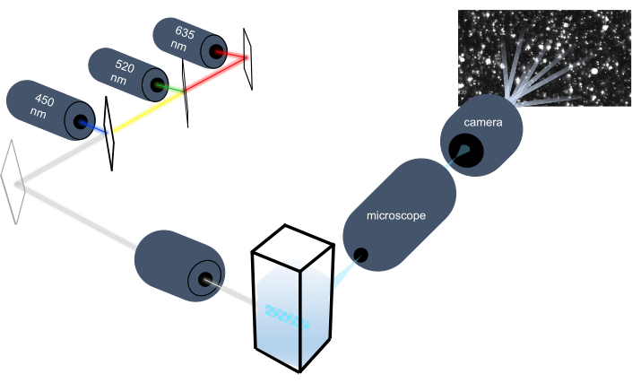
그림 1: 입자 추적 기기 광학 시스템. NTA 계측기는 450nm, 520nm, 635nm의 세 레이저를 사용하여 입자를 조명합니다. 개별 입자에서 흩어져 있는 빛의 비디오 녹화는 큐벳에서 90° 방향의 디지털 비디오 카메라에 의해 감지되고 추적됩니다. 이 그림의 더 큰 버전을 보려면 여기를 클릭하십시오.

Protocol

이러한 샘플의 모든 작업은 기관 동물 관리 및 사용 위원회 및 기관 검토 위원회 지침에 따라 수행되었습니다. NTA 메서드에 대한 회로도 개요는 도 2에묘사됩니다. 그림 2: 입자 추적 기기를 사용하는 NTA 메서드개요입니다. 샘플은 …

Representative Results

이 데모 전에, 계측기의 교정은 폴리스티렌 비드 표준의 크기 분포를 측정하여 획득 된 데이터의 유효성을 보장하기 위해 먼저 테스트되었습니다. 기본 기록 매개 변수및 이 프로토콜에 권장되는 처리 설정을 사용하여 100nm 및 400 nm 구슬의 크기 분포를 테스트했습니다(그림8). 100 nm 폴리스티렌 비드 표준의 경우 4.205 x 10⁷ 입자 /mL의 농도를 측정하…

Discussion

여기서, 우리는 다양한 입자 크기의 크기 분포를 동시에 측정하고 다분산 샘플에서 총 EV 농도를 측정하는 NTA의 NTA 프로토콜을 시연한다. 이 연구에서는 마우스 페리고나달 지방 조직과 인간 혈장이 EV의 공급원으로 사용되었습니다. 그러나, 혈청, 소변, 타액, 모유, 양수 및 세포 배양 과 같은 다른 조직 또는 생물학적 유체로부터 분리된 전기자동차도 NTA에 사용될 수 있다. 폴리스티렌 비드 표준의…

Divulgations

The authors have nothing to disclose.

Acknowledgements

이 작품은 국립 보건원 (ES030973-01A1, R01ES025225, R01DK066525, P30DK026687, P30DK063608)에 의해 지원되었습니다. 우리는 제프리 바디콤, 호리바 인스트루먼트 박사, 악기 를 보정하는 그의 도움을 위해 통합 인정합니다.

Materials

1X dPBS VWR 02-0119-1000 To dilute samples
100 nm bead standard Thermo Scientific 3100A To test ViewSizer 3000 calibration
400 nm bead standard Thermo Scientific 3400A To test ViewSizer 3000 calibration
Centrifugal Filter Unit Amicon UFC901024 To filter PBS diluent
Collection tubes, 2 mL Qiagen 19201 For isolation of human plasma extracellular vesicles
Compressed air duster DustOff DPSJB-12 To clean cuvettes
Cuvette insert HORIBA Scientific Provided with purchase of ViewSizer 3000
Cuvette jig HORIBA Scientific To align magnetic stir bar while placing inserts inside cuvette; Provided with purchase of ViewSizer 3000
De-ionized water VWR 02-0201-1000 To clean cuvettes
Desktop computer with monitor, keyboard, mouse, and all necessary cables Dell Provided with purchase of ViewSizer 3000
Ethanol (70-100%) Millipore Sigma To clean cuvettes
ExoQuick ULTRA System Biosciences EQULTRA-20A-1 For isolation of human plasma extracellular vesicles
Glass scintillation vials with lids Thermo Scientific B780020 To clean cuvettes
"Hook" tool Excelta Provided with purchase of ViewSizer 3000
Lint-free microfiber cloth Texwipe TX629 To clean cuvettes and cover work surface
Microcentrifuge tubes, 2 mL Eppendorf 22363344 For isolation of human plasma extracellular vesicles
Stir bar Sp Scienceware F37119-0005
Suprasil Quartz cuvette with cap Agilent Technologies AG1000-0544 Initially provided with purchase of ViewSizer 3000
ViewSizer 3000 HORIBA Scientific Nanoparticle tracking instrument

References

  1. Colombo, M., Raposo, G., Théry, C. Biogenesis, secretion, and intercellular interactions of exosomes and other extracellular vesicles. Annual Review of Cell and Developmental Biology. 30, 255-289 (2014).
  2. Hessvik, N. P., Llorente, A. Current knowledge on exosome biogenesis and release. Cellular and molecular life sciences: CMLS. 75, 193-208 (2018).
  3. Johnstone, R. M., Adam, M., Hammond, J. R., Orr, L., Turbide, C. Vesicle formation during reticulocyte maturation. Association of plasma membrane activities with released vesicles (exosomes). The Journal of Biological Chemistry. 262, 9412-9420 (1987).
  4. Théry, C., Ostrowski, M., Segura, E. Membrane vesicles as conveyors of immune responses. Nature Reviews Immunology. 9, 581-593 (2009).
  5. Yáñez-Mó, M., et al. Biological properties of extracellular vesicles and their physiological functions. Journal of Extracellular Vesicles. 4, 27066 (2015).
  6. Lo Cicero, A., Stahl, A., Raposo, G. Extracellular vesicles shuffling intercellular messages: for good or for bad. Current Opinion in Cell Biology. 35, 69-77 (2015).
  7. Raposo, G., Stoorvogel, W. Extracellular vesicles: exosomes, microvesicles, and friends. The Journal of Cell Biology. 200, 373-383 (2013).
  8. Mathivanan, S., Ji, H., Simpson, R. J. Exosomes: extracellular organelles important in intercellular communication. Journal of Proteomics. 73, 1907-1920 (2010).
  9. Zhang, M., et al. Methods and technologies for exosome isolation and characterization. Small Methods. 2, 1800021 (2018).
  10. Szatanek, R., et al. The methods of choice for extracellular vesicles (EVs) characterization. International Journal of Molecular Sciences. 18, (2017).
  11. Erdbrügger, U., Lannigan, J. Analytical challenges of extracellular vesicle detection: A comparison of different techniques. Cytometry. Part A: The Journal of the International Society for Analytical Cytology. 89, 123-134 (2016).
  12. Konoshenko, M. Y., Lekchnov, E. A., Vlassov, A. V., Laktionov, P. P. Isolation of Extracellular Vesicles: General Methodologies and Latest Trends. BioMed Research International. 2018, 1-27 (2018).
  13. Cvjetkovic, A., Lötvall, J., Lässer, C. The influence of rotor type and centrifugation time on the yield and purity of extracellular vesicles. Journal of Extracellular Vesicles. 3, (2014).
  14. Taylor, D. D., Shah, S. Methods of isolating extracellular vesicles impact down-stream analyses of their cargoes. Methods. 87, 3-10 (2015).
  15. Desdín-Micó, G., Mittelbrunn, M. Role of exosomes in the protection of cellular homeostasis. Cell Adhesion & Migration. 11, 127-134 (2017).
  16. Kanemoto, S., et al. Multivesicular body formation enhancement and exosome release during endoplasmic reticulum stress. Biochemical and Biophysical Research Communications. 480, 166-172 (2016).
  17. Benedikter, B. J., et al. Cigarette smoke extract induced exosome release is mediated by depletion of exofacial thiols and can be inhibited by thiol-antioxidants. Free Radical Biology & Medicine. 108, 334-344 (2017).
  18. Saeed-Zidane, M., et al. Cellular and exosome mediated molecular defense mechanism in bovine granulosa cells exposed to oxidative stress. PloS One. 12, 0187569 (2017).
  19. Wang, K., et al. Mechanical stress-dependent autophagy component release via extracellular nanovesicles in tumor cells. ACS Nano. 13, 4589-4602 (2019).
  20. King, H. W., Michael, M. Z., Gleadle, J. M. Hypoxic enhancement of exosome release by breast cancer cells. BMC Cancer. 12, 421 (2012).
  21. Bonzini, M., et al. Short-term particulate matter exposure induces extracellular vesicle release in overweight subjects. Environment Research. 155, 228-234 (2017).
  22. Neri, T., et al. Particulate matter induces prothrombotic microparticle shedding by human mononuclear and endothelial cells. Toxicology In Vitro. 32, 333-338 (2016).
  23. Flaherty, S. E., et al. A lipase-independent pathway of lipid release and immune modulation by adipocytes. Science. 363, 989-993 (2019).
  24. van Niel, G., D’Angelo, G., Raposo, G. Shedding light on the cell biology of extracellular vesicles. Nature Reviews. Molecular Cell Biology. 19, 213-228 (2018).
  25. Dragovic, R. A., et al. Sizing and phenotyping of cellular vesicles using Nanoparticle Tracking Analysis. Nanomedicine: Nanotechnology, Biology, and Medicine. 7, 780-788 (2011).
  26. Saveyn, H., et al. Accurate particle size distribution determination by nanoparticle tracking analysis based on 2-D Brownian dynamics simulation. Journal of Colloid and Interface Science. 352, 593-600 (2010).
  27. Vander Meeren, P., Kasinos, M., Saveyn, H. Relevance of two-dimensional Brownian motion dynamics in applying nanoparticle tracking analysis. Methods in Molecular Biology. , 525-534 (2012).
  28. Filipe, V., Hawe, A., Jiskoot, W. Critical evaluation of Nanoparticle Tracking Analysis (NTA) by NanoSight for the measurement of nanoparticles and protein aggregates. Pharmaceutical Research. 27, 796-810 (2010).
  29. Bachurski, D., et al. Extracellular vesicle measurements with nanoparticle tracking analysis – An accuracy and repeatability comparison between NanoSight NS300 and ZetaView. Journal of Extracellular Vesicles. 8, 1596016 (2019).
  30. Varga, Z., et al. Hollow organosilica beads as reference particles for optical detection of extracellular vesicles. Journal of Thrombosis and Haemostasis. 16, 1646-1655 (2018).
  31. Serrano-Pertierra, E., et al. Extracellular vesicles: Current analytical techniques for detection and quantification. Biomolecules. 10, (2020).
  32. Maguire, C. M., Rösslein, M., Wick, P., Prina-Mello, A. Characterisation of particles in solution – a perspective on light scattering and comparative technologies. Science and Technology of Advanced Materials. 19, 732-745 (2018).
  33. Bohren, C. F., Huffman, D. R. . Absorption and Scattering of Light by Small Particles. , (1983).
check_url/fr/62447?article_type=t

Play Video

Citer Cet Article
Comfort, N., Cai, K., Bloomquist, T. R., Strait, M. D., Ferrante Jr., A. W., Baccarelli, A. A. Nanoparticle Tracking Analysis for the Quantification and Size Determination of Extracellular Vesicles. J. Vis. Exp. (169), e62447, doi:10.3791/62447 (2021).

View Video