Open globe eye injuries may go untreated for multiple days in rural or military-relevant scenarios, resulting in blindness. Therapeutics are needed to minimize loss of vision. Here, we detail an organ culture open globe injury model. With this model, potential therapeutics for stabilizing these injuries can be properly evaluated.
Open globe injuries have poor visual outcomes, often resulting in permanent loss of vision. This is partly due to an extended delay between injury and medical intervention in rural environments and military medicine applications where ophthalmic care is not readily available. Untreated injuries are susceptible to infection after the eye has lost its watertight seal, as well as loss of tissue viability due to intraocular hypotension. Therapeutics to temporarily seal open globe injuries, if properly developed, may be able to restore intraocular pressure and prevent infection until proper ophthalmic care is possible. To facilitate product development, detailed here is the use of an anterior segment organ culture open globe injury platform for tracking therapeutic performance for at least 72 h post-injury. Porcine anterior segment tissue can be maintained in custom-designed organ culture dishes and held at physiological intraocular pressure. Puncture injuries can be created with a pneumatic-powered system capable of generating injury sizes up to 4.5 mm in diameter, similar to military-relevant injury sizes. Loss of intraocular pressure can be observed for 72 h post-injury confirming proper injury induction and loss of the eye's watertight seal. Therapeutic performance can be tracked by application to the eye after injury induction and then tracking intraocular pressure for multiple days. Further, the anterior segment injury model is applicable to widely used methods for functionally and biologically tracking anterior segment physiology, such as assessing transparency, ocular mechanics, corneal epithelium health, and tissue viability. Overall, the method described here is a necessary next step toward developing biomaterial therapeutics for temporarily sealing open globe injuries when ophthalmic care is not readily available.
Open globe (OG) injuries can result in permanent loss of vision when not treated or at least stabilized following injury1. Delays, however, are prevalent in remote areas where access to ophthalmic intervention is not readily available, such as in rural areas or on the battlefield in military scenarios. When treatment is not readily available, the current standard of care is to protect the eye with a rigid shield until medical intervention is possible. In military medicine, this delay is currently up to 24 h, but it is anticipated to increase up to 72 h in future combat operations in urban environments where air evacuation is not possible2,3,4. These delays can be even longer in rural, remote civilian applications where access to ophthalmic intervention is limited5,6. An untreated OG injury is highly susceptible to infection and loss of intraocular pressure (IOP) due to the watertight seal of the eye being compromised7,8. Loss of IOP can impact tissue viability, making any medical intervention unlikely to restore vision if the delay between injury and therapeutic is too long9.
To enable the development of easy-to-apply therapeutics for sealing OG injuries until an ophthalmic specialist can be reached, a benchtop OG injury model was previously developed10,11. With this model, high-speed injuries were created in whole porcine eyes while IOP was captured by pressure transducers. Therapeutics can then be applied to assess their ability to seal the OG injury site12. However, as this model uses whole porcine eyes, it can only assess immediate therapeutic performance with no way of tracking longer-term performance across the possible 72 h window in which the therapeutic must stabilize the injury site until the patient reaches specialty care. As a result, an anterior segment organ culture (ASOC) OG injury model was developed and detailed in this protocol as a platform for tracking long-term therapeutic performance13.
ASOC is a widely used technique for maintaining avascular tissue of the anterior segment, such as the cornea, for multiple weeks post-enucleation14,15,16,17. The anterior segment is maintained under physiological IOP by perfusing fluid at physiological flow rates and preserving the trabecular meshwork outflow region, the tissue responsible for regulating IOP, during ASOC setup18,19. The ASOC platform can maintain tissue physiologically, induce an OG injury using a pneumatic-powered device, apply a therapeutic, and track injury stabilization for at least 72 h post-injury13.
Here, the protocol provides a step-by-step methodology for using the ASOC platform. First it details how to set up and fabricate the ASOC platform. Next, the protocol details how to aseptically dissect the anterior segment and maintain the trabecular meshwork, followed by setting up anterior segment tissue in custom-built organ culture dishes. Then, it details how to create open globe injuries and apply therapeutic immediately following injury. Lastly, the protocol provides an overview on characterization parameters that are possible for use with this method that assesses functional, mechanical, and biological properties of the eye and how well the injury was stabilized. Overall, this model provides a much-needed platform to accelerate product development for stabilizing and treating open globe injuries and improve the poor vision prognosis following injury.
Before performing this protocol, be aware that there are legal and ethical requirements in place for the use of animals in research and training. If live animals are used for the source of ocular tissue, seek approval by the local ethical or legal authority (IACUC or Ethics committee, etc.) before beginning. If there is any question in obtaining approval for the use of animals, do not proceed. We previously determined and reported that fresh porcine eyes obtained and used within 24 h post-mortem compared closest to in vivo physiology and fared well for these studies (Animal Technologies, Tyler, TX, USA)10,13. No live animals were used throughout this protocol, using a tissue vendor to obtain tissue within 24 h.
NOTE: Prior to tissue arrival, fabricate the organ culture dishes (Supplementary Protocol 1), clamping rings (Supplementary Protocol 1), dish stands (Supplementary Protocol 1), pressure transducer data collection setup (Supplementary Protocol 2), and pneumatic puncture platform (Supplementary Protocol 3). Sterilize the dishes, tools, and supplies and prepare the work areas. It's useful to have a non-sterile area to perform gross dissection on the eyes, as they usually come with connective, extra orbital tissue attached. Execute these first steps on an open, clean work surface, and then transfer the eyes aseptically into a BSC II cabinet for micro-dissection (cabinet #1). Optimally, the BSC II cabinet utilized for micro-dissection is separated from the dish assembly BSC II cabinet (cabinet #2) to minimize airflow and maximize workspace. Set up the micro-dissection cabinet with a dissecting microscope and a way to visualize the work surface (camera or eyepieces protruding from the cabinet).
1. Sterilization steps, supplies (see Table of Materials for more details), and setup
2. Dissection of tissue
3. Setting up anterior segments in organ culture dishes
4. Starting anterior segment organ culture
5. Daily maintenance of ASOC
6. OG injury induction with pneumatic-powered puncture device
NOTE: Construction of the pneumatic puncture device is detailed in Supplementary Protocol 3. OG injuries are induced after IOP has stabilized, which normally occurs after 3 days in culture. Acceptable IOP values are 5-20 mmHg based on physiological IOP, which can be determined by evaluating the IOP data files or setting LED indicators in the pressure measurement system as described in Supplementary Protocol 2.
7. Removing ASOC from culture
NOTE: Depending on endpoint analysis (see Representative Results for possible endpoint methods), the AS needs to remain in the ASOC dish inflated while other methods require AS tissue isolated from the culture chamber. The below methodology describes how to take AS out of the organ culture dishes and to remove the rest of the setup.
8. IOP data analysis
Images captured via Optical Coherence Tomography (OCT) are shown for OG injured eyes to illustrate how a successful injury induction looks. Figure 3 shows images for control and OG injured AS tissue immediately after injury and 72 h later. Two views are shown: cross-sectional images through the injury site and top-down maximum intensity projection (MIPs) to visualize the surface area of the image. Control eyes show no noticeable disruption in the cornea, while clear injuries can be located that cross the entire cornea after OG injury. From MIPs, it is evident that injuries are irregular in shape and size, but the injury size does decrease over 72 h. Previously, this effect has shown to be significant for a number of injury sizes tested13.
The primary data output for the OG injury model described in this protocol is intraocular pressure over the course of the experimental setup. Data is recorded in units of millivolts as an output from each pressure transducer which can be converted into mmHg via calibration (Supplementary Protocol 4). Example IOP data vs the experimental time course is provided for eyes that are considered acceptable and others that would not be considered usable (Figure 4A). From the pressure trace data, eyes were attached to sensors after 24 h in culture, but IOP continues to fluctuate over the first 72 h in culture. Physiological IOP for AS tissue in organ culture is approximately 8-10 mmHg, so 2x and ½x range was decided upon as a gate for usable IOP values after values have stabilized (5-20 mmHg). Only eyes that were in that range would be allowable for use with the remainder of the protocol. From prior experiments, we had a 90% success rate that was achieved in ASOC setup for eyes stabilizing in the required range (Figure 4B).
The results for how IOP changes due to OG injury and therapeutic intervention are also provided (Figure 4C,D). After OG injury induction, pressure should significantly drop and remain that way until the tissue is removed from ASOC (Figure 4C). If an eye after injury induction does not decrease in pressure, this indicates that a successful injury was not induced as IOP should be reduced if the watertight seal of the eye is compromised. However, smaller injury sizes may self-heal, which could result in IOP being restored. If therapeutic is applied to the eye after OG injury induction, restoration of IOP can be tracked during ASOC. This concept is demonstrated with data showing a Dermabond adhesive applied to 2.4 mm OG injuries (Figure 4D). Average results for five separate ASOC experiments with and without therapeutic are shown and it is evident the therapeutic is increasing IOP. This method can measure the efficacy of the therapeutic for restoring IOP and track whether that pressure is restored across the key 72 h post-OG injury.
Further, the ASOC protocol is adaptable for use with a wide range of characterization endpoints to meet the end user's experimental requirements. During culture, outflow media leaving the eye can be collected on a daily or even hourly basis which can be utilized for tracking protein level changes occurring during ASOC, after OG injury induction, or after therapeutic is applied. For instance, gelatin zymography has been previously performed to detect matrix metalloproteinase levels to track wound healing and tissue remodeling20. Further biological endpoints are possible after removing tissue from culture via traditional immunohistochemistry methods for assessing tissue viability21,22, tracking pathophysiological changes to the cornea23,24, or antibody-based staining for any protein of interest25,26.
Functional corneal metrics can also be obtained from eyes maintained in ASOC. Corneal epithelium integrity can be assessed via a fluorescein eye stain and image acquisition using a blue light source27,28. After removal from culture, corneal tissue can be assessed for transparency through simple image acquisition13. Traditional ocular imaging can also be performed to assess tissue structure with or without therapeutic intervention. OCT images, as shown in Figure 3, can create cross sectional images through the cornea and can be captured non-invasively, potentially allowing image collection while maintaining tissue in culture. Other imaging modalities such as slit-lamp microscopy, ultrasound, or in vivo confocal microscopy can also be adapted for acquiring further anatomical information.
Lastly, assessment of mechanical properties of the anterior segment can be captured to understand the effect of the OG injury or subsequent therapeutic on the underlying tissue. While IOP data collection alone highlights how the integrity of the watertight seal of the eye has been compromised, we have previously shown that additional test metrics can be measured to tease out additional mechanical features10,11. Ocular compliance, a lumped mechanical property describing how intraocular pressure changes due to inflation (change in volume/change in pressure), can be measured with a syringe pump to inject sudden small volumes of fluid into the eye and recording the resulting pressure increase with a pressure transducer. Higher compliance indicates the tissue is less stiff and can be used to track how therapeutic material properties differ from the underlying corneal tissue. Leak rate from the eye or a traditional outflow facility can be measured and calculated to determine the precise fluidic flow rate leaving the eye per unit of pressure20,29. Lastly, with regards to therapeutic testing, burst pressure can be measured to determine the maximum pressure the eye can hold prior to the therapeutic failing. This can be used to compare performance to uninjured eyes or to track changes in performance with time12,13.
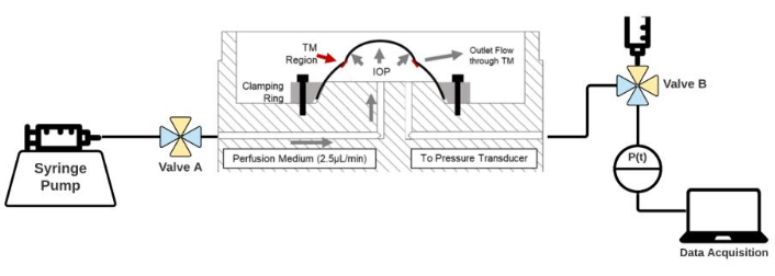
Figure 1: Diagram of the ASOC setup. Eyes are held in custom-built organ culture dishes and held in place with a clamping ring. ASOC media is infused via syringe pump through Valve A and connected to a pressure transducer, and subsequent data acquisition with Valve B. Open ports in each valve are highlighted in blue while yellow indicates closed channels. Please click here to view a larger version of this figure.
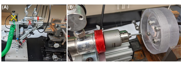
Figure 2: Overview of the OG injury setup. (A) Pneumatic powered injury device setup. From left to right, compressed air is introduced to the device via a compressed air line, which passes through a regulator to set pressure at 50 psi as measured by the pressure gauge. Two solenoid valves are connected to a linear actuator to direct expansion/retraction of the drill chuck holding the puncture object. Vise is positioned in front of the puncture device to hold the eye at the appropriate x, y, z positioning. (B) Representative ASOC is placed in front of the injury induction device. Further details of the device and its construction are detailed in Supplementary Protocol 3. Please click here to view a larger version of this figure.
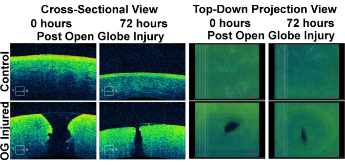
Figure 3: Optical Coherence Tomography Images of ASOC OG injury experiments. Images are shown for control eyes (uninjured) and OG injured eyes immediately post-injury and 72 h post-injury. Views are shown as cross-sections through the cornea (left side) and top-down maximum intensity projection views of the corneal surface (right side). The figure has been adapted with permission from Snider et al.13. Please click here to view a larger version of this figure.
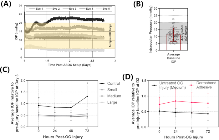
Figure 4: Representative IOP results for ASOC experiments. (A) Raw IOP data for the first 72 h of ASOC setup. Eyes are punctured at 72 h so the first 3 days of data are assessed to determine whether IOP stabilizes in the acceptable IOP range (5-20 mmHg). From the representative results, three of the five eyes fall within the acceptable IOP range, while one has IOP too high and one has IOP too low (falling outside of the highlighted yellow region on the plot). (B) Stabilized IOP for n = 50 ASOC setups from previous experiments to demonstrate the typical success rate with the ASOC method. (C) IOP for uninjured eyes compared to three different OG injury sizes after injury induction for 72 h. The loss of IOP is evident, with no signs of recovery. (D) Injured IOP results compared to injuries treated with a Dermabond adhesive. While the error rate is high due to some eyes being sealed and others not, the method can track changes to IOP over the 72 h period post-injury. The figure has been adapted with permission from Snider et al.13. Please click here to view a larger version of this figure.
Supplemental Files. Please click here to download these files.
There are critical steps with the ASOC OG injury platform that should be highlighted to improve the likelihood of success when using the methodology. First, during the anterior segment dissection, preserving the trabecular meshwork is essential but challenging to do correctly. If the TM is disrupted, the eye will not maintain physiological pressure and will not meet eligibility criteria for experimental use. It is recommended to practice the dissection process under normal conditions first rather than introducing the additional aseptic technique challenges until proper dissections are obtained. Second, when setting the eyes in the ASOC dishes, it is imperative that they are tight enough to prevent fluid from leaking but loose enough to prevent damaging the ASOC dishes. If the eye is not secured tightly, fluid will leak out from the eye through non-physiological means resulting in little or no IOP. However, the clamping ring holding the eye down is plastic and can be easily broken if overtightened. It is essential to clamp the eyes down over 2 days as the scleral tissue under the ring will compress and loosen the tissue during the first 24 h. It is recommended to tighten the rings just until resistance to tightening is felt on day 1 and follow this up by re-tightening to similar levels after 24 h in culture for best results.
Third, it is critical to fully understand where fluid flow is directed at all times when using this model. Each ASOC dish is connected to multiple three-way valves to direct fluid flow from the syringe pump or 10 mL syringe reservoir and connect to pressure transducers. Different instances of the setup process require valves to be positioned in such a way so as to flush air bubbles from the eye or to protect pressure transducers from over-pressurization. Care should still be taken to understand what is open/close at all times prior to critical protocol steps. Lastly, maintaining sterility throughout the ASOC OG injury protocol is critical but easy to lose across the multi-step, multi-day process. Perfusion media contains high levels of antibiotics and antimycotics to prevent this, and eyes are submerged in betadine prior to set up to prevent contaminations, but there are still critical steps where mistakes are most likely. During the initial setup in the dish, avoid contact with the eyes while tightening clamping rings in place and keep lids on the dishes at all times when not in use. A more likely exposure step is during the day-to-day ASOC maintenance. It is important to do these routine steps in a biosafety cabinet, even if it seems they can be quickly accomplished without removing the eyes from the incubator. Carefully following the protocol and maintaining good aseptic technique should minimize contamination risks across the 6-day ASOC experiments.
Overall, the ASOC OG injury platform is unique from other methodologies looking at open globe injuries due to two key criteria. First is the injury induction method. The high-speed pneumatic injury device utilized induces injuries with a high force amount. This allows for inducing injuries with objects that are not especially sharp nor with a small diameter. This more closely mimics injuries that are irregular in shape; high-speed shrapnel injures resulting from explosive devices30,31. The pneumatic device can easily be fitted with irregular-shaped shrapnel mimicking objects to create injuries more challenging to heal compared to previous methods using lasers, needles, or scalpel blades to create clean, precise injury geometries32,33,34. Second, the ASOC methodology allows for tracking injury progress and therapeutic performance beyond initial injury induction. Being able to track out to 72 h was not possible in the previously developed benchtop OG injury platform10,11,12 and was the motivation behind developing this protocol. In fact, cell viability remained high in the corneal endothelium for at least 1 week in ASOC13. ASOC is the only means this long-term characterization can be accomplished without transitioning into costly in vivo experiments.
The main applications for the ASOC platform are two-fold. First, the model can be utilized for further characterizing open globe injuries, especially considering how they change with time. In the previous study, OG injuries were characterized in this manner and wound healing was observed over 72 h following injury13. Further tracking different injury sizes, shapes, locations for 72 h or even longer with regards to biological changes occurring will inform critical medical decisions that have to be made following OG injuries. Certain injury parameters may allow for self-healing by the cornea, or other parameters may be more severe if the intervention is not applied within the first 24 h. This information will be invaluable for triaging patients when limited medical supplies or evacuation resources are available.
Second, the ASOC OG platform can be used for developing and testing product development. For this application, the organ culture platform can fill a number of roles. During initial product development, shorter time frames can be tested with a range of product formulations to determine what is most effective. The organ culture system can be configured for even greater high-throughput for this application with additional syringe pumps to move beyond the ten simultaneous experiments possible with the system detailed here. For more refined products, longer time points can be evaluated to assess performance for 72 h or potentially even longer. Lastly, wound healing evaluation may be possible when evaluating biologically active products that may permanently treat OG injuries rather than temporary stabilization.
However, there are limitations with the ASOC OG platform that should be taken into consideration. First, while the model allows for longer-term assessment of therapeutics, it is missing all tissue of the eye outside of the corneoscleral shell, such as the iris and lens. These additional tissues are likely to be influenced by the OG injury and may play a role in injury progression. Similarly, an isolated anterior segment is missing immune response elements that would be included when transitioning from the ASOC model to subsequent animal testing. Next, the model is only suitable for creating corneal OG injures and potentially limbal OG injuries. Scleral or posterior OG injuries cannot be induced with this method. However, many of these injury types result in damage to the retina, making any temporary stabilization therapeutic unlikely to prevent loss of vision35,36. Lastly, injuries with the model out to 72 h post-injury were only tracked. ASOC has been utilized in other applications out to 2 weeks, so the model can likely be utilized for these applications, but it has not been tested at this time37,38,39.
The authors have nothing to disclose.
This material is based upon work supported by the United States Department of Defense through an interagency agreement (#19-1006-IM) with the Temporary Corneal Repair acquisition program (United States Army Medical Materiel Development Agency).
10-32 Polycarbonate straight plug, male threaded pipe connector | McMaster-Carr | 51525K431 | |
10-32 Socket cap screw, ½" | McMaster-Carr | 92196A269 | |
10 mL syringe | BD | 302995 | |
20 mL syringe | BD | 302830 | |
Anti-Anti | Gibco | 15240-096 | |
Ball-End L key | McMaster-Carr | 5020A25 | |
Betadine | Fisher Scientific | NC1696484 | |
BD Intramedic PE 160 Tubing | Fisher Scientific | 14-170-12E | |
Cotton swabs | Puritan | 25-8061WC | |
DMEM media | ATCC | 30-2002 | |
FBS | ATCC | 30-2020 | |
Fine forceps | World Precision Instruments | 15914 | |
Gauze | Covidien | 8044 | |
Gentamicin | Gibco | 15710-064 | |
Glutamax | Gibco | 35050-061 | |
High temperature silicone O-ring, 2 mm wide, 4 mm ID | McMaster-Carr | 5233T47 | |
Large forceps | World Precision Instruments | 500365 | |
Large surgical scissors | World Precision Instruments | 503261 | |
Medium toothed forceps | World Precision Instruments | 501217 | |
Nail (puncture object) | McMaster-Carr | 97808A503 | |
Nylon syringe filters | Fisher | 09-719C | |
PBS | Gibco | 10010-023 | |
Petri dish (100 mm) | Fisher | FB0875713 | |
Polycarbonate, three-way, stopcock with male luer lock | Fisher | NC9593742 | |
Razor blade | Fisher | 12-640 | |
Stainless steel 18 G 90 degree angle dispensing needle | McMaster-Carr | 75165A81 | |
Stainless steel 18 G straight ½'’ dispensing needle | McMaster-Carr | 75165A675 | |
Sterile 100 mL beakers with lids | VWR | 15704-092 | |
Vannas scissors | World Precision Instruments | WP5070 |