We present a protocol for determining multicellular chirality in vitro, using the micropatterning technique. This assay allows for automatic quantification of the left-right biases of various types of cells and can be used for screening purposes.
Chirality is an intrinsic cellular property, which depicts the asymmetry in terms of polarization along the left-right axis of the cell. As this unique property attracts increasing attention due to its important roles in both development and disease, a standardized quantification method for characterizing cell chirality would advance research and potential applications. In this protocol, we describe a multicellular chirality characterization assay that utilizes micropatterned arrays of cells. Cellular micropatterns are fabricated on titanium/gold-coated glass slides via microcontact printing. After seeding on the geometrically defined (e.g., ring-shaped), protein-coated islands, cells directionally migrate and form a biased alignment toward either the clockwise or the counterclockwise direction, which can be automatically analyzed and quantified by a custom-written MATLAB program. Here we describe in detail the fabrication of micropatterned substrates, cell seeding, image collection, and data analysis and show representative results obtained using the NIH/3T3 cells. This protocol has previously been validated in multiple published studies and is an efficient and reliable tool for studying cell chirality in vitro.
Left-right (LR) asymmetry of the cell, also known as cellular handedness or chirality, describes the cell polarity in the LR axis and is recognized to be a fundamental, conserved, biophysical property1,2,3,4,5. Cell chirality has been observed both in vivo and in vitro at multiple scales. Previous findings revealed chiral swirling of actin cytoskeleton in single cells seeded on circular islands6, biased migration and alignment of cells within confined boundaries7,8,9,10,11, and asymmetrical looping of chicken heat tube12.
At the multicellular level, cell chirality can be determined from directional migration or alignment, cellular rotation, cytoskeletal dynamics, and cell organelle positioning7,8,9,10,11,12,13. We have established a micropatterning-based14 assay to efficiently characterize the chiral bias of adherent cells7,8,9,10. With the ring-shaped micropatterns geometrically confining cell clusters, the cells collectively exhibit directional migration and biased alignment. A MATLAB program was developed to automatically detect and measure cell alignment in phase-contrast images of the ring. The direction of local cell alignment is quantified with a biased angle, depending on its deviation from the circumferential direction. Following statistical analysis, the ring pattern of cells is designated either as counterclockwise (CCW) biases or clockwise (CW) biases.
This assay has been used to characterize the chirality of multiple cell phenotypes (Table 1), and the LR asymmetry of cells has been found to be phenotype-specific7,11,15. Moreover, disruption to actin dynamics and morphology can result in a reversal of chiral bias7,8, and oxidative stress can alter cell chirality as well9. Because of the simplicity of the procedure and the robustness of the approach7,8,9,10, this 2D chirality assay provides an efficient and reliable tool for determining and studying multicellular chirality in vitro.
The purpose of this protocol is to demonstrate the use of this method to characterize cell chirality. This protocol describes how to fabricate patterned cellular arrays via microcontact printing technique and conduct chirality analysis in an automated fashion using the MATLAB program.
1. Fabrication of polydimethylsiloxane (PDMS) stamps16
2. Coating of glass slides
3. Microcontact printing
4. Seeding cells onto micropatterned slides
5. Image collection
6. Cell chirality characterization (Figure 2)
Fifteen minutes after the seeding of NIH/3T3 cells, cell adhesion on the ring pattern was visually confirmed by phase-contrast imaging. After subsequent culture of 24 h, cells on the patterns became confluent and elongated with clearly asymmetrical alignments, biased towards the clockwise direction (Figure 2). Directional migration of attached cells is recorded by time-lapse imaging, cell motility and morphogenesis can be quantified with further analyses of the video. To conduct chirality analysis, high-resolution phase-contrast images are taken after fixation (Figure 2A–C) and fed into the MATLAB program. The program detects intensity gradients and calculates corresponding cell alignment directions on the ring. Then cell chirality is determined based on the circular statistics of cell alignment deviating from the circumferential direction of the rings. The cells can, therefore, be designated as clockwise (CW), counterclockwise (CCW), or non-chiral (NC) (Figure 2D). After processing, the analysis showed that the majority of rings have a dominant CW bias, indicating 3T3 cells have a strong CW chirality. Circular statistics generated also provide additional information of biased angles for further analyses if needed (Figure 2E).
Cell chirality depends on its phenotype. The ring chirality assay has been verified to be compatible with multiple cell-types7,8,9,10, including both cell lines and primary cells, such as fibroblasts, myoblast, endothelial cells, and stem cells (Table 1). Interestingly, because cell chirality is originated from the functionalities of actin cytoskeleton, alterations in actin dynamics may impact the chiral bias of cells. With mouse myoblast C2C12 cells, we found that by disrupting actin polymerization with 50 nM Latrunculin A treatment, the cells exhibited an alteration of CCW chiral bias into CW (Figure 3A). In addition, human umbilical vascular endothelial cells (hUVECs) treated with a small-molecule drug, 12-o-tetradecanoylphorbol-13-acetate (TPA), to activate the protein kinase c displayed a dose-dependent shift of cell chirality from CW to CCW (Figure 3B). These findings demonstrate the utility of the developed ring pattern chirality assay experimentally as well as the sensitivity of this assay to alterations in the cytoskeleton.
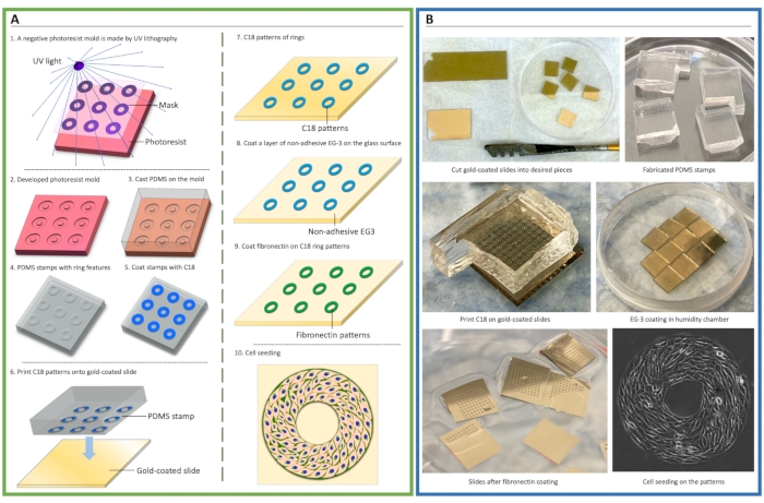
Figure 1. Schematic of cellular micropatterning. (A) Procedure of microfabrication and microcontact printing for cell patterning. A negative photoresist mold was made by ultraviolet (UV) crosslinking of photoresist via a mask containing micropatterning features (1-2). Polydimethylsiloxane (PDMS) elastomeric prepolymers were cast onto the mold to create stamps (3-4). Then, an adhesive self-assembly monolayer (SAM), octa-decanethiol (C18), was coated onto the stamp and transferred onto gold-coated glass slides via microcontact printing (5-7), followed by coating of non-adhesive ethylene glycol-terminated SAM, HS-(CH2)11-EG3 (EG3) (8), and fibronectin (9). Cells were then seeded to attach to the patterns (10). (B) Photos demonstrate the key steps of cell micropatterning. Please click here to view a larger version of this figure.
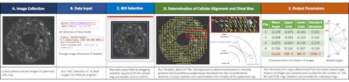
Figure 2. Workflow of imaging analyses. (A) Image collection. Acquire phase-contrast images of each ring. (B) Input data into the MATLAB program by running "ROI_Selection.m" file setting directory and image size. (C) Select regions of interest (ROIs) by dragging the selection square to fit the cellular ring and double click to confirm. (D) Determine cell alignment and chiral biases by running the "Analysis_Batch.m" file. (E) Example outputs with a summary of biased ring numbers and circular statistics for each ring. Please click here to view a larger version of this figure.
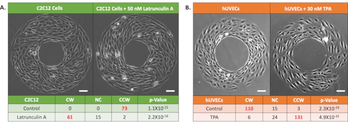
Figure 3. Representative results of the chirality of cells under drug treatment. (A) Phase-contrast images and chirality characterization results of mouse myoblast C2C12 cells: Control (left) and 50 nM Latrunculin A treated groups (right). Bold red font indicates dominant chirality at p < 0.05 by rank test. Scale bars: 100 µm. (B) Phase-contrast images and chirality characterization results of human umbilical vascular endothelial cells (hUVECs) on micro-patterned rings: Control (left) and 30 nM TPA treated groups (right). Bold red font indicates dominant chirality at p < 0.05 by rank test. Scale bars: 100 µm. Please click here to view a larger version of this figure.
CW Biased | CCW Biased |
· NIH/3T3 cells (ATCC CRL-1658) | · C2C12 (ATCC CRL-1772) |
· MC3T3-E1 cells (ATCC CRL-2593) | · Human skeletal muscle cells (Lonza CC-2661) |
· Rat cardiac fibroblasts | · A human skin cancer fibroblast line (ATCC CRL-7762) |
· Human primary skin fibroblasts (ATCC PCS-201-012) | · Madin-Darby canine kidney epithelial cells (ATCC CCL-34) |
· Human adipose-derived stem cells | |
· Human mesenchymal stem cells | |
· Human umbilical vascular endothelial cells (Lonza CC-2935) | |
· Human brain microvascular endothelial cells (Cell systems ACBRI-376) |
Table 1. Chiral biases of different cell types characterized by the micropatterning assay. CW: clockwise; CCW: counter-clockwise.
Supplementary File 1: MATLAB code files for chirality characterization. Please click here to download this File.
Supplemental Coding Files. Please click here to download this File.
The ring-shaped patterning assay described here provides an easy-to-use tool for quantitative characterization of multicellular chirality, capable of producing highly reliable and repeatable results. Rapid generation of identical defined microenvironments and unbiased analysis enables automated high-throughput processing of large size of samples. This protocol discusses the fabrication of the ring micropatterns, cell patterning, and automatic analysis of the biased cell alignment and directional motion. This method is compatible with live-imaging techniques to study cell motility and chiral morphogenesis and can be potentially integrated into other planforms for toxicity detection9 or drug screening applications.
There are a few additional tips to share. First, in the microcontact printing step, the stamp needs to be made on the gold side of the glass (it might be difficult to distinguish after the substrate is cut into pieces), and double print should be avoided. Second, for the cells that require other proteins for proper attachment, the preferred proteins can be used at step 3.17. Third, for cell culture, cell density should not be too high, and the cells should not be grown over-confluence on the patterns. Otherwise, cells might breach the inner boundary and fill the ring into a solid circle. Fourth, if cell patterns are broken or incomplete after seeding, it may be due to an incomplete transfer of pattern of C18 during contact printing (steps 3.7-3.8). The PDMS stamps should be examined for flaws. Applying slightly higher force when stamping onto slides can also be helpful. Finally, if cells attach to the entire surface of the glass slide without visible patterns, the force applied at step 3.8 may be too high, resulting in the entire surface being stamped with C18. It is also possible that the slides are defective or have been stored in PBS for too long. Patterns of self-assembly monolayers (i.e., C18 and EG3) should be used within a month after printing.
Although the proposed assay provides a convenient tool for robust chirality characterization, there are some limitations. First, the patterning assay is only compatible with adherent cells. The chirality of non-adherent cells, such as circulating tumor cells, adipocytes, or non-mammalian cells, could be possibly analyzed using the 3D Matrigel bilayer method18,19. Second, for cell types that have a natural cobble shape and do not show significant elongation, the analysis accuracy might be decreased. However, the chirality of these cell types can be characterized by conducting live imaging and tracking the migration bias7. Finally, the micropatterning assay is an endpoint analysis method, not intended for long-term cell culture. Experiments such as drug treatment should not exceed 72 h after seeding onto patterns. For applications that require prolonged treatment time, consider pretreating cells prior to seeding.
The authors have nothing to disclose.
This work was funded by the National Institutes of Health (OD/NICHD DP2HD083961 and NHBLI R01HL148104). Leo Q. Wan is a Pew Scholar in Biomedical Sciences (PEW 00026185), supported by the Pew Charitable Trusts. Haokang Zhang is supported by American Heart Association Predoctoral Fellowship (20PRE35210243).
200 proof ethanol | Koptec | DSP-MD-43 | |
BZX microscope system | Keyence | BZX-600 | |
Dulbecco's modified eagle medium (DMEM), high glucose | Gibco | 11965092 | |
Electron beam evaporator | Temscal | BJD-1800 | Gold-titanum film coating |
Fetal bovine serum | VWR | 89510-186 | |
Fibronectin from bovine plasma | Sigma | F1141-5MG | |
Glass microscope slides | VWR | 10024-048 | |
Glass tweezers | Exelta | 390BSAPI | |
Gold evaporation pellets | International Advanced Materials | AU18 | |
HS-(CH2)11-EG3-OH (EG3) | Prochimia | TH 001-m11.n3-0.2 | |
MATLAB | Mathworks | MATLAB_R2020b | |
NIH/3T3 cells | ATCC | CRL-1658 | |
OAI contact aligner | OAI | 200 | UV photolithography |
Octadecanethiol (C18) | Sigma | O1858-25ML | |
Orbital shaker | VWR | 89032-088 | |
Phosphate buffered saline (PBS) | Research product international | P32080-100T | |
Polydimethylsiloxane Sylgard 184 | Dow Corning | DC4019862 | |
Silicon Wafer | University Wafer | ID#809 | |
Sodium pyruvate | Thermo fisher scientific | 11360-070 | |
SU-8 3050 photoresist | MicroChem | Y311075 0500L1GL | |
Titanium evaporation pellets | International Advanced Materials | TI14 | |
Transparency mask (with feature) | Outputicity.com | N/A | Mask printing service |
Trypsin-EDTA (0.25%) | Thermo fisher scientific | 25200-072 |