Two recent technologies-tattoo and textiles-have demonstrated promising results in cutaneous sensing. Here, we present the fabrication and evaluation methods of tattoo and textile electrodes for cutaneous electrophysiological sensing. These electronic interfaces made of conductive polymers outperform the existing standards in terms of comfort and sensitivity.
Wearable electronic devices are becoming key players in monitoring the body signals predominantly altered during physical activity tracking. Considering the growing interest in telemedicine and personalized care driven by the rise of the Internet of Things era, wearable sensors have expanded their field of application into healthcare. To ensure the collection of clinically relevant data, these devices need to establish conformable interfaces with the human body to provide high-signal-quality recordings and long-term operation. To this end, this paper presents a method to easily fabricate conformable thin tattoo- and soft textile-based sensors for their application as wearable organic electronic devices in a broad spectrum of surface electrophysiological recordings.
The sensors are developed through a cost-effective and scalable process of cutaneous electrode patterning using poly(3,4-ethylenedioxythiophene)-poly(styrenesulfonate) (PEDOT:PSS), the most popular conductive polymer in bioelectronics, on off-the-shelf, wearable substrates. This paper presents key steps in electrode characterization through impedance spectroscopy to investigate their performance in signal transduction when coupled with the skin. Comparative studies are required to position the performance of novel sensors with respect to the clinical gold standard. To validate the fabricated sensors’ performance, this protocol shows how to perform various biosignal recordings from different configurations through a user-friendly and portable electronic setup in a laboratory environment. This methods paper will allow multiple experimental initiatives to advance the current state of the art in wearable sensors for human body health monitoring.
Noninvasive biopotential recording is performed through skin-contact electrodes, providing a vast amount of data on the physiological status of the human body in fitness and healthcare1. Novel types of wearable biomonitoring devices have been developed from the latest technological advances in electronics through the downscaling of integrated controlling and communicating components to portable dimensions. Smart monitoring devices pervade the market daily, offering multiple monitoring capabilities with the ultimate goal of providing sufficient physiological content to enable medical diagnostics2. Therefore, safe, reliable, and robust interfaces with the human body present critical challenges in the development of legitimate wearable technologies for healthcare. Tattoo and textile electrodes have recently appeared as reliable and stable interfaces perceived as innovative, comfortable devices for wearable biosensing3,4,5.
Tattoo sensors are dry and thin interfaces that, owing to their low thickness (~1 µm), ensure adhesive-free, conformable skin contact. They are based on a commercially available tattoo paper kit composed of a layered structure, which allows the release of an ultrathin polymeric layer on the skin6. The layered structure also allows for easy handling of the thin polymeric layer during the sensor's fabrication process and its transfer to the skin. The final electrode is fully conformable and almost imperceptible to the wearer. Textile sensors are electronic devices obtained from fabric functionalization with electroactive materials7. They are mainly integrated or simply sewed into clothes to ensure the user's comfort due to their softness, breathability, and evident affinity with garments. For almost a decade, textile and tattoo electrodes have been assessed in surface electrophysiological recordings3,8,9, showing good results both in wearability and signal quality recordings and reporting high signal-to-noise ratio (SNR) in short- and long-term evaluations. They are also conceived as a potential platform for wearable biochemical sweat analysis1,10.
The growing interest in tattoo, textile, and, in general, flexible thin film technologies (e.g., those made of plastic foils such as parylene or different elastomers) is mainly promoted by the compatibility with low-cost and scalable fabrication methods. Screen printing, inkjet printing, direct patterning, dip coating, and stamp transfer have been successfully adopted to produce such kinds of electronic interfaces11. Among these, inkjet printing is the most advanced digital and fast prototyping technique. It is mainly applied to the patterning of conductive inks in a non-contact, additive fashion under ambient conditions and on a large variety of substrates12. Although multiple wearable sensors have been fabricated through noble metal ink patterning13, metal films are brittle and undergo cracking when mechanically stressed. Different research groups have adopted different strategies to endow metals with the property of mechanical compatibility with skin. These strategies include reducing the film thickness and using serpentine designs or wrinkled and prestretched substrates14,15,16. Soft and intrinsically flexible conductive materials, such as conductive polymers, found their application in flexible bioelectronic devices. Their polymeric flexibility is combined with electric and ionic conductivity. PEDOT:PSS is the most used conductive polymer in bioelectronics. It is characterized by softness, biocompatibility, sustainability, and printing processability17, which make it compatible with the widespread production of biomedical devices.
Devices, such as planar electrodes connected to an acquisition system, allow the recording of biopotentials in health monitoring. Human body biopotentials are electrical signals generated by electrogenic cells that propagate through the body up to the skin surface. According to where the electrodes are placed, it is possible to acquire data related to the electrical activity of the brain (EEG), muscles (EMG), heart (ECG), and skin conductivity (e.g., bioimpedance or electrodermal activity, EDA). The quality of the data is then assessed to evaluate the usability of the electrodes in clinical applications. A high SNR defines their performance18, which is typically compared with state-of-the-art Ag/AgCl electrode recordings. Although the Ag/AgCl electrodes also have high SNR, they lack long-term operationality and conformable wearability. High-quality biosignal recordings provide insights into human health status related to a particular organ's function. Thus, these benefits of comfortable tattoo or textile interfaces indicate their promise for long-term applications that can enable real-life mobile health monitoring and pave the way for the development of telemedicine19.
This paper reports how to fabricate and assess tattoo and textile electrodes in health biomonitoring. After its fabrication, a novel electrode must be characterized. Typically, electrochemical impedance spectroscopy (EIS) is adopted to study the electrical performance of the electrode with respect to a target interface (e.g., skin) in terms of the transfer function. EIS is used to compare the impedance characteristics of multiple electrodes and perform tests under different conditions (e.g., varying the electrode design or studying long-term responses). This paper shows the recording of surface biosignals through an easy setup and reports a user-friendly method to record different types of biosignals applicable to any novel fabricated electrode that needs to be validated for cutaneous biopotential recordings.
NOTE: Experiments involving human subjects did not involve the collection of identifiable private information related to the individual's health status and are only used here for technological demonstration. Data were averaged over three different subjects. The electrophysiological recordings were extracted from previously published data6,21.
1. Inkjet-printed PEDOT:PSS electrode fabrication
NOTE: The following protocol has been used to fabricate electrodes for electrophysiology on commercial, flexible substrates-tattoo paper6 and textile21. The same approach has been largely adopted to make electrodes on flexible substrates such as thin plastic foils22. In all cases, an inkjet printer was used for the patterning of PEDOT:PSS (see the Table of Materials).
2. Electrode characterization using electrochemical impedance spectroscopy
3. Surface electrophysiological recordings
NOTE: The following section describes the electrode placement for each biosignal of interest. Once the electrodes are correctly placed and well attached to the skin, they can be connected to the portable acquisition system to start the recordings. The video content of this article shows an example of electrophysiological monitoring using commercially available Ag/AgCl electrodes and a portable electronic unit.
This paper shows the fabrication of comfortable skin-contact electrodes by inkjet printing and a method to characterize them and perform electrophysiology recordings. We reported the fabrication steps of PEDOT:PSS inkjet printing directly on different substrates, such as fabric (Figure 1A), PEN (Figure 1B), and tattoo paper (Figure 1C,D) for reference. The proposed designs in protocol step 1.2.1. and step 1.3.1.5. define a circular sensing area of 1 cm2 to compare electrodes with the state-of-the-art Ag/AgCl mainly adopted in clinics.
To characterize the electrodes' performance, their impedances were measured through the three-electrode EIS setup (Figure 2A,B). This method allows the study of skin-electrode impedance when performing on-body measurements with electrodes placed on the arm. As an example, the representative impedance of textile electrodes is reported in Figure 2C, where the impedance modulus is reported in the Bode plot. Textile electrodes exhibit slightly higher but comparable impedances than Ag/AgCl electrodes, the gold standard in electrophysiology. The shape of the impedance modulus (Figure 2C) indicates a slightly higher resistive behavior in the case of the textile electrodes, whereas the standard Ag/AgCl shows typical resistive-capacitive behavior24. All three types of electrodes, tattoo, textile, and thin-foils, have been studied via EIS, enabling the characterization of their interface with the skin25.
By placing the electrodes on the skin in different body areas, as shown in Figure 3, we have access to multiple biosignals (e.g., EEG, ECG, EMG, and EDA). Biosignal recordings can be easily obtained by connecting the electrodes to appropriate portable or lab-scale instrumentation. Figure 3A displays the EEG tracing-the electrical activity recording of populations of active neurons. One of the basic groups of brain waves is the alpha waves (8-13 Hz). The alpha waves reflect the state of the brain under relaxation and can be induced by asking the subject to close their eyes26. The grey vertical dashed line (Figure 3A) marks the moment in the recording when the volunteer was asked to open their eyes. In the ECG tracing in Figure 3B, the polarization and depolarization of the atria and ventricles of the heart are represented by the characteristic pattern consisting of the P wave, the QRS complex, and a T wave27. In Figure 3B, the QRS complex is identifiable, and the R peaks show the highest amplitude and are used to calculate the heart rate by considering the time between two consecutive ones.
Figure 3C shows the EMG tracing while the volunteer progressively increased the force of their arm muscles. The intensified muscle activity is quantified by the increased amplitude of the voltage peaks. In an EMG tracing, spikes with amplitude from a few microvolts to a few millivolts, in the frequency range of 10-1,000 Hz, reflect the muscle fiber activity driven by the motor unit action potentials. Figure 3D shows the EDA tracing typically composed of tonic and phasic components. The tonic component reflects the skin conductance level and corresponds to the background signal. The phasic component reflects the response of the subject to a specific stimulus and is detectable by a change in the skin conductance value28. This tracing is used to evaluate human stress levels and body hydration.
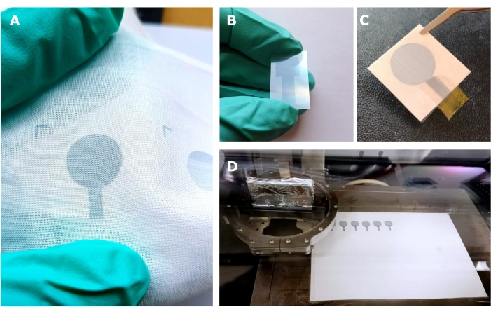
Figure 1: PEDOT:PSS inkjet-printed electrodes. Electrodes printed on (A) 100% cotton fabric, (B) PET foil, and (C) temporary tattoo paper. (D) Photograph of the inkjet printer while printing multiple PEDOT:PSS electrodes on tattoo paper substrate. Abbreviations: PET = polyethylene terephthalate; PEDOT:PSS = poly(3,4-ethylenedioxythiophene)-poly(styrenesulfonate). Please click here to view a larger version of this figure.
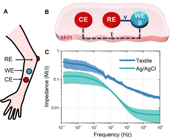
Figure 2: EIS measurements. (A) Schematic of the electrode configuration for on-body EIS measurement; the working electrode is placed 3 cm apart from the counter Ag/AgCl electrode; the reference Ag/AgCl is placed on the elbow of the volunteer. (B) Scheme of the three-electrode setup for EIS measurements on the skin. A current is applied between the counter and working electrodes, and the voltage is measured between the reference and the sense electrodes. (C) Impedance modulus of Ag/AgCl and PEDOT:PSS-ionic liquid gel textile electrodes (blue and green curves, respectively). Impedance was measured with a three-electrode setup on the arm. This figure has been modified from Bihar et al.21. Abbreviations: EIS = electrochemical impedance spectroscopy; CE = counter electrode; WE = working electrode; RE = reference electrode; S = sense electrode; PEDOT: PSS = poly(3,4-ethylenedioxythiophene)-poly(styrenesulfonate). Please click here to view a larger version of this figure.
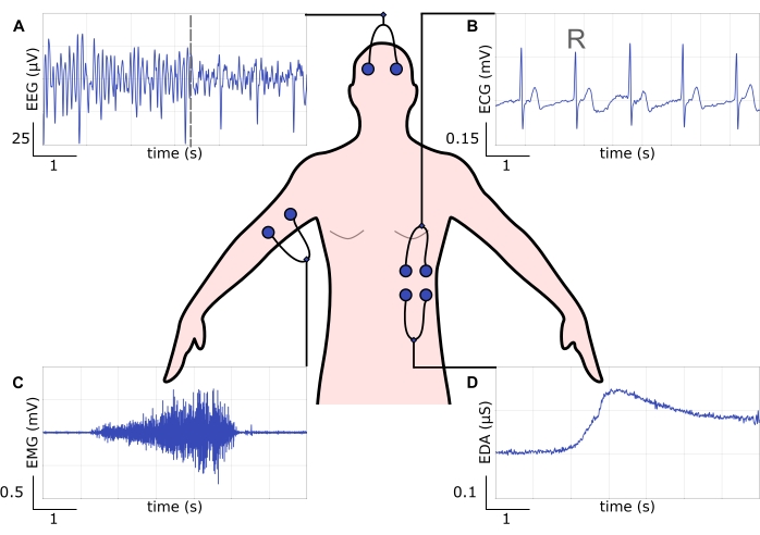
Figure 3: Electrode body positioning schematic with the respective electrophysiological recording tracings. (A) EEG tracing. The dashed vertical line indicates the transition from a state with alpha waves to a state without, which coincides with when the volunteer was asked to open their eyes. (B) ECG tracing. The upper spikes represent the R peaks that belong to the QRS complex. (C) EMG tracing.The muscle activity is represented by a voltage signal whose amplitude increases with the increasing activity of the muscle evoked by the volunteer. (D) EDA tracing. During the first 2 s, the signal represents the tonic component, while its following amplitude increase indicates the phasic component, which mirrors the volunteer's response to a stimulus. All the recordings were performed with Ag/AgCl electrodes on a healthy volunteer. Abbreviations: EEG = electroencephalography; ECG = electrocardiography; EMG = electromyography; EDA = electrodermal activity. Please click here to view a larger version of this figure.
Supplemental Figure S1: Tattoo paper layered structure scheme. A backing paper sheet supports the releasable nanofilm made with a polyurethane and other polymers mixture. Two water-soluble polyvinylalcohol (PVA) layers cover both sides of the film. Please click here to download this File.
This paper describes an easy and scalable process to fabricate wearable electrodes and demonstrates a method for recording electrophysiological biosignals. It uses three examples of wearable substrates, such as tattoo, textile, and thin films. It introduces how to build a sensor on these substrates and characterize its performance prior to its application. To make the electrodes here, we employed PEDOT:PSS, a conductive polymer that stands out from metal-based conductors due to its cost-effectiveness, versatile processability, biocompatibility, softness, and sustainability for its compatibility with green processing29. PEDOT:PSS patterning on off-the-shelf substrates was achieved via an inkjet printing technique that allows precise control of the ink deposition with design freedom (Figure 1).
Inkjet printing is a non-contact technique that allows the selective functionalization of flexible and unconventional substrates that are chemically and physically incompatible with traditional photolithography microfabrication processes. Compared to screen printing, another technique often used for electrode fabrication, inkjet does not require masks, thus resulting in lower ink waste and simple customization30. The inkjet technology controls the thickness rigorously by multiple layer deposition (inkjet: <1 µm vs. screen: >a few µm). Indeed, when printing on tattoo paper (Figure 1D), one PEDOT:PSS-printed layer (thickness of 240 nm ± 30 nm) is sufficient to get a homogeneous conductive film (Figure 1C), with a sub-micrometer thickness that naturally self-adheres to the skin following its rugosity31. However, when printing on fabrics, the ink drops over the 3D porous structures created by knitted or waved yarns (Figure 1A). Multiple layers are necessary to get electrical connection among the coated fibers, functionalizing the textile matter in a controlled and customized fashion32.
When printing on new and atypical substrates, it is critical to find the optimal number of printed layers, considering the tradeoff between the performance and speed of the fabrication process. For textile electrode fabrication, attention must be paid to keeping the substrate flat during the printing (see protocol section 1.3.). Therefore, the printing strategy should consider optimizing the printing layout in multilayer deposition and a possibility for alignment in the deposition of consecutive materials.
However, it is important to point out some limitations of these electrodes and their fabrication. Textile electrodes might need additional printing steps of a gel electrolyte. It has been demonstrated that it plays a key role in decreasing the skin-electrode contact impedance, thus providing high-quality biosignal recordings33 Moreover, the washability of textile wearable sensors is a critical aspect when envisaging full integration into clothes. The physicochemical properties of the textile substrate and the conductive polymer ink affect the final device's compliance with washing cycles. Therefore, one should exhaustively investigate the aforementioned aspect to fully assess their long-term performance.
In tattoo sensor fabrication, a delicate step is to find the best electrical interconnection between the tattoo sensor and the acquisition system (see protocol section 1.3.). Indeed, tattoo technology has gained interest due to the thin film format that makes tattoo electrodes imperceptible. Therefore, their manipulation requires particular care when mechanical stress is applied, particularly to the interconnection part. It is also important to remember the transfer mechanism of tattoos onto the skin that requires wetting the supporting paper with water. Although this method is straightforward, any abrupt contact between water and the already-transferred tattoo sensor will delaminate the latter. While the conformability of ultrathin tattoos is a key advantage for wearable technology, the vulnerability to water and rubbing mechanical stresses narrow the tattoo sensor operation period to a couple of days.
When a new type of electrode is introduced, EIS helps provide the primary assessment of the electrode's performance compared with the benchmark (the Ag/AgCl electrodes) before moving forward with an application. Protocol section 2 described the EIS measurements of the fabricated electrodes when directly placed onto the human body to get insights into how they are electrically coupled with the skin. The three-electrode configuration (Figure 2A,B) evaluates the signal transfer capability through the skin-electrode interface. The novel electrode to be investigated is the one connected to the WE and S of the EIS. The other two electrodes are used as the CE and RE. EIS is performed in a potentiostatic mode, where a small (0.1 V) sinusoidal current (0.1-100 Hz) is applied between the CE and WE, while the potential variation is measured across the RE-S couple. The impedance is then computed at each frequency. The measured impedance consists of two contributions: the skin impedance and the skin-electrode contact impedance.
The capacitive and resistive behaviors of an electrode are defined from the EIS plots (Figure 2C). By developing equivalent circuits to fit the experimental data, it is possible to understand how an electrode transduces biosignals and what kind of interface it establishes with the skin34. While tattoo electrodes are dry and adherent to the skin, their impedances differ slightly from the standard gelled Ag/AgCl electrodes. The presence of a gel interface between the skin and the electrode promotes signal transduction and lowers contact impedance.
Mechanical strength is another key characteristic of wearables. Textile PEDOT:PSS electrodes have been demonstrated to withstand stretching stress33. Combined with printed ionic liquid gels, they offer stable electrical contact with the skin and mechanical robustness in wearable conditions. The stretchability, softness, and structural porosity, which endow the capability to pass perspiration due to contact with the human body, drive this type of electrode to be the most suitable technology for wearable electronics. Once again, the interconnection with electronic systems remains delicate. Therefore, these systems can be directly deposited into the fabric.
The ultimate validation of cutaneous sensors can only be performed on subjects. Cutaneous sensors are conditioned by the skin variability between subjects and various dynamic factors and environmental conditions, which directly affect their performance. Here, we have demonstrated how to obtain meaningful EEG, ECG, EMG, and EDA tracings through a fully portable platform. Electrode placement plays an important role in getting reliable and accurate information during monitoring. The analysis of the recordings shown in Figure 3 can confirm the electrode's capability in electrophysiological recordings and obtain valuable body monitoring outcomes. The recording capability varies from extremely weak neural activity (Figure 3A) to high power muscle contractions (Figure 3C).
In Figure 3B and Figure 3D, the cardiac activity and the electrodermal responses demonstrate the resolution and sensitivity of the fabricated electrodes. Biosignal recording provides useful data about the user's body health, performance under specific conditions, and response to specific internal or external stimuli, expanding their application to a variety of biomedical studies. Multiple portable electronics front-ends exist to acquire biosignals such as ECG, EMG, EEG, and EDA. Examples are the portable electrophysiology amplifier chips RHD2216 from Intan Technologies, the Shimmer wearable, the DueLite device from OT Bioelettronica, the PLUX wireless device in the advanced version (named Biosignal PLUX), or the DIT version (named BITalino).
To conclude, multiple sensors can be fabricated with the presented protocols for a variety of health-monitoring applications. For instance, tattoo-based PEDOT:PSS multielectrode arrays (MEAs) have been successfully employed for facial EMG as they do not impair natural facial movements and allow biosignal recording free from alteration25,35. However, thin and stretchable electrodes have been fabricated by inkjet printing PEDOT:PSS on low-cost, stretchable pantyhose substrate, obtaining high-quality ECG recordings, both under resting and movement conditions, with minimal discomfort for the user33. With this protocol, we obtained soft, conformable, and comfortable skin sensors through the patterning of conductive ink on off-the-shelf substrates. Inkjet printing is a low-cost and scalable technique that stands out from traditional microelectronic fabrication processes. The proposed method describes how to acquire electrophysiological signals, which vary from weak neural activity to high power muscle contractions. These signals allow insights into the user's body's physiological status to be obtained. Overall, we present initial steps on the feasibility of seamless wearable electronic devices for a variety of biomedical applications, which extend from fitness to healthcare monitoring.
The authors have nothing to disclose.
This work was supported by the French National Research Agency through the ANR JCJC OrgTex project (ANR-17-CE19-0010). It has also received funding from the European Union's Horizon 2020 research and innovation program under the Marie Sklodowska-Curie grant agreement no. 813863. E.I. wishes to thank the CMP cleanroom staff at the Centre Microelectronics in Provence for their technical support during the development of the project.
Biosignalplux – Plux wireless device for electrophysiological recordings | PLUX Wireless Biosignals S.A | EEG, ECG, EMG, EDA sensors | |
Covidien Kendal Disposable electrodes, medical grade disposable electrodes (Pregelled, 24 mm) | Covidien / Kendal (formally Tyco) ARBO electrodes | H124SG | Commercial Ag/AgCl electrodes for electrophysiology |
Dimatix inkjet printer | Fujifilm | DMP 2800 | Inkjet printer |
Laser Cutter | Universal Laser Systems | VLS 3.50, 50 W | Laser cutter to cut the glue sheet for tattoo electrodes fabrication |
NOVA | Metrohm Autolab | NOVA 2.1 | Electrochemistry software to control Autolab instruments |
OpenSignals | 2020 PLUX wireless biosignals, S.A. | Software suite for real-time biosignals visualisation, capable of direct interaction with PLUX devices | |
PEDOT:PSS inkjet printable ink | Heraeus Deutschland GmbH & Co. KG | CLEVIOS Pjet 700 | |
Polyethylene naphthalene (PEN) foil | Goodfellow | thickness 1.3 μm | Used for tattoo electrodes interconnection fabrication |
Polyimide tape | 3M | Kapton tape by 3 M, thickness 50 μm | Used for tattoo electrodes interconnection fabrication |
Potentiostat | Metrohm Autolab | Autolab potentiostat B.V. | Used for EIS measurements |
Silhouette temporary tattoo paper kit | Silhouette Americ, Inc, US | Substrate for tattoo-based electrodes | |
Wowen textile 100% cotton and commercially available pantyhose | Substrate for textile-based electrodes |