Summary

כלים לטיפול בפני השטח של מיקרואלקטרודות תוך-קורטיקליות תוך-קורטיקליות מסיליקון

Published: June 08, 2022
doi:

Summary

הפרוטוקול הנוכחי מתאר כלים לטיפול במיקרו-אלקטרוניקה תוך-קורטיקלית מישורית של סיליקון במהלך טיפולים לשינוי פני השטח באמצעות תצהיר גז ותגובות תמיסה מימיות. ההרכבה של הרכיבים המשמשים לטיפול במכשירים לאורך ההליך מוסברת בפירוט.

Abstract

מיקרואלקטרודות תוך-קורטיקליות מחזיקות בפוטנציאל טיפולי רב. אבל הם מאותגרים עם ירידה משמעותית בביצועים לאחר משכי השתלה צנועים. תרומה משמעותית לירידה הנצפית היא הפגיעה ברקמה העצבית הפרוקסימלית של השתל והתגובה הנוירו-דלקתית שלאחר מכן. המאמצים לשפר את אורך החיים של המכשיר כוללים שינויים כימיים או יישומי ציפוי על פני השטח של המכשיר כדי לשפר את תגובת הרקמה. פיתוח של טיפולי פני שטח כאלה נעשה בדרך כלל באמצעות בדיקות “דמה” לא פונקציונליות שחסרות את הרכיבים החשמליים הנדרשים ליישום המיועד. תרגום להתקנים פונקציונליים דורש התייחסות נוספת בהתחשב בשבריריות של מערכי מיקרו-אלקטרוניקה תוך-קורטיקליים. כלי הטיפול מקלים מאוד על טיפולי פני השטח במכשירים מורכבים, במיוחד עבור שינויים הדורשים זמנים פרוצדורליים ארוכים. כלי הטיפול המתוארים כאן משמשים לטיפולי פני השטח המיושמים באמצעות תצהיר פאזה גזית וחשיפה לתמיסה מימית. אפיון הציפוי מתבצע באמצעות אליפסומטריה וספקטרוסקופיית פוטו-אלקטרונית של קרני רנטגן. השוואה של הקלטות ספקטרוסקופיה של עכבה חשמלית לפני ואחרי הליך הציפוי במכשירים פונקציונליים אישרה את שלמות המכשיר לאחר השינוי. ניתן להתאים בקלות את הכלים המתוארים להתקני אלקטרודות חלופיים ולשיטות טיפול השומרות על תאימות כימית.

Introduction

מכשירים נוירופרוסטטיים שואפים לשחזר יכולות חושיות ומוטוריות לקויות או נעדרות במגוון רחב של אוכלוסיות מטופלים, כולל אלה עם פגיעה בחוט השדרה, טרשת אמיוטרופית צידית (ALS), שיתוק מוחין וקטיעות 1,2,3. מיקרו-אלקטרודות תוך-קורטיקליות (IMEs) יכולות ליצור מסלול תקשורת בין נוירונים בקליפת המוח לבין המכשירים המשמשים לשליטה בנוירו-פרוסטטיקה. יתרון מובהק של מיקרו-אלקטרוניקה תוך-קורטיקלית הוא יכולתם להקליט אותות עצביים ברזולוציה המרחבית והזמנית הגבוהה, המועדפת על עיבוד אותות ובקרה הבאים של ממשקי מוח-מחשב 4,5. למרבה הצער, הביצועים של מיקרו-אלקטרוניקה תוך-קורטיקלית פוחתים באופן דרמטי תוך חודשים עד שנה לאחר ההשתלה 2,6,7,8. אובדן איכות האות ויציבותו משפיע לרעה על יישום הטכנולוגיה.

תרומה משמעותית לירידה שנצפתה בביצועים היא התגובה הביוטית לנזק לרקמות הקשורות להשתלה ולדלקת עצבית כרונית 9,10,11. השתלה של IMEs גורמת נזק לרקמת המוח, וכתוצאה מכך לשחרור מולקולות איתות היוזמות מפלים של תהליכי הגנה תאיים ריאקציוניים. התממשקות כרונית מחריפה את תגובת הגוף הזר, ומובילה לדלקת עצבית מתמשכת הפוגעת ברקמות הפרוקסימליות למכשיר; מוכרים לעתים קרובות כתסמינים של דלקת עצבית, הצטלקות וניוון עצבי מקומי התורמים לירידה ברישום איכות האות 12,13,14,15. הצלקת, המורכבת מקונגלומרט צפוף של אסטרוציטים עם מיקרוגליה מופעלת ומקרופאגים, יוצרת סביבה מקומית שלילית עם הובלת חומרים מופחתת והצטברות מקומית של גורמים דלקתיים 16,15,16,16,17,18.

מחקרים רבים תיארו את תגובת המוח למיקרו-אלקטרוניקה תוך-קורטיקלית או לגישות למיתון התגובה7. מחקר ופיתוח לשיפור תגובת הרקמה כללו מגוון אסטרטגיות, כולל שינויים במבנה הכולל, בטופולוגיית פני השטח, בחומרים וביישום הציפויים. מאמצים אלה מתכוונים למזער את הנזק שנגרם מאירוע ההשתלה, להציג ממשק נוח יותר בין המכשיר לתאים הפרוקסימליים, או להפחית את עומס הרקמה לאחר השתלת המכשירים7. שיטות המכוונות באופן ספציפי לתגובה הביולוגית הכרונית הובילו למספר ציפויים ביו-אקטיביים שמטרתם לייצב את אתר ההשתלה ולקדם כימית את בריאות התא. דוגמאות לכך כוללות פולימרים מוליכים כגון פולי(אתילן דיאוקסיתיופין) (PEDOT)19,20, ננו-צינוריות פחמן21, הידרוג’לים22, והוספת מולקולות ותרופות ביו-אקטיביות כדי להתמקד בתהליכים תאיים ספציפיים 23,24,25. קבוצת המחקר שלנו, בפרט, בחנה מנגנונים רבים לקידום הפחתה של התגובה הדלקתית למיקרו-אלקטרוניקה מושתלת, כולל, אך לא רק, מזעור הטראומה הקשורה להשתלת המכשיר26, מזעור חוסר ההתאמה של נוקשות המכשיר/רקמה 27,28,29,30,30,31,32,33, אופטימיזציה של העיקור נהלים34,35, הפחתת עקה/נזק חמצוני 28,36,37,38,39,40,41,42, חקירת חומרי אלקטרודה חלופיים43 וחיקוי הננו-ארכיטקטורה של המטריצה החוץ-תאית הטבעית 44,45,46 . העניין האחרון הוא פיתוח של ציפויי משטח ביומימטיים כדי למתן את התגובה הנוירו-דלקתית בממשק רקמת המיקרו-אלקטרוד ישירות39.

שינוי הממשק מציע את היתרון הייחודי של מיקוד ישיר של הפצע והרקמה הפרוקסימלית הדרושה להקלטת אותות. טיפול על פני השטח המקדם ריפוי מבלי להחמיר את התגובה החיסונית יכול להועיל לכל החיים של רישום איכותי ולהסיר מגבלות במימוש הפוטנציאל הטיפולי והמחקרי של מיקרו-אלקטרוניקה תוך-קורטיקלית. העבודה המוצגת מפרטת שיטות ליישום טיפולי פני שטח על מערכי מיקרו-אלקטרוניקה הדורשים זמני תגובה ארוכים יותר תוך התאמה לשבריריות המכשירים. הטכניקה המוצגת נועדה לשתף שיטות לשינוי פני השטח עם מכשירים פונקציונליים שבהם לא ניתן לטפל במכשיר לאורך כל יישום הטיפול. הכלים מוצגים לטיפול בבדיקות דמה לא פונקציונליות ובמערכי מיקרו-אלקטרוניקה מישוריים פונקציונליים מסיליקון.

הגישה המוצגת לשינוי משטח האלקטרודה מאפשרת השעיה מאובטחת של בדיקות דמה לא פונקציונליות או מערכי אלקטרודות מישוריות סיליקון פונקציונליות לתצהיר פאזה גזית ותגובה עם תמיסות מימיות. מספר חלקים מודפסים בתלת-ממד משמשים לטיפול בהתקנים השבריריים האלה (איור 1 ואיור 2). דוגמה לכך ניתנת הליך המשתמש הן בשלבי פאזה של גז והן בשלבי תמיסה לשינוי פני השטח עם ציפוי נוגד חמצון הכולל אימוביליזציה של פורפירין Mn(III)tetrakis (4-חומצה בנזואית) (MnTBAP). MnTBAP הוא מטאלופורפירין סינתטי בעל תכונות נוגדות חמצון עם תיווך מוכח של דלקת47,48. הדוגמה שסופקה על מערכי אלקטרודות מישוריות סיליקון פונקציונליות מאמתת עדכון לפרוטוקול שדווח בעבר עבור התקנים לא פונקציונליים40. ההתאמה של טכניקת תצהיר פאזת גז מ- Munief et al. תומכת בתאימות הפרוטוקול לאלקטרודות פונקציונליות49. תצהיר פאזת הגז מנוצל לאמין כדי לתפקד את פני השטח כהכנה לתגובה המימית הכרוכה בכימיה של קרוסלינקר קרבודימיד כדי לשתק את ה-MnTBAP הפעיל. מתודולוגיית הטיפול שפותחה כאן ניתנת כפלטפורמה הניתנת לשינוי כך שתתאים לציפויים אחרים והתקנים דומים.

הפרוטוקול ממחיש את הגישה באמצעות בדיקות דמה לא פונקציונליות הכוללות שוק סיליקון ולשונית מודפסת בתלת-ממד עם ממדים דומים למערכי האלקטרודות המישוריות הפונקציונליות של הסיליקון. אריזת המחבר של המכשיר נחשבת מקבילה לכרטיסייה המודפסת בתלת-ממד של גשושית הדמה הלא פונקציונלית בהוראה שסופקה.

Figure 1
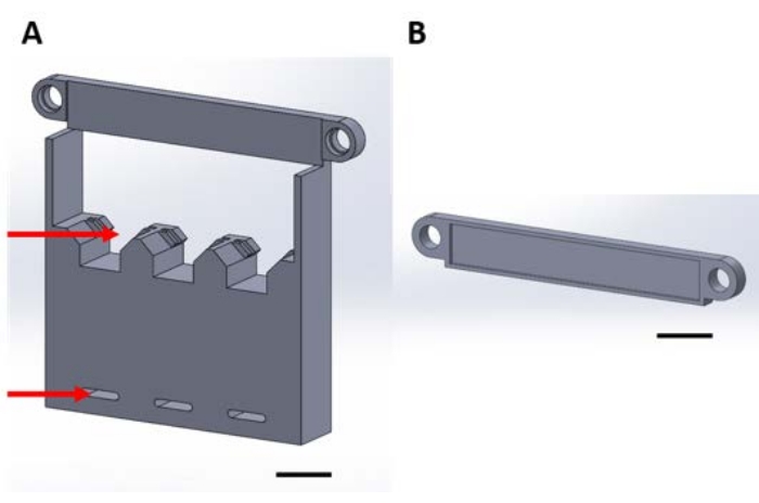
איור 1: חלקים מודפסים בתלת-ממד לטיפול בהתקנים פונקציונליים במהלך תצהיר פאזת הגז במעש ואקום. (A) בסיס המבנה כולל מחזיקים עבור ריבועי סיליקון לדוגמה בגודל 1 ס”מ על 1 ס”מ (חץ עליון) וחורים לאבטחת לוח ייבוש (חץ תחתון). (ב) הלוחית משמשת לאבטחת ההשעיה של המכשירים. מכאן והלאה, כל חתיכה באיור זה תיקרא או חתיכה 1A או 1B. סרגל קנה מידה = 1 ס”מ. אנא לחץ כאן כדי להציג גרסה גדולה יותר של נתון זה.

Figure 2
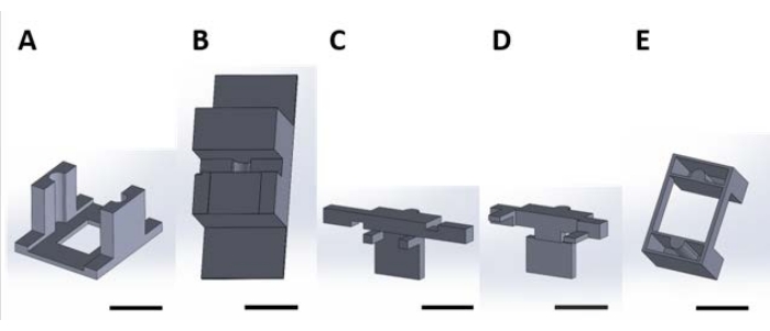
איור 2: חלקים מודפסים בתלת-ממד לטיפול בהתקנים פונקציונליים לתגובת פני השטח המתרחשת בתמיסה המימית. (B) חלקי ספסל המשמשים לייצוב חלקים (C) ו-(D) בעת ההרכבה. (C) ו-(D) מאבטחים יחד את ההשעיה של המכשירים למיקום בלוח הבאר, ו-(E) מאבטחים עוד יותר חלקים (C) ו-(D) למכסה לוחית הבאר. מכאן והלאה, חלקים בודדים בכל לוח של איור זה ייקראו מספרי חתיכות המתאימים למספר הלוח של נתון זה. סרגל קנה מידה = 1 ס”מ. אנא לחץ כאן כדי להציג גרסה גדולה יותר של נתון זה.

Protocol

כל קבצי הקידוד להדפסה בתלת-ממד מסופקים בקבצי קידוד משלימים 1-16. הניתוח המופיע בתוצאות הייצוגיות מתואר באמצעות מערכי אלקטרודות סיליקון מישוריות פונקציונליות שנרכשו באופן מסחרי (ראו טבלת חומרים). 1. טיפול במכלול לתצהיר פאזה של גז במייבש ואקום <p class…

Representative Results

כדי להדגים את השימוש ברכיבי הטיפול, יושמה המתודולוגיה המתוארת כדי להתאים את השתק של מתווך מחמצן לסיליקון מופעל. היישום של כימיה זו על IMEs להפחתת עקה חמצונית פותח על ידי Potter-Baker et al. והודגם על בדיקות דמה סיליקון לא פונקציונליות40. טיפול זה בפני השטח משתק את נוגד החמצון, MnTBAP, למשטח ס…

Discussion

הפרוטוקול המתואר תוכנן לטיפול בפני השטח של מערכי מיקרו-אלקטרוניקה מישוריים מסיליקון. הכלים המודפסים בתלת-ממד מותאמים אישית למערכי מיקרו-אלקטרוניקה בסגנון מישיגן עם מחברים בפרופיל נמוך50. בדיקות לא פונקציונליות הורכבו על ידי הדבקת גשושית סיליקון לכרטיסיות מודפסות בתלת-ממד ב…

Divulgations

The authors have nothing to disclose.

Acknowledgements

מחקר זה נתמך בחלקו על ידי פרס Merit Review IRX002611 (Capadona) ופרס מדען קריירה מחקרית IK6RX003077 (Capadona) משירות המחקר והפיתוח של המחלקה לענייני חיילים משוחררים של ארצות הברית (ארה”ב). בנוסף, עבודה זו נתמכה בחלקה גם על ידי המכון הלאומי לבריאות, המכון הלאומי להפרעות נוירולוגיות ושבץ מוחי R01NS110823 (Capadona /Pancrazio), ותוכנית עמיתי המחקר לתארים מתקדמים של הקרן הלאומית למדע (קרבס).

Materials

1-[3-(Dimethylamino)propyl]-3-ethylcarbodiimide methiodide (EDC) Sigma-Aldrich 165344-1G Solid, stored desiccated at -20 °C
15 mL Conical Centrifuge Tubes Fisher Scientific 14-959-70C
18 Pound Solid Nylon Cable/Zip Ties Cole-Parmer EW-06830-66 Length 4 inches
2-(N-Morpholino)ethanesulfonic acid (MES) Sigma-Aldrich 4432-31-9 Solid
3-aminopropyltriethoxysilane (APTES) Sigma-Aldrich 440140-100ML Liquid, container with Sure/Seal
50 mL Conical Centrifuge Tubes Fisher Scientific 14-959-49A
Aluminum foil Fisher Scientific 01-213-103
Aluminum weighing dishes Fisher Scientific 08-732-102 Diameter 66 mm
Bel-Art Vacuum Desiccator Fisher Scientific 08-594-15B
Corning Costar TC-Treated Multiple Well Plates Millipore Sigma CLS3527-100EA 24-well plate, polystyrene
Cyanoacrylate Adhesive LocTite N/A
Digital Microscope Keyence VHX-S750E
Disco DAD3350 Dicing Saw Disco DAD3350 Used to cut silicon wafer into 1 cm x 1 cm samples
Double-Sided Polyimide Tape Kapton Tape PPTDE-1/4 ¼” x 36 yds.
EP21LVMed – low viscosity, two component epoxy compound Masterbond EP21LVMed Meets USP Class VI certification, Passes ISO 10993-5 for cytotoxicity
Epilog Fusion Pro 48 Laser Machine Epilog N/A CO2 laser
Foam tape XFasten N/A 1/8" Thick
Gamry Interface 1010E Potentiostat Gamry 992-00129
High precision 45° curved tapered very fine point tweezers/forceps Fisher Scientific 12-000-131
Lab tape Fisher Scientific 15-901-10L
Mn(III)tetrakis (4-benzoic acid) porphyrin (MnTBAP) EMD Millipore 475870-25MG Solid, stored at -20 °C
N-Hydroxysulfosuccinimide sodium salt, ≥98% (HPLC) Sigma-Aldrich 56485-250MG Solid, stored desiccated at 4°C
Platinum clad niobium mesh anode Technic N/A Clad with 125μ” of platinum on one side, framed in titanium with (1) 1” x 6” titanium strap centered on one 6” dimension
Silicon Planar Microelectrode Array, 16 Channel NeuroNexus A1x16-3mm-100-177-CM16LP Electrode site material is iridium, shank thickness is 15 μm
Silicon Wafer University Wafer 1575 Diameter 100 mm, p-type, boron-doped, 100 oriented, resistivity 0.01-0.02 Ohm-cm, thickness 525 um, single side polished, prime grade
Silver/silver Chloride reference electrode Gamry Instruments 930-00015
Solidworks N/A
Stainless Steel Phillips Flat Head Screws McMaster Carr 96877A629 #8-32, 1 1/2", fully threaded
Type I deionized water ChemWorld CW-DI1-20
Ultimaker 3 3D printer Ultimaker  N/A
Ultimaker Cura Ultimaker N/A 3D printing software
Ultimaker NFC ABS Filament Dynamism, Inc. 1621 2.85 mm
Ultimaker NFC PLA Filament Dynamism, Inc. 1609 2.85 mm
Vacuum Gauge Vacuum Gauge Measureman Direct N/A Glycerin Filled, 2-1/2” Dial Size, ¼”NPT, -30” Hg/-100kpa-0
Wing nuts Everbilt 934917 #8-32, zinc plated

References

  1. Donoghue, J. Bridging the brain to the world: A perspective on neural interface systems. Neuron. 60 (3), 511-521 (2008).
  2. Ajiboye, A. B., et al. Restoration of reaching and grasping movements through brain-controlled muscle stimulation in a person with tetraplegia: a proof-of-concept demonstration. The Lancet. 398 (10081), 1821-1830 (2017).
  3. Ereifej, E. S., et al. Neural engineering: the process, applications, and its role in the future of medicine. Journal of Neural Engineering. 16 (6), 063002 (2019).
  4. Nicolas-Alonso, L. F., Gomez-Gil, J. Brain computer interfaces, a review. Sensors (Basel). 12 (2), 1211-1279 (2012).
  5. Leuthardt, E. C., Schalk, G., Moran, D., Ojemann, J. G. The emerging world of motor neuroprosthetics: a neurosurgical perspective. Neurosurgery. 59 (1), 1-14 (2006).
  6. Barrese, J. C., et al. Failure mode analysis of silicon-based intracortical microelectrode arrays in non-human primates. Journal of Neural Engineering. 10 (6), 066014 (2013).
  7. Jorfi, M., Skousen, J. L., Weder, C., Capadona, J. R. Progress towards biocompatible intracortical microelectrodes for neural interfacing applications. Journal of Neural Engineering. 12 (1), 011001 (2015).
  8. Prasad, A., et al. Comprehensive characterization and failure modes of tungsten microwire arrays in chronic neural implants. Journal of Neural Engineering. 9 (5), 056015 (2012).
  9. Hermann, J. K., Capadona, J. R. Understanding the role of innate immunity in the response to intracortical microelectrodes. Critical Reviews in Biomedical Engineering. 46 (4), 341-367 (2018).
  10. Ravikumar, M., et al. The roles of blood-derived macrophages and resident microglia in the neuroinflammatory response to implanted intracortical microelectrodes. Biomaterials. 35 (28), 8049-8064 (2014).
  11. Sawyer, A. J., et al. The effect of inflammatory cell-derived MCP-1 loss on neuronal survival during chronic neuroinflammation. Biomaterials. 35 (25), 6698-6706 (2014).
  12. Prasad, A., Sanchez, J. C. Quantifying long-term microelectrode array functionality using chronic in vivo impedance testing. Journal of Neural Engineering. 9 (2), 026028 (2012).
  13. Salatino, J. W., Ludwig, K. A., Kozai, T. D. Y., Purcell, E. K. Glial responses to implanted electrodes in the brain. Nature Biomedical Engineering. 1 (11), 862-877 (2017).
  14. McConnell, G. C., et al. Implanted neural electrodes cause chronic, local inflammation that is correlated with local neurodegeneration. Journal of Neural Engineering. 6 (5), 056003 (2009).
  15. Rennaker, R. L., Miller, J., Tang, H., Wilson, D. A. Minocycline increases quality and longevity of chronic neural recordings. Journal of Neural Engineering. 4 (2), 1-5 (2007).
  16. Carnicer-Lombarte, A., Chen, S. T., Malliaras, G. G., Barone, D. G. Foreign body reaction to implanted biomaterials and its impact in nerve neuroprosthetics. Frontiers in Bioengineering and Biotechnology. 9, 622524 (2021).
  17. Roitbak, T., Sykova, E. Diffusion barriers evoked in the rat cortex by reactive astrogliosis. Glia. 28 (1), 40-48 (1999).
  18. Polikov, V. S., Tresco, P. A., Reichert, W. M. Response of brain tissue to chronically implanted neural electrodes. Journal of Neuroscience Methods. 148 (1), 1-18 (2005).
  19. Cui, X., Martin, D. C. Electrochemical deposition and characterization of poly(3,4-ethylenedioxythiophene) on neural microelectrode arrays. Sensors and Actuators B: Chemical. 89 (1), 92-102 (2003).
  20. Ludwig, K. A., Uram, J. D., Yang, J., Martin, D. C., Kipke, D. R. Chronic neural recordings using silicon microelectrode arrays electrochemically deposited with a poly(3,4-ethylenedioxythiophene) (PEDOT) film. Journal of Neural Engineering. 3 (1), 59-70 (2006).
  21. Keefer, E. W., Botterman, B. R., Romero, M. I., Rossi, A. F., Gross, G. W. Carbon nanotube coating improves neuronal recordings. Nature Nanotechnology. 3 (7), 434-439 (2008).
  22. Kim, D. -. H., Wiler, J. A., Anderson, D. J., Kipke, D. R., Martin, D. C. Conducting polymers on hydrogel-coated neural electrode provide sensitive neural recordings in auditory cortex. Acta Biomaterialia. 6 (1), 57-62 (2010).
  23. He, W., McConnell, G. C., Bellamkonda, R. V. Nanoscale laminin coating modulates cortical scarring response around implanted silicon microelectrode arrays. Journal of Neural Engineering. 3 (4), 316-326 (2006).
  24. Azemi, E., Lagenaur, C. F., Cui, X. T. The surface immobilization of the neural adhesion molecule L1 on neural probes and its effect on neuronal density and gliosis at the probe/tissue interface. Biomaterials. 32 (3), 681-692 (2011).
  25. Zhong, Y., Bellamkonda, R. V. Controlled release of anti-inflammatory agent alpha-MSH from neural implants. Journal of Controlled Release. 106 (3), 309-318 (2005).
  26. Shoffstall, A. J., et al. Potential for thermal damage to the blood-brain barrier during craniotomy: implications for intracortical recording microelectrodes. Journal of Neural Engineering. 15 (3), 034001 (2018).
  27. Bedell, H. W., et al. Understanding the effects of both CD14-meditated innate immunity and device/tissue mechanical mismatch in the neuroinflammatory response to intracortical microelectrodes. Frontiers in Neuroscience. 12, 772 (2018).
  28. Nguyen, J. K., et al. Influence of resveratrol release on the tissue response to mechanically adaptive cortical implants. Acta Biomaterialia. 29, 81-93 (2016).
  29. Sridharan, A., Nguyen, J. K., Capadona, J. R., Muthuswamy, J. Compliant intracortical implants reduce strains and strain rates in brain tissue in vivo. Journal of Neural Engineering. 12 (3), 036002 (2015).
  30. Nguyen, J. K., et al. Mechanically-compliant intracortical implants reduce the neuroinflammatory response. Journal of Neural Engineering. 11, 056014 (2014).
  31. Harris, J. P., et al. In vivo deployment of mechanically adaptive nanocomposites for intracortical microelectrodes. Journal of Neural Engineering. 8 (4), 046010 (2011).
  32. Shoffstall, A. J., et al. Characterization of the neuroinflammatory response to Thiol-ene/Acrylate shape memory polymer coated intracortical microelectrodes. Micromachines. 10, 486 (2018).
  33. Simon, D. M., et al. Design and demonstration of an intracortical probe technology with tunable modulus. Journal of Biomedical Materials Research. Part A. 105 (1), 159-168 (2017).
  34. Ravikumar, M., et al. The effect of residual endotoxin contamination on the neuroinflammatory response to sterilized intracortical microelectrodes. Journal of Materials Chemistry. B. 2 (17), 2517-2529 (2014).
  35. Ecker, M., et al. Sterilization of thiol-ene/acrylate based shape memory polymers for biomedical applications. Macromolecular Materials and Engineering. 302 (2), 160331 (2017).
  36. Ereifej, E. S., et al. Implantation of neural probes in the brain elicits oxidative stress. Frontiers in Bioengineering and Biotechnology. 6 (9), 1-12 (2018).
  37. Potter, K. A., et al. The effect of resveratrol on neurodegeneration and blood brain barrier stability surrounding intracortical microelectrodes. Biomaterials. 34 (29), 7001-7015 (2013).
  38. Potter, K. A., et al. Curcumin-releasing mechanically adaptive intracortical implants improve the proximal neuronal density and blood-brain barrier stability. Acta Biomaterialia. 10 (5), 2209-2222 (2014).
  39. Potter-Baker, K. A., Capadona, J. R. Reducing the "stress": Antioxidative therapeutic and material approaches may prevent intracortical microelectrode failure. ACS Macro Letters. 4 (3), 275-279 (2015).
  40. Potter-Baker, K. A., et al. Development of superoxide dismutase mimetic surfaces to reduce accumulation of reactive oxygen species for neural interfacing applications. Journal of Materials Chemistry B. 2 (16), 2248-2258 (2014).
  41. Potter-Baker, K. A., et al. Implications of chronic daily antioxidant administration on the inflammatory response to intracortical microelectrodes. Journal of Neural Engineering. 12 (4), 046002 (2015).
  42. Kim, Y., et al. Ventricular delivery of resveratrol improves microelectrode recording performance and reduces oxidative stress. Micromachines. 12, 1446 (2021).
  43. Deku, F., et al. Amorphous silicon carbide ultramicroelectrode arrays for neural stimulation and recording. Journal of Neural Engineering. 15 (1), 016007 (2018).
  44. Ereifej, E. S., et al. The neuroinflammatory response to nanopatterning parallel grooves into the surface structure of intracortical microelectrodes. Advanced Functional Materials. 28 (12), 1704420 (2018).
  45. Kim, Y., et al. Nano-architectural approaches for improved intracortical interface technologies. Frontiers in Neuroscience. 12, 456 (2018).
  46. Mahajan, S., et al. Towards standardization of electrophysiology and computational tissue strain in rodent intracortical microelectrode models. Frontiers in Bioengineering and Biotechnology. 8, 416 (2020).
  47. Suresh, M. V., et al. The protective role of MnTBAP in oxidant-mediated injury and inflammation in a rat model of lung contusion. Surgery. 154 (5), 980-990 (2013).
  48. Liu, D., Shan, Y., Valluru, L., Bao, F. Mn (III) tetrakis (4-benzoic acid) porphyrin scavenges reactive species, reduces oxidative stress, and improves functional recovery after experimental spinal cord injury in rats: comparison with methylprednisolone. BMC Neuroscience. 14 (1), 23 (2013).
  49. Munief, W. M., et al. Silane deposition via gas-phase evaporation and high-resolution surface characterization of the ultrathin siloxane coatings. Langmuir. 34 (35), 10217-10229 (2018).
  50. Hoogerwerf, A. C., Wise, K. D. A three-dimensional microelectrode array for chronic neural recording. IEEE Transactions on Biomedical Engineering. 41 (12), 1136-1146 (1994).
  51. Staros, J. V., Wright, R. W., Swingle, D. M. Enhancement by N-hydroxysulfosuccinimide of water-soluble carbodiimide-mediated coupling reactions. Analalytical Biochemistry. 156 (1), 220-222 (1986).
  52. Yuan, X., Wolf, N., Mayer, D., Offenhausser, A., Wordenweber, R. Vapor-phase deposition and electronic characterization of 3-Aminopropyltriethoxysilane self-assembled monolayers on silicon dioxide. Langmuir. 35 (25), 8183-8190 (2019).
  53. Montgomery, D. C. . Design and Analysis of Experiments. Eighth edition. , (2013).
  54. Shoffstall, A. J., Capadona, J. R. Bio-inspired materials and systems for neural interfacing. Current Opinions in Biomedical Engineering. 6, 110-119 (2018).
  55. Skousen, J. L., Tresco, P. A. . Neuroprosthetics. Theory and Practice 2nd Edition. , 259-299 (2017).
  56. Michelson, N. J., et al. multi-modal analysis uncovers complex relationship at the brain tissue-implant neural interface: new emphasis on the biological interface. Journal of Neural Engineering. 15 (3), 033001 (2018).
  57. Hofmann, U. G., Capadona, J. R. Editorial: Bridging the gap in neuroelectronic interfaces. Frontiers in Neuroscience. 14, 457 (2020).
  58. Usoro, J., Sturgill, B., Musselman, K., Capadona, J. R., Pancrazio, J. J. On the definition of ‘chronic’ for intracortical microelectrode array applications. Micromachines. 12 (8), 972 (2021).
  59. Thompson, C. H., Saxena, A., Heelan, N., Salatino, J., Purcell, E. K. Spatiotemporal patterns of gene expression around implanted silicon electrode arrays. Journal of Neural Engineering. 18 (4), 1741 (2021).
  60. Golabchi, A., Woeppel, K. M., Li, X., Lagenaur, C. F., Cui, X. T. Neuroadhesive protein coating improves the chronic performance of neuroelectronics in mouse brain. Biosensors and Bioelectronics. 155, 112096 (2020).
  61. Zheng, X. S., et al. A superoxide scavenging coating for improving tissue response to neural implants. Acta Biomaterialia. 99, 72-83 (2019).
  62. Lee, H. C., et al. Foreign body response to intracortical microelectrodes is not altered with dip-coating of Polyethylene Glycol (PEG). Frontiers in Neuroscience. 11, 513 (2017).
  63. Boehler, C., et al. Actively controlled release of Dexamethasone from neural microelectrodes in a chronic in vivo study. Biomaterials. 129, 176-187 (2017).
  64. Hess, A. E., et al. Development of a stimuli-responsive polymer nanocomposite toward biologically optimized, MEMS-based neural probes. Journal of Micromechanics and Microengineering. 21 (5), 054009 (2011).
check_url/fr/63500?article_type=t

Play Video

Citer Cet Article
Krebs, O. K., Mittal, G., Ramani, S., Zhang, J., Shoffstall, A. J., Cogan, S. F., Pancrazio, J. J., Capadona, J. R. Tools for Surface Treatment of Silicon Planar Intracortical Microelectrodes. J. Vis. Exp. (184), e63500, doi:10.3791/63500 (2022).

View Video