生きているゼブラフィッシュ幼虫の眼球除去による神経支配依存性の成長と視覚系の発達を調べる
Summary
この記事では、網膜入力が視蓋の成長と発達にどのように影響するかを調査するための第一歩として、生きているゼブラフィッシュの幼虫から外科的に目を取り除く方法について説明します。さらに、この記事では、幼虫の麻酔、固定、および脳解剖に関する情報を提供し、続いて免疫組織化学および共焦点イメージングを行う。
Abstract
ゼブラフィッシュは生涯にわたる顕著な成長と再生能力を示します。例えば、胚発生中に確立された特殊な幹細胞ニッチは、眼および脳の両方において、視覚系全体の継続的な成長をサポートする。網膜と視蓋の間の協調的な成長は、新しいニューロンが目と脳に追加されるにつれて、正確な網膜トピックマッピングを保証します。網膜軸索が、生存、増殖、および/または分化などの眼底幹および前駆細胞の挙動を調節するための重要な情報を提供するかどうかに対処するには、同じ動物内および動物間で神経支配および脱神経化されたテクタルローブを比較できることが必要である。
生きている幼虫ゼブラフィッシュから片目を外科的に除去し、続いて視蓋骨を観察することで、この目標を達成します。付属のビデオは、幼虫を麻酔し、タングステン針を電気的に鋭くし、それらを使用して片目を除去する方法を示しています。次に、固定されたゼブラフィッシュ幼虫から脳を解剖する方法を示します。最後に、このビデオでは、免疫組織化学のプロトコルの概要と、顕微鏡検査のために染色された胚を低融点アガロースにマウントする方法のデモンストレーションを提供します。
Introduction
この方法の目的は、網膜入力がゼブラフィッシュ脳の視覚処理センターである視蓋骨の成長と発達にどのように影響するかを調べることです。片目を取り除いた後、視蓋の両側を比較することで、同一試料内のテクタル変化を観察・正規化することができ、複数の検体間での比較が可能となります。この技術と組み合わせた現代の分子アプローチは、視覚系の成長と発達、ならびに軸索の変性と再生の根底にあるメカニズムへの洞察をもたらすでしょう。
視覚、聴覚、体性感覚などの感覚系は、外部器官から情報を収集し、その情報を中枢神経系に中継し、中脳全体の外界の「マップ」を生成します1,2。視覚は、多くの魚を含むほぼすべての脊椎動物にとって支配的な感覚様式です。眼の神経組織である網膜は、網膜の投影ニューロンである視受容体、双極性細胞、網膜神経節細胞(RGC)を主体とする神経回路で情報を収集します。RGCは長い軸索を持ち、網膜の内面を横切って視神経頭に向かい、そこで脳を魅了して一緒に移動し、最終的に背側中脳の視覚処理センターで終わる。この構造は、魚類や他の哺乳類以外の脊椎動物では視蓋骨と呼ばれ、哺乳類の優れた丘陵地帯と相同である3。
視蓋骨は、背側中脳における両側対称の多層構造である。ゼブラフィッシュおよび他のほとんどの魚類では、視蓋の各葉は対側眼からのみ視覚入力を受け取るため、左視神経は右眼房で終了し、右視神経は左眼球4で終了する(図1)。哺乳類の対応物である上甲骨と同様に、オプティックテクタムは視覚情報をオーディションや身体感覚などの他の感覚入力と統合し、視覚的注意の変化やサッケードなどの眼球運動を制御します1,5,6。しかし、哺乳類の上斜膜とは異なり、視蓋骨は、テクタル増殖ゾーン7と呼ばれるテクタルローブの内側および尾縁付近の特殊な幹細胞ニッチから新しいニューロンおよびグリアを連続的に生成する。視蓋および中枢神経系の他の領域における増殖性前駆細胞の維持は、部分的には、ゼブラフィッシュ8に文書化された顕著な再生能力に寄与する。
盲目または片目の魚の脳を調べた以前の研究は、視蓋の大きさがそれが受ける網膜神経支配の量に正比例することが明らかになった 9,10,11.初期胚発生で目が退化する成体の洞窟魚類では、オプティックテクタムは、近縁の、目の見える表面魚9のそれよりも著しく小さい。洞窟魚眼の変性は、胚発生時に内因性レンズを表面魚由来のレンズと交換することによって阻止することができる。これらの片目の洞窟魚が成体期まで飼育されると、神経支配されたテクタルローブは、非神経化されたテクタルローブ9よりも約10%多くの細胞を含む。同様に、同じ個体内で異なるサイズの目を生成するために化学的処置を用いて孵化した幼虫キリフィッシュにおいて、より神経支配されたテクタムの側面はより大きく、より多くのニューロン10を含んでいた。成体金魚における視神経圧潰実験からの証拠は、神経支配が増殖を促進し、神経支配が破壊されるとテクタル細胞増殖が減少することを示している11。
これらの古典的な研究を確認し、拡張して、いくつかの最近の報告は、神経支配に応答した増殖が、少なくとも部分的には、BDNF-TrkB経路12、13によって調節されることを示唆するデータを提供する。発達中の感覚系が損傷および軸索変性にどのように対処するか、どの細胞および分子シグナルが網膜入力が視蓋成長を調節することを可能にするか、これらのメカニズムがいつアクティブになるか、神経支配に関連した増殖および分化が網膜およびその標的組織が成長速度を調整し、正確な網膜トピックマッピングを確実にすることを可能にするかどうかなど、眼テクタムの成長および発達に関する多くの未解決の疑問が残っている。さらに、活動依存性の発達については、以下に説明するような外科的アプローチでゼブラフィッシュ視覚系を尋問することによって対処できるはるかに大きな質問がある。
神経活動、特に視覚入力からの神経活動が細胞の生存と増殖を変化させる細胞および分子のメカニズムを調べるために、記載されたアプローチは、個々のゼブラフィッシュ幼虫内の神経支配および脱神経化されたテクタルローブ(図1)を直接比較する。この方法は、視蓋におけるRGC軸索変性の文書化および有糸分裂細胞の数が神経支配と相関することを確認することを可能にする。

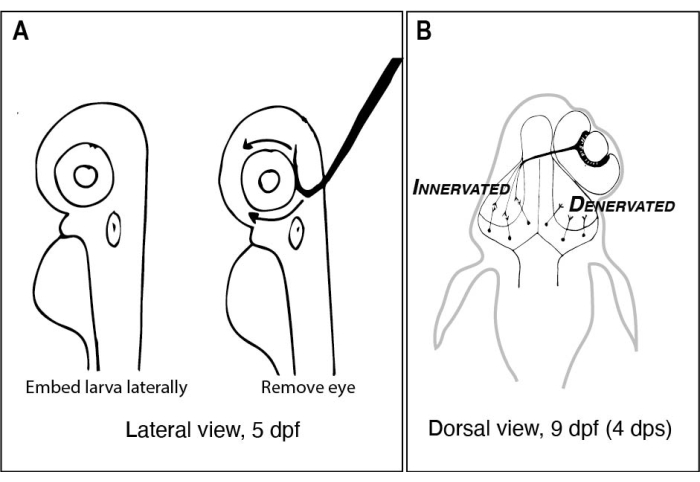
図1:片側眼除去前後のゼブラフィッシュ幼虫のスケッチ 。 (A)解剖顕微鏡下で見た5 dpf幼虫の図面。各幼虫は、低融点アガロースに埋め込まれ、鋭い引っ掛けられた先端を有するタングステン針が上を向いて眼をすくい取るために使用される前に横方向に向けられる(この例では左目)。(B) Aに描かれた手術から生じた9dpf幼虫の背部図の図面。右目から高度に図式化された3つのRGC軸索のみが、左蓋葉のニューロンと脱魅惑的で連結していることが示されている。略語: dpf = 受精後日数;dps = 手術後の日数;RGC = 網膜神経節細胞。 この図の拡大版を表示するには、ここをクリックしてください。
Protocol
Representative Results
Discussion
この論文で説明する技術は、ゼブラフィッシュにおける脊椎動物の視覚系発達を研究するための多くのアプローチの1つを示しています。他の研究者は、胚性網膜を解剖し、遺伝子発現解析19 を実行する方法、または視蓋骨30におけるニューロン活性を視覚化する方法を発表した。この論文は、差動網膜入力が視蓋内の細胞挙動にどのように影響するかを探?…
Divulgations
The authors have nothing to disclose.
Acknowledgements
この研究のための資金は、主にリードカレッジからKLCへのスタートアップ資金、OLHへのヘレンスタッフォードリサーチフェローシップ資金、およびYKへのリードカレッジサイエンスリサーチフェローシップによって支えられました。このプロジェクトは、Wellcome Trust Studentship(2009-2014)の支援を受けたHRとのコラボレーションとして、Steve Wilsonの研究室で始まりました。このプロジェクトに関する最初の議論をしてくれたMáté Varga、Steve Wilson、およびWilson研究室の他のメンバーに感謝し、特にKLCにアガロースに胚をマウントし、ゼブラフィッシュの脳解剖を行う方法を最初に教えたFlorencia CavodeassiとKate Edwardsに感謝します。また、タングステン針研ぎ装置の組み立てに協力してくれたグレタ・グローバーとジェイ・ユーイングにも感謝します。
Materials
| Equipment and supplies: | |||
| Breeding boxes | Aquaneering | ZHCT100 | |
| Dow Corning high vacuum grease | Sigma or equivalent supplier | Z273554 | |
| Erlenmeyer flasks (125 mL) | For making Marc's Modified Ringers (MMR) with antibiotics for post-surgery incubation | ||
| Fine forceps – Dumont #5 | Fine Science Tools (FST) | 11252-20 | |
| Glass Pasteur pipettes | DWK Lifescience | 63A53 & 63A53WT | For pipetting embryos and larvae |
| Glass slides for microscopy | VWR or equivalent supplier | 48311-703 | Standard glass microscope slides can be ordered from many different laboratory suppliers. |
| Glassware including graduated bottles and graduated cylinders | For making and storing solutions | ||
| 2-part epoxy resin | ACE Hardware or other equivalent supplier of Gorilla Glue or equivalent | 0.85 oz syringe | https://www.acehardware.com/departments/paint-and-supplies/tape-glues-and-adhesives/glues-and-epoxy/1590793 |
| Microcentrifuge tube (1.7 mL) | VWR or equivalent supplier | 22234-046 | |
| Nickel plated pin holder (17 cm length) | Fine Science Tools (FST) | 26018-17 | To hold tungsten wire while sharpening and performing surgeries/dissections. |
| Nylon mesh tea strainer or equivalent | Ali Express or equivalent | For harvesting zebrafish eggs after spawning; https://www.aliexpress.com/item/1005002219569756.html | |
| Paper clip | For Tungsten needle sharpening device. | ||
| Petri dishes 100 mm | Fischer Scientific or equivalent supplier | 50-190-0267 | |
| Petri dishes 35 mm | Fischer Scientific or equivalent supplier | 08-757-100A | |
| Pipette pump | SP Bel-Art or equivalent | F37898-0000 | |
| Potassium hydroxide (KOH) | Sigma | 909122 | For Tungsten needle sharpening device. Make a 10% w/v solution of KOH in the hood by adding pellets to deionized water. |
| Power supply (variable voltage) | For Tungsten needle sharpening device. Any power supply with variable voltage will work (even one used for gel electrophoresis). | ||
| Sylgard 184 Elastomer kit | Dow Corning | 3097358 | |
| Tungsten wire (0.125 mm diameter) | World Precision Instruments (WPI) | TGW0515 | Sharpen to remove eye and dissect larvae. |
| Variable temperature heat block | The Lab Depot or equivalent supplier | BSH1001 or BSH1002 | Set to 40-42 °C ahead of experiments. |
| Wide-mouth glass jar with lid (e.g., clean jam or salsa jar) | For Tungsten needle sharpening device. | ||
| Wires with alligator clip leads | For Tungsten needle sharpening device. | ||
| Microscopes: | |||
| Dissecting microscope | Any type will work but having adjustable transmitted light on a mirrored base is preferred. | ||
| Laser scanning confocal microscope | High NA, 20-25x water dipping objective lens is recommended. Microscope control and image capture software (Elements) is used here but any confocal microscope will work. |
||
| Reagents for surgeries and dissections: | |||
| Calcium chloride dihydrate | Sigma | C7902 | For Marc's Modified Ringers (MMR) and embryo medium (E3). |
| HEPES | Sigma | H7006 | For Marc's Modified Ringers (MMR). |
| Low melting point agarose | Invitrogen | 16520-050 | Make 1% in embryo medium (E3) or Marc's Modified Ringers (MMR). |
| Magnesium chloride hexahydrate | Sigma | 1374248 | For embryo medium (E3). |
| Magnesium sulfate | Sigma | M7506 | For Marc's Modified Ringers (MMR). |
| Paraformaldehyde | Electron Microscopy Sciences | 19210 | Dilute 8% (w/v) stock with 2x concentrated PBS (diluted from 10x PBS stock). |
| Penicillin/Streptomycin | Sigma | P4333-20ML | Dilute 1:100 in Marc's Modified Ringers. |
| Phosphate buffered saline (PBS) tablets | Diagnostic BioSystems | DMR E404-01 | Make 10x stock in deionized water, autoclave and store at room temperature. Dilute to 1x working concentration. |
| Potassium chloride | Sigma | P3911 | For Marc's Modified Ringers (MMR) and embryo medium (E3). |
| Sodium chloride | Sigma | S9888 | For Marc's Modified Ringers (MMR) and embryo medium (E3). |
| Sodium hydroxide | Sigma | S5881 | Make 10 M and use to adjust pH of MMR to 7.4. |
| Sucrose | Sigma | S9378 | |
| Tricaine-S | Pentair | 100G #TRS1 | Recipe: https://zfin.atlassian.net/wiki/spaces/prot/pages/362220023/TRICAINE |
| Reagents for immunohistochemistry: | |||
| Alexafluor 568 tagged Secondary antibody to detect rabbit IgG | Invitrogen | A-11011 | Use at 1:500 dilution for wholemount immunohistochemistry. |
| DAPI or ToPro3 | Invitrogen | 1306 or T3605 | Make up 1 mg/mL solutions in DMSO; 1:5,000 dilution for counterstaining. |
| Dimethyl sulfoxide (DMSO) | Sigma | D8418 | A component of immunoblock buffer. |
| Methanol (MeOH) | Sigma | 34860 | Mixing MeOH with aqueous solutions like PBST is exothermic. Make the MeOH/PBST solutions at least several hours ahead of time or cool them on ice before using. |
| Normal goat serum | ThermoFisher Scientific | 50-062Z | A component of immunoblock buffer. Can be aliquoted in 1-10 mL volumes and stored at -20 °C. |
| Primary antibody to detect phosphohistone H3 | Millipore | 06-570 | Use at 1:300 dilution for wholemount immunohistochemistry. |
| Primary antibody to detect Red Fluorescent Protein (RFP; detects dsRed derivatives) | MBL International | PM005 | Use at 1:500 dilution for wholemount immunohistochemistry. |
| Proteinase K (PK) | Sigma | P2308-10MG | Make up 10 mg/mL stock solutions in PBS and use at 10 µg/mL. |
| Triton X-100 | Sigma | T8787 | Useful to make a 20% (v/v) stock solution in PBS. |
| Software for data analysis | |||
| ImageJ (Fiji) | freeware for image analysis; https://imagej.net/software/fiji/ | ||
| Rstudio | freeware for statistical analysis and data visualization; https://www.rstudio.com/products/rstudio/download/ | ||
| Adobe Photoshop or GIMP | Proprietary image processing software (Adobe Photoshop and Illustrator) are often used to compose figures). A freeware alternative is Gnu Image Manipulation Program (GIMP; https://www.gimp.org/) | ||
| Zebrafish strains | available from the Zebrafish International Resource Centers in the US (https://zebrafish.org/home/guide.php) or in Europe (https://www.ezrc.kit.edu/). Specialized transgenic strains that have not yet been deposited in either resource center can be requested from individual labs after publication. |
References
- Butler, A. B., Hodos, W. Optic tectum. Comparative Vertebrate Neuroanatomy: Evolution and Adaptation. , 311-340 (2005).
- Cang, J., Feldheim, D. A. Developmental mechanisms of topographic map formation and alignment. Annual Review of Neuroscience. 36 (1), 51-77 (2013).
- Basso, M. A., Bickford, M. E., Cang, J. Unraveling circuits of visual perception and cognition through the superior colliculus. Neuron. 109 (6), 918-937 (2021).
- Burrill, J. D., Easter, S. S. Development of the retinofugal projections in the embryonic and larval zebrafish (Brachydanio rerio). The Journal of Comparative Neurology. 346 (4), 583-600 (1994).
- Regeneration in the goldfish visual system. Webvision: The Organization of the Retina and Visual System Available from: https://webvision.med.utah.edu/book/part-x-repair-and-regeneration-in-the-visual-system/regeneration-in-the-goldfish-visual-system/ (2021)
- Regeneration in the visual system of adult mammals. Webvision: The Organization of the Retina and Visual System Available from: https://webvision.med.utah.edu/book/part-x-repair-and-regeneration-in-the-visual-system/regeneration-in-the-visual-system-of-adult-mammals/ (2021)
- Cerveny, K. L., Varga, M., Wilson, S. W. Continued growth and circuit building in the anamniote visual system. Developmental Neurobiology. 72 (3), 328-345 (2012).
- Lindsey, B. W., et al. Midbrain tectal stem cells display diverse regenerative capacities in zebrafish. Scientific Reports. 9 (1), 4420 (2019).
- Soares, D., Yamamoto, Y., Strickler, A. G., Jeffery, W. R. The lens has a specific influence on optic nerve and tectum development in the blind cavefish Astyanax. Developmental Neuroscience. 26 (5-6), 308-317 (2004).
- White, E. L. An experimental study of the relationship between the size of the eye and the size of the optic tectum in the brain of the developing teleost, Fundulus heteroclitus. Journal of Experimental Zoology. 108 (3), 439-469 (1948).
- Raymond, P., Easter, S., Burnham, J., Powers, M. Postembryonic growth of the optic tectum in goldfish. II. Modulation of cell proliferation by retinal fiber input. The Journal of Neuroscience. 3 (5), 1092-1099 (1983).
- Sato, Y., Yano, H., Shimizu, Y., Tanaka, H., Ohshima, T. Optic nerve input-dependent regulation of neural stem cell proliferation in the optic tectum of adult zebrafish. Developmental Neurobiology. 77 (4), 474-482 (2017).
- Hall, Z. J., Tropepe, V. Visual experience facilitates BDNF-dependent adaptive recruitment of new neurons in the postembryonic optic tectum. The Journal of Neuroscience. 38 (8), 2000-2014 (2018).
- Nusslein-Volhard, C., Dahm, R. . Zebrafish. , (2002).
- Cold Spring Harbor Protocols. Marc’s modified Ringer’s (MMR) (10X, pH 7.4). Cold Spring Harbor Protocols. , (2009).
- Turner, K. J., Bracewell, T. G., Hawkins, T. A. Anatomical dissection of zebrafish brain development. Brain Development. 1082, 197-214 (2014).
- Brady, J. A simple technique for making very fine, durable dissecting needles by sharpening tungsten wire electrolytically. Bulletin of the World Health Organization. 32 (1), 143-144 (1965).
- . ZFIN Tricaine recipe Available from: https://zfin.atlassian.net/wiki/spaces/prot/pages/362220023/TRICAINE (2018)
- Zhang, L., Leung, Y. F. Microdissection of zebrafish embryonic eye tissues. Journal of Visualized Experiments: JoVE. (40), e2028 (2010).
- . ZFIN protocols Available from: https://zfin.atlassian.net/wiki/spaces/prot/overview (2021)
- Engerer, P., Plucinska, G., Thong, R., Trovò, L., Paquet, D., Godinho, L. Imaging subcellular structures in the living zebrafish embryo. Journal of Visualized Experiments: JoVE. (110), e53456 (2016).
- ImageJ with batteries included. Fiji Available from: https://figi.sc/ (2021)
- O’Brien, J., Hayder, H., Peng, C. Automated quantification and analysis of cell counting procedures using ImageJ plugins. Journal of Visualized Experiments: JoVE. (117), e54719 (2016).
- Poggi, L., Vitorino, M., Masai, I., Harris, W. A. Influences on neural lineage and mode of division in the zebrafish retina in vivo. The Journal of Cell Biology. 171 (6), 991-999 (2005).
- Karlstrom, R. O., et al. Zebrafish mutations affecting retinotectal axon pathfinding. Development. 123 (1), 427-438 (1996).
- Harvey, B. M., Baxter, M., Granato, M. Optic nerve regeneration in larval zebrafish exhibits spontaneous capacity for retinotopic but not tectum specific axon targeting. PLOS ONE. 14 (6), 0218667 (2019).
- Robles, E., Filosa, A., Baier, H. Precise lamination of retinal axons generates multiple parallel input pathways in the tectum. Journal of Neuroscience. 33 (11), 5027-5039 (2013).
- Vargas, M. E., Barres, B. A. Why Is Wallerian degeneration in the CNS so slow. Annual Review of Neuroscience. 30 (1), 153-179 (2007).
- Hughes, A. N., Appel, B. Microglia phagocytose myelin sheaths to modify developmental myelination. Nature Neuroscience. 23 (9), 1055-1066 (2020).
- de Calbiac, H., Dabacan, A., Muresan, R., Kabashi, E., Ciura, S. Behavioral and physiological analysis in a zebrafish model of epilepsy. Journal of Visualized Experiments: JoVE. (176), e58837 (2021).
- Adams, S. L., Zhang, T., Rawson, D. M. The effect of external medium composition on membrane water permeability of zebrafish (Danio rerio) embryos. Theriogenology. 64 (7), 1591-1602 (2005).
- Fredj, N. B., et al. Synaptic activity and activity-dependent competition regulates axon arbor maturation, growth arrest, and territory in the retinotectal projection. Journal of Neuroscience. 30 (32), 10939-10951 (2010).
- Alberio, L., et al. A light-gated potassium channel for sustained neuronal inhibition. Nature Methods. 15 (11), 969-976 (2018).
- Kay, J. N., Finger-Baier, K. C., Roeser, T., Staub, W., Baier, H. Retinal ganglion cell genesis requires lakritz, a zebrafish atonal homolog. Neuron. 30 (3), 725-736 (2001).
- Gnuegge, L., Schmid, S., Neuhauss, S. C. F. Analysis of the activity-deprived zebrafish mutant macho reveals an essential requirement of neuronal activity for the development of a fine-grained visuotopic map. The Journal of Neuroscience. 21 (10), 3542-3548 (2001).
- Jeffery, W. R. Astyanax surface and cave fish morphs. EvoDevo. 11 (1), 14 (2020).
- Sieger, D., Peri, F. Animal models for studying microglia: The first, the popular, and the new. Glia. 61 (1), 3-9 (2013).
- Svahn, A. J., et al. Development of ramified microglia from early macrophages in the zebrafish optic tectum. Developmental Neurobiology. 73 (1), 60-71 (2013).
- Herzog, C., et al. Rapid clearance of cellular debris by microglia limits secondary neuronal cell death after brain injury in vivo. Development. 146 (9), (2019).
- Chen, J., Poskanzer, K. E., Freeman, M. R., Monk, K. R. Live-imaging of astrocyte morphogenesis and function in zebrafish neural circuits. Nature Neuroscience. 23 (10), 1297-1306 (2020).

