The present work describes an experimental protocol of transesophageal atrial burst pacing for efficient induction of atrial fibrillation (AF) in rats. The protocol can be used in rats with healthy or remodeled hearts, allowing the study of AF pathophysiology, identification of novel therapeutic targets, and evaluation of new therapeutic strategies.
Animal studies have brought important insights into our understanding regarding atrial fibrillation (AF) pathophysiology and therapeutic management. Reentry, one of the main mechanisms involved in AF pathogenesis, requires a certain mass of myocardial tissue in order to occur. Due to the small size of the atria, rodents have long been considered ‘resistant’ to AF. Although spontaneous AF has been shown to occur in rats, long-term follow-up (up to 50 weeks) is required for the arrhythmia to occur in those models. The present work describes an experimental protocol of transesophageal atrial burst pacing for rapid and efficient induction of AF in rats. The protocol can be successfully used in rats with healthy or remodeled hearts, in the presence of a wide variety of risk factors, allowing the study of AF pathophysiology, identification of novel therapeutic targets, and evaluation of novel prophylactic and/or therapeutic strategies.
Atrial fibrillation (AF) is the most common sustained cardiac arrhythmia encountered in clinical practice and its incidence and prevalence continue to increase dramatically worldwide1. This arrhythmia affects up to 4% of the world population according to recent studies2. However, given that paroxysmal AF can be asymptomatic and may therefore escape detection, the true prevalence of AF is likely to be much higher than that presented in the literature.
The pathophysiology of AF has been intensely studied. Nevertheless, the underlying mechanisms of this complex arrhythmia remain incompletely elucidated and this reflects in the limited therapeutic options, with questionable efficacy. Animal studies have brought important insights into our understanding regarding AF pathophysiology and therapeutic management. Reentry, one of the main mechanisms involved in AF pathogenesis3, requires a certain mass of myocardial tissue in order to occur. Thus, large animals have generally been preferred in AF studies, whereas, due to the small size of their atria, rodents have long been considered 'resistant' to AF. However, the use of large animals is hampered mostly by handling difficulties. Meanwhile, although spontaneous AF has been shown to occur in rats4, long-term follow-up (up to 50 weeks) is required for the arrhythmia to occur in those models5. Models that ensure rapid AF occurrence in small rodents have also been developed. Most often, those models use acute electrical stimulation, often in the presence of other favoring conditions, such as concomitant parasympathetic stimulation or asphyxia, to artificially induce AF6,7. Although efficient, such models do not allow the evaluation of critical AF-related features, such as the progressive electrical, structural, autonomic, or molecular remodeling of the atria, nor the effects of conventional or non-conventional antiarrhythmic drugs on the atrial substrate or on the risk of ventricular pro-arrhythmia8,9.
The present work describes an experimental protocol of long-term transesophageal atrial burst pacing for rapid and efficient induction of AF in rats. The protocol is suitable for both acute and long-term studies and can be successfully used in rats with healthy or remodeled hearts, in the presence of a wide variety of risk factors, allowing the study of AF pathophysiology, identification of novel therapeutic targets, and evaluation of novel prophylactic and/or therapeutic strategies.
Procedures involving animal subjects were approved by the Ethics Committee of the University of Medicine, Pharmacy, Science and Technology "George Emil Palade" of Târgu Mureș, by the Romanian National Sanitary Veterinary and Food Safety Authority and complied with the International Council for Laboratory Animal Science guidelines (Directive 2010/63/EU).
1. Transesophageal atrial burst pacing protocol
In a proof-of-concept study, 22 adult male Wistar rats (200-400 g) were randomly assigned into two groups: STIM (n = 15) and SHAM (n = 7). All animals were housed individually in polycarbonate cages, in a climate-controlled room (21-22 °C), having free access to water and dry food throughout the study. The transesophageal stimulation protocol described above was applied to all animals for 10 days, 5 days per week. All animals underwent the same protocol, except that the rats in the SHAM group did not receive active electrical stimulation.
As expected, no episodes of AF were induced in the SHAM animals throughout the protocol. Hence, no other parameters (i.e., the duration of AF episodes and the presence of 'persistent' AF episodes) could be evaluated in this group.
On the first day of stimulation, 12 (80%) of the 15 STIM animals presented AF episodes (Relative Risk = 3.33, p < 0.001 vs. the SHAM group using Fisher's exact test). In the STIM rats, out of 164 stimulation cycles applied on the first day of stimulation, 42 were followed by AF episodes (median inducibility of 20% [interquartile range of 6.67-72.22] vs. 0% in the SHAM group) (Figure 5).
During the 10 days of protocol, AF was efficiently induced in all animals (Figure 6). An average of 15.6 ± 8.7 episodes of AF was induced in the STIM animals during the entire duration of the protocol. Of the total number of stimulation cycles applied, 20.05% were followed by AF, and 41 (17.30%) episodes of AF lasted more than 600 s. The average duration of AF episodes lasting less than 600 s is 40.12 s (Table 1).
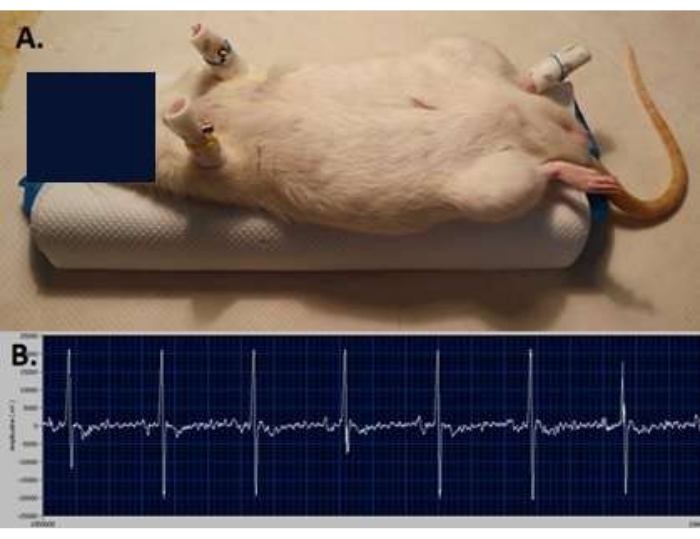
Figure 1: Surface ECG recording. (A) ECG electrodes positioning—two at the level of the forelimbs and one on the left hindlimb of the animal. (B) Surface ECG tracing recorded before applying electrical stimulation. Please click here to view a larger version of this figure.
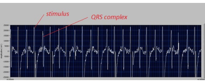
Figure 2: ECG tracing confirming capture of the atria. The ECG tracing confirms the correct position of the catheter, i.e., a narrow QRS complex is observed after each electrical stimulus at a frequency of 400 stimuli/minute. Please click here to view a larger version of this figure.
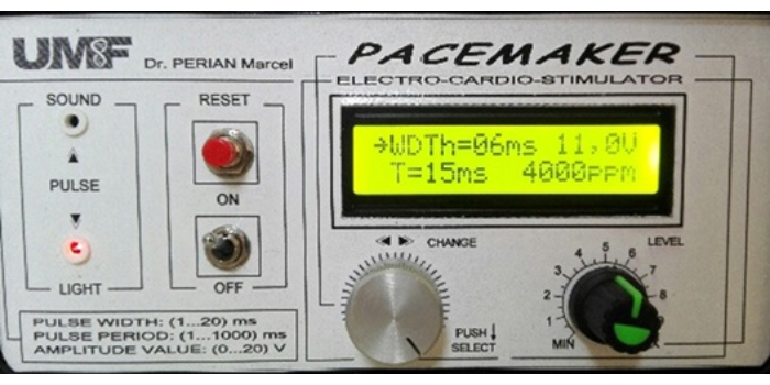
Figure 3: Microcontroller-based cardiac pacemaker settings. Stimulation parameters are set at a frequency of 4,000 stimuli/minute (ppm: pulses per minute), stimulus duration of 6 ms (WDTh: width), and tension of 11 V (i.e., 3 V above the diastolic threshold). Please click here to view a larger version of this figure.
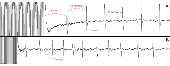
Figure 4: ECG tracings confirming the effectiveness of the stimulation protocol. (A) The sinus node recovery time (SNRT). Note that the time interval at the cessation of stimulation (SNRT) is longer than the cycle length recorded during sinus rhythm (RR interval, i.e., the interval between the R-waves of two consecutive QRS complexes, representing the duration of a cardiac cycle). (B) The appearance of an atrial fibrillation episode after completion of the atrial electrical stimulation cycle. Note the irregular, narrow QRS complexes, the absence of P waves, and the small, distorted "f" waves. Please click here to view a larger version of this figure.
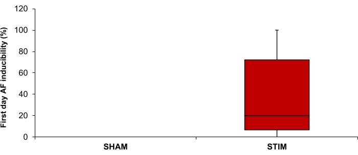
Figure 5: Inducibility of atrial fibrillation (AF) on the first day of stimulation in the STIM (n = 15) and SHAM (n = 7) groups. Data are expressed as median and interquartile range. Please click here to view a larger version of this figure.
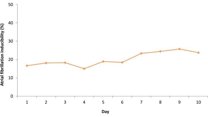
Figure 6: Mean daily inducibility of atrial fibrillation during the 10 days of the stimulation protocol in the STIM rats. Please click here to view a larger version of this figure.
Electrically-induced atrial fibrillation episodes (n = 237) | Number (%) | Mean duration (seconds) |
duration ≥600 seconds | 41 (17.30%) | – |
duration < 600 seconds | 196 (82.70%) | 40.12 |
Table 1: Temporal parameters of electrically-induced 'persistent' and 'non-persistent' atrial fibrillation episodes in the STIM group.
The present paper describes an experimental protocol of long-term transesophageal atrial burst pacing for rapid and efficient induction of AF in rats, suitable for both acute and long-term AF studies. The 10-day stimulation protocol described herein has been successfully used to develop a ‘secondary spontaneous AF model’ (i.e., a model in which, following a period of AF induction by electrical stimulation, AF develops spontaneously)10. However, the duration of the protocol can vary depending on the exact purpose of the study.
Other parameters, such as the size of the stimulation catheter, can also be adjusted, depending on the size of the animals. However, care should be taken to avoid the use of excessively large catheters, as they may cause pressure on the trachea and impede normal breathing. For 200-400 g rats, 5-6 F catheters cause negligible pressure on the trachea and allow the protocol to be implemented without the need for endotracheal intubation.
A key step of the protocol is the correct positioning of the stimulation catheter inside the esophagus, at the level of the atria (step 1.7). This step should only be performed after careful check of the depth of the anesthesia, as lack of effective anesthesia increases the risk of cardio-respiratory arrest during the following steps of the protocol. Monitoring the electrical activity of the heart using surface ECG generally provides sufficient data to confirm that the catheter is correctly positioned inside the esophagus (i.e., stimulation at a frequency higher than the intrinsic heart rate illustrates the overdrive suppression phenomenon on the sinus node and each stimulus is followed by a narrow QRS complex). However, performing esophageal electrocardiogram recordings could be used to further confirm the correct position of the stimulation catheter.
Considering the normal baseline heart rate of Wistar rats12, it is important to perform the initial stimulation at a frequency higher than the rats’ own heart rate (i.e., >400 stimuli/minute), to ensure constant capture of the atria (step 1.8). During this step, stimulation frequency should be adapted to each animal’s baseline heart rate. In the presence of a correct stimulation rate, lack of constant atrial capture could be due to either incorrect positioning of the catheter or to stimulation at a voltage below the diastolic threshold (step 1.9). Both scenarios can result in inefficient stimulation and protocol failure. Given that variations in body temperature can promote cardiac arrhythmias13, attention should be given to maintaining constant body temperature (37 °C) during the entire procedure.
The technique described herein also has a number of limitations. Given the anatomical proximity of the vagus nerve to the esophagus, concomitant electrical stimulation of the vagus nerve can occur during the protocol, increasing the risk of cardio-respiratory arrest. In addition, one should keep in mind that parasympathetic stimulation is likely to also contribute to AF occurrence in this model and that other models may be more adequate for studies aiming to evaluate and/or manipulate the autonomic nervous system.
Animal models continue to play an important role in unraveling the pathophysiological mechanisms that underlie AF and in improving therapeutic strategies. An ideal animal AF model should be fast and easy to recreate, reproducible, and should mimic as much as possible the pathology observed in humans14. In rodents, most AF models consist in acute AF induction, most commonly in the presence of other favoring factors, in addition to electrical stimulation of the atria6,15. However, such models cannot assess the role of progressive atrial remodeling in AF pathophysiology, cannot test the long-term effects of various antiarrhythmic drugs, and cannot assess the ventricular proarrhythmic risk associated with chronic antiarrhythmic treatment8,9. In other studies16, a single stimulation protocol was applied to chronically remodeled, AF-prone atria. Although this strategy overcomes some of these disadvantages, it does not take into account the impact of the AF per se on atrial proarrhythmic remodeling and on future AF occurrence8,9. Meanwhile, prolonged (e.g., 10 days) application of the transesophageal atrial pacing protocol described above induces progressive atrial proarrhythmic remodeling and creates the atrial environment required for the spontaneous occurrence of AF, after the stimulation protocols are completed10.
The experimental model described herein can therefore be efficiently used not only for assessing acute AF induction, but also for creating a model of (secondary) spontaneous AF. This model therefore brings a number of major advantages, creating the premises for a better understanding of the mechanisms involved in the occurrence and maintenance of AF, as well as for identifying and testing new therapeutic strategies.
The authors have nothing to disclose.
This work was supported by a grant of the Romanian Ministry of Education and Research, CNCS – UEFISCDI, project number PN-III-P1-1.1-TE-2019-0370, within PNCDI III.
Antisedan (Atipamezole Hydrochloride) 5mg / mL, solution for injection | Orion Corporation | 06043/4004 | for Rats use 1 mg / kg |
Dormitor (Medetomidine Hydrochloride) 1 mg / mL, solution for injection | Orion Corporation | 06043/4003 | for Rats use 0.5 mg / kg |
E-Z Anesthesia Single Animal System | E-Z Systems Inc | EZ-SA800 | Allows the manipulation of one animal at a time |
Isoflurane 99.9%, 100 mL | Rompharm Company | N01AB06 | |
Ketamine 10%, 25 mL | for Rats use 75 mg / kg | ||
Microcontroller-based cardiac pacemaker for small animals | Developed in our laboratory (See Reference number 10 in the manuscript) | ||
Surface ECG recording system | Developed in our laboratory (See Reference number 10 in the manuscript) |