This article presents the integration of a spectral-focusing module and a dual-output pulse laser, enabling rapid hyperspectral imaging of gold nanoparticles and cancer cells. This work aims to demonstrate the details of multimodal nonlinear optical techniques on a standard laser scanning microscope.
Probing gold nanoparticles (AuNPs) in living systems is essential to reveal the interaction between AuNPs and biological tissues. Moreover, by integrating nonlinear optical signals such as stimulated Raman scattering (SRS), two-photon excited fluorescence (TPEF), and transient absorption (TA) into an imaging platform, it can be used to reveal biomolecular contrast of cellular structures and AuNPs in a multimodal manner. This article presents a multimodal nonlinear optical microscopy and applies it to perform chemically specific imaging of AuNPs in cancer cells. This imaging platform provides a novel approach for developing more efficient functionalized AuNPs and determining whether they are within vasculatures surrounding the tumor, pericellular, or cellular spaces.
Gold nanoparticles (AuNPs) have shown great potential as biocompatible imaging probes, for example, as effective surface-enhanced Raman spectroscopy (SERS) substrates in various biomedical applications. Major applications include fields such as biosensing, bioimaging, surface-enhanced spectroscopies, and photothermal therapy for cancer treatment1. Furthermore, probing AuNPs in living systems is crucial to assessing and understanding the interaction between AuNPs and biological systems. There are various analytical techniques, including Fourier transform infrared (FTIR) spectroscopy2, laser ablation inductively coupled mass spectrometry (LA-ICP-MS)3, and magnetic resonance imaging (MRI)4 that have been successfully used to investigate the distribution of AuNPs in tissues. Nevertheless, these methods suffer from several drawbacks such as being time-consuming and involving complex sample preparation3, requiring long acquisition times, or the lack of sub-micron spatial resolution2,4.
Compared to conventional imaging techniques, nonlinear optical microscopy offers several advantages for probing live cells and AuNPs: The nonlinear optical microscopy achieves deeper imaging depth and provides intrinsic 3D optical sectioning capability with the use of near-IR ultrafast lasers. With the significant improvement of imaging speed and detection sensitivity, two-photon excited fluorescence (TPEF)5,6,7 and second harmonic generation (SHG)8,9,10 microscopy have been demonstrated to further improve non-invasive imaging of endogenous biomolecules in living cells and tissues. Moreover, utilizing novel pump-probe nonlinear optical techniques such as transient absorption (TA)11,12,13,14 and stimulated Raman scattering (SRS)15,16,17,18, it is possible to derive label-free biochemical contrast of cellular structures and AuNPs. Visualizing AuNPs without the use of extrinsic labels is of great importance since chemical perturbations of the nanoparticles will modify their physical properties and hence their uptake in cells.
This protocol presents the implementation of a Spectral Focusing Timing and Recombination Unit (SF-TRU) module for a dual-wavelength pulse laser, enabling fast multimodal imaging of AuNPs and cancer cells. This work aims to demonstrate the details of integrated TPEF, TA, and SRS techniques on a laser scanning microscope.
1. Switching on the laser system
2. Switching on the Spectral Focusing Timing and Recombination Unit (SF-TRU)
3. Modulating the Stokes beam for SRS imaging
4. Switching on the lock-in amplifier
5. Operating on the laser scanning microscope
6. Mounting a sample in the microscope stage
7. Changing the Raman shift and collecting a hyperspectral data stack
NOTE: The Raman shift at which the imaging takes place is dependent on the delay stage position in the SF-TRU box and the wavelength of the pump beam from the laser system.
The Spectral Focusing Timing and Recombination Unit (SF-TRU) module is introduced between the dual-output femtosecond laser and the modified laser scanning microscope. The tunable ultrafast laser system used in this study has two output ports delivering one beam at a fixed 1,045 nm wavelength and the other beam tunable in the range of 680–1,300 nm. A detailed schematic of the SF-TRU module and multimodal imaging platform is depicted in Figure 1. The SF-TRU is employed to chirp two femtosecond laser beams to picosecond pulses and overlaps two beams spatially and temporally. Hyperspectral stimulated Raman scattering (SRS) imaging is performed by scanning the delay between the picosecond pump and Stokes pulses.
All the samples are illuminated by pulsed lasers through a high numerical aperture (NA) objective on a modified inverted microscope. The SRS and TPEF signals are collected in the forward direction with a high NA condenser. For SRS microscopy, the tunable (680–1,300 nm) and fixed (1,045 nm) laser outputs are used as the pump and the Stokes beams, respectively. The combination of a large-area photodiode and lock-in amplifier is used to perform SRS imaging, while the Stokes beam is blocked with two filters (890/210 band-pass and 950 nm short-pass filters). An important advantage of the spectral focusing SRS approach over the picosecond laser-based systems is the ability to tune the Raman shift of interest by simply controlling the time delay between the chirped pump and Stokes pulses. This approach allows fast probing of Raman shifts within a range of several hundred wavenumbers without changing the pump and Stokes wavelengths, allowing much faster hyperspectral imaging.
To validate the performance of spectral resolution and chemical selectivity, the sample of polystyrene (PS) microspheres is imaged first (Figure 2). The laser powers at the sample (after 60x NA 1.2 water immersion objective) are 10 mW for the 1,045 nm Stokes beam and 20 mW for the 802 nm pump beam. The SRS spectrum can be extracted at every pixel in the frames. Figure 2B shows the hyperspectral SRS spectrum of PS beads. For comparison, the spontaneous Raman spectrum of PS beads is shown in Figure 2A. The SRS and spontaneous Raman spectra of PS microspheres are almost identical except for relative intensity differences at the sides. These spectroscopic measurements allow converting optical delay line stage (mm) to Raman wavenumbers (cm-1) for SRS imaging.
Since the Raman spectra of major biomolecules in the carbon-hydrogen stretching vibrational band is in the range of 2,800–3,050 cm−1, the SRS imaging of 4T1 cancer cells is performed at 2,852 cm−1 (lipid), 2,930 cm−1 (protein), 2,968 cm−1 (DNA), and the overlay images as shown in Figure 3. SRS images of different Raman bands are acquired with a pixel dwell time of 4 μs at a frame size of 512 x 512 pixels.
Finally, this method has further extended to multimodal imaging of 4T1 cancer cells dosed with gold nanoparticles (AuNPs), allowing us to image the AuNPs’ distributions in cancer cells more precisely. The acquired SRS (CH2 and CH3 channels), TPEF (LysoTracker fluorescent probes), and TA (off-resonance SRS channel) images are depicted in Figure 4.
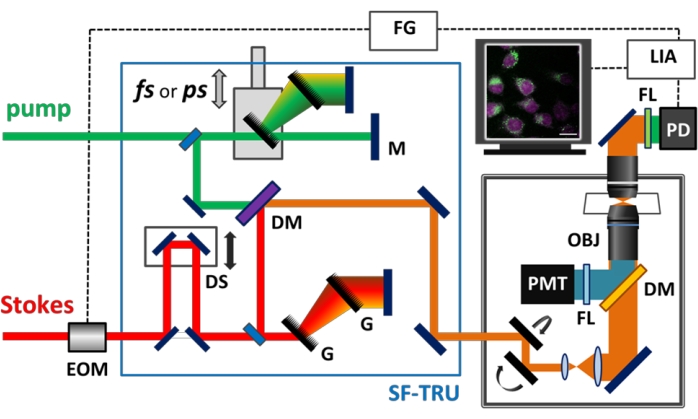
Figure 1: Schematic of the hyperspectral multimodal imaging platform. Illustration of multimodal nonlinear optical microscopy. DM, dichroic mirror; DS, delay stage; EOM, electro-optical modulator; FG, function generator; FL, filter; G, grating; LIA, lock-in amplifier; M, mirror; OBJ, objective; PD, photodiode; PMT, photomultiplier tube; SF-TRU, Spectral Focusing Timing, and Recombination Unit. Please click here to view a larger version of this figure.
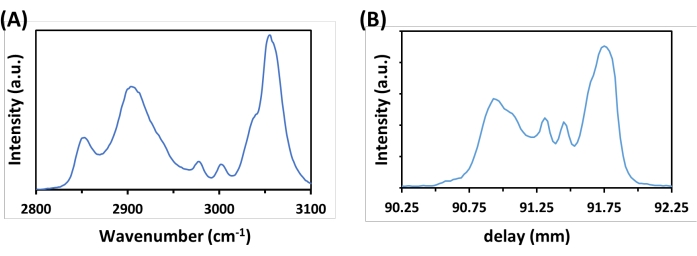
Figure 2: Spectra of polystyrene (PS) microspheres. The spontaneous (A) Raman spectrum of PS microspheres shows good agreement with the (B) hyperspectral SRS spectrum. Please click here to view a larger version of this figure.
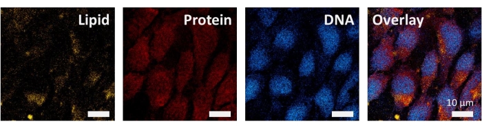
Figure 3: Spectroscopic SRS imaging of 4T1 cancer cells. SRS images at 2,852 cm−1, 2,930 cm−1, 2,968 cm−1, and overlay image. Scale bar: 10 µm. Please click here to view a larger version of this figure.
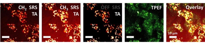
Figure 4: Multimodal imaging of gold nanoparticles uptake by 4T1 cancer cells. Hyperspectral SRS images at 2,852 cm−1 (CH2), 2,928 cm−1 (CH3), 3,080 cm−1 (off-resonance, only TA signal left), TPEF, and overlay image. Scale bar: 10 µm. Please click here to view a larger version of this figure.
This study has presented the combination of SF-TRU module and ultrafast dual-output laser system demonstrated its applications for multimodal microspectroscopy. With its ability to investigate gold nanoparticles’ (AuNPs’) uptake by cancer cells, the multimodal imaging platform can visualize the cellular responses to hyperthermic cancer treatments when laser beams are absorbed by AuNPs.
Moreover, rapid chemically specific imaging and high spectral resolution are achieved by employing the spectral focusing technique, using two sets of grating pairs to control the chirps in each laser beam. Compared to the usage of fixed-length glass rods for chirping, grating pairs allow matching the spectral resolution and the line widths of the Raman lines of interest19.
Furthermore, this system can be easily switched from femtosecond to chirped picosecond regimes by utilizing specially designed manual sliders to remove the gratings from the beam paths without affecting alignment inside the microscope. In order to achieve high sensitivity and spectral resolution, SRS is typically implemented using the narrowband picosecond pump and Stokes pulses. However, picosecond lasers are not sufficient for multiphoton excitation that require higher peak power levels, which are achievable with femtosecond lasers. The multimodal capabilities allow using the method for a variety of applications such as multiphoton imaging20, coherent Raman scattering21, and pump- probe spectroscopy.
There are several strategies that can be used to further improve the performance of the demonstrated device. For example, a faster-motorized delay line stage and data-acquisition software offer a higher imaging rate, which can increase the throughput of the multimodal imaging platform. In addition, the current setup covered ~300 cm−1 of spectral range with a spectral resolution up to 5 cm−1. To further enlarge the spectral range, a modified laser system with a broader spectral bandwidth has been employed for hyperspectral SRS microscopy22.
Collectively, this multimodal and non-invasive imaging platform provides new insights into nanomedicine and opens a new avenue to high-content multimodal molecular imaging of cells, biological tissues, and nanoparticles23,24,25.
The authors have nothing to disclose.
This research was supported by EPSRC Grants: Raman Nanotheranostics (EP/R020965/1) and CONTRAST facility (EP/S009957/1).
APE SRS Detection Unit | APE (Angewandte Physik & Elektronik GmbH) | APE Lock-in Module | Combined system containing a large area Si photo-diode for detecting the pump beam along with a Lock-In amplifier for detecting the beam modulations |
Confocal Scanning Unit | Olympus | FV 3000 | Confocal scanning unit used for imaging |
CML Latex Beads, 4% w/v, 1.0 µm | Invitrogen | C37483 | Polystyrene microspheres |
Coverslips | Thorlabs | CG15CH2 | 22 mm x 22 mm coverslips for seeding cells |
FBS | Gibco | 10500-064 | Foetal Bovine Serum (Heat Inactivated) |
Flouview | Olympus | FV31S-SW | Laser scanning microscope control software |
Function Generator | BX precision | 40543 | Used to generate square wave function which is fed to EOM in SF-TRU to produce modulations in the stokes beam |
FV3000 | Olympus | IX83P2ZF | Other microscope frames can be used. |
Gold Nanoparticles | Nanopartz | A11-60 | Spherical gold nanoparticles, 60 nm diameter |
Input Output Interface | Olympus | FV30 ANALOG | This unit allows voltage readouts from PMT and LockIn to be fed into the confocal scanning software and allows timing pulses to be sent between the olympus microscope and the SF-TRU unit. |
InSight X3 | Newport | Spectra-Physics | Dual-output femtosecond pulsed laser. Tunable (680–1300 nm) and fixed (1045 nm) laser outputs with the repetition rate of 80 MHz. |
Microscope Frame | Olympus | IX83 | Inverted microscope |
Mouse 4T1 cells | ATCC | CRL-2539 | Mouse breast cancer cells |
NA 1.2 Water Immersion Objective | Olympus | UPLSAPO60XW/IR | The multiphoton 60x Objective has a 0.28 mm working distance. Other similar objectives can be used. |
NA 1.4 Condenser | Nikon | CSC1003 | Other condensers with NA higher than the excitation objective can also be used. |
PMT | Hamamatsu | R3896 | PMT used for detecting anti-stokes photos for CARS micrsocopy |
PMT Connector | Hamamatsu | C13654-01-Y002 | Connector for PMT |
Power Supply | RS | RSPD-3303 C | Programmable power supply which is used for providing the correct voltage to the PMT |
RPMI-1640 | Gibco | A10491-01 | Roswell Park Memorial Institute (RPMI) 1640 Medium has since been found suitable for a variety of mammalian cells. |
SF-TRU | Newport Spectra Physics | SF-TRU | System designed for controlling the time delay and dispersion of the 2 laser outputs and for performing the beam modulations required for SRS |