L’utilisation de porcs dans la recherche a augmenté ces dernières années. Néanmoins, les porcs sont caractérisés par une anatomie difficile des voies respiratoires. En démontrant comment effectuer une intubation endotrachéale guidée par endoscopie, le présent protocole vise à accroître davantage la sécurité des animaux de laboratoire afin d’éviter la souffrance animale et la mort inutile.
Un abonnement JoVE est requis pour visualiser ce contenu. Connectez-vous ou commencez votre période d'essai gratuite.
Article de méthode
Intubation endotrachéale utilisant un endoscope d’intubation flexible comme modèle normalisé pour la gestion sécuritaire des voies respiratoires chez les porcs
Dans cet article
Avis d’erratum
Important: There has been an erratum issued for this article. View Erratum Notice
Résumé
Résumé
L’intubation endotrachéale est souvent une exigence de base pour la recherche translationnelle dans des modèles porcins pour diverses interventions qui nécessitent une voie respiratoire sécurisée ou des pressions de ventilation élevées. L’intubation endotrachéale est une compétence difficile, nécessitant un nombre minimum d’intubations endotrachéales réussies pour atteindre un taux de réussite élevé dans des conditions optimales, ce qui est souvent irréalisable pour les chercheurs non anesthésiologistes. En raison de l’anatomie spécifique des voies respiratoires porcines, on peut généralement supposer une voie respiratoire difficile. L’impossibilité d’établir une voie aérienne sûre peut entraîner des blessures, des événements indésirables ou la mort de l’animal de laboratoire. En utilisant une approche d’évaluation prospective, randomisée et contrôlée, il a été démontré que l’intubation endotrachéale assistée par fibre optique prend plus de temps, mais a un taux de réussite de premier passage plus élevé que l’intubation conventionnelle sans provoquer de baisses cliniquement pertinentes de la saturation en oxygène. Ce modèle présente un schéma posologique normalisé pour l’intubation endotrachéale guidée par endoscopie, fournissant une voie respiratoire sécurisée, en particulier pour les chercheurs qui sont inexpérimentés dans la technique d’intubation endotrachéale par laryngoscopie directe. Cette procédure devrait réduire au minimum la souffrance animale et les pertes inutiles d’animaux.
Introduction
L’intubation endotrachéale est souvent une exigence de base pour la recherche translationnelle dans des modèles porcins pour diverses interventions qui nécessitent des voies respiratoires sécurisées ou des pressions de ventilation élevées (comme la ventilation pendant la réanimation cardiorespiratoire1 ou le syndrome de détresse respiratoire aiguë2) ou qui exigent que le flux sanguin cérébral ne soit pas compromis par la compression interne par des dispositifs des voies respiratoires supraglottiques3 , qui sont parfois multipliés comme solutions de remplacement dans le contexte d’une difficulté an....
Accès restreint. Veuillez vous connecter ou commencer un essai pour afficher ce contenu.
Protocole
Les expériences de ce protocole ont été approuvées par le Comité national et institutionnel pour la protection des animaux (Landesuntersuchungsamt Rheinland-Pfalz, Coblence, Allemagne; approbation n° G20-1-135). Les expériences ont été menées conformément aux directives ARRIVE. Dans l’ensemble, 10 porcs mâles anesthésiés (Sus scrofa domestica) pesant en moyenne 30 kg ± 2 kg et âgés de 12 à 16 semaines ont été utilisés pour la présente étude.
1. Préparation des animaux
- Maintenez un environnement normal pour les animaux afin de minimiser le stress. Retenir la nourriture 6 h avant l’expérience prévue pour ré....
Accès restreint. Veuillez vous connecter ou commencer un essai pour afficher ce contenu.
Résultats
L’intubation endotrachéale a été réalisée sur 10 porcs mâles (âgés de 12 à 16 semaines, poids 30 kg ± 3 kg) dans le cadre d’une étude prospective, randomisée et contrôlée. Les porcs ont été randomisés en deux groupes : l’un était intubé par laryngoscopie conventionnelle (groupe IC) et l’autre groupe était intubé assisté par un endoscope à intubation flexible tel que décrit dans le protocole (groupe FIE). L’affectation de groupe a été faite en tirant des enveloppes scellées. L’enquêteur a été assigné au hasard su.......
Accès restreint. Veuillez vous connecter ou commencer un essai pour afficher ce contenu.
Discussion
Dans des études antérieures, notre groupe de recherche a déjà décrit des détails spécifiques concernant les avantages translationnels du modèle porcin 2,27,32,33. En règle générale, la réduction du niveau de stress de l’animal et de la douleur inutile devrait faire partie intégrante de tout protocole d’étude et est primordiale pour générer des données reproductibles de manière fiable. Par consé.......
Accès restreint. Veuillez vous connecter ou commencer un essai pour afficher ce contenu.
Déclarations de divulgation
L’endoscope à intubation flexible et ses accessoires ont été fournis sans condition par le fabricant à des fins de recherche uniquement. Les auteurs ne déclarent plus de conflits d’intérêts financiers ou autres.
Remerciements
Les auteurs tiennent à remercier Dagmar Dirvonskis pour son excellent soutien technique.
....Accès restreint. Veuillez vous connecter ou commencer un essai pour afficher ce contenu.
Matériaux
| Nom | Entreprise | Numéro de catalogue | Commentaires |
|---|---|---|---|
| Ambu aScope Regular | Ambu GmbH, Medizinprodukte, Bad Nauheim, Allemagne | Fibre optique jetable diamètre extérieur 5 mm | |
| Ambu aView Monitor | Ambu GmbH, Medizinprodukte, Bad Nauheim, Allemagne | moniteur | |
| Atracurium Hikma 50 mg/5mL | Hikma Pharma GmbH, Martinsried | atracurium | |
| Azaperone (Stresnil) 40mg/mL | Lilly Deutschland GmbH, Bad Homburg, Allemagne | azaperone | |
| BD Discardit II Spritze 2, 5, 10, 20 mL | Becton Dickinson S.A. Carretera, Mequinenza Fraga, Espagne | seringue | |
| BD Luer Connecta | Becton Dickinson Infusion Therapy, AB Helsingborg, Schweden | 3-way-stopcock | |
| BD Microlance 3 20 G | Becton Dickinson S.A. Carretera, Mequinenza Fraga, Espagne | canule | |
| Curafix i.v. classics | Lohmann & Rauscher International GmbH & Co. KG, Rengsdorf, Allemagne | Pansement de rétention de canule | |
| Engströ ; m Carestation | GE Heathcare, Madison USA | ventilateur | |
| Fentanyl-Janssen 0,05 mg/mL | Janssen-Cilag GmbH, Neuss | fentanyl | |
| Fü ; hrungsstab, Durchmesser 4.3 | Rü ; | sch introducteur de sonde endotrachéale | |
| IBM SPSS Statistics pour Windows, version 20 | IBM SPSS Statistics pour Windows, version 20.0. Armonk, NY : IBM Corp.) | Logiciel statistique | |
| Incetomat-line 150 cm | Fresenius, Kabi Deutschland, GmbH | ligne perfusor | |
| Intrafix Primeline | B. Braun Melsungen AG, Melsungen, Allemagne | Ligne d’infusion | |
| JOZA Einmal Nitril Untersuchungshandschuhe | JOZA, Mü ; nchen, Germany | gants jetables | |
| Laryngoscope, 45.48.50, KL 2000 | Medicon | Poignée de laryngoscope | |
| Littmann Classic III Stethoscope | 3M Deutschland GmbH, Neuss, Germany | stéthoscope | |
| Luer Lock | B.Braun Melsungen AG, Germany | ||
| Minjureded Vlieskompresse | Maged GmbH, Neuenkirchen, Germany | Compresse polaire pour fixer la languette | |
| Masimo LNCS Adtx SpO2 sensor | Masimo Corporation Irvine, Ca 92618 USA | clip de saturation pour la queue | |
| Masimo LNCS TC-I SpO2 capteur de clip d’oreille | Masimo Corporation Irvine, Ca 92618 USA | Clip de saturation pour l’oreille | |
| Masimo Radical 7 | Masimo Corporation Irvine, Ca 92618 États-Unis | saturation périphérique en oxygène | |
| Midazolam 15 mg/3 mL | Hameln Pharma GmbH, Hameln, Allemagne | midazolam | |
| Midmark Masque canin petit plastique avec diaphragme FRSCM-0005 | Midmark Corp., Dayton, Ohio, États-Unis | masque de ventilation pour chien | |
| Octeniderm farblos | Schü ; lke & Mayr GmbH, Nordenstedt, Allemagne | Désinfectant alcoolique | |
| Original Seringue Perfusor 50 mL | B.Braun Melsungen AG, Allemagne | Seringue | |
| Perfusor FM Braun | B.Braun Melsungen AG, Allemagne | Pousse-seringue | |
| Propofol 2 % 20 mg/mL (flacons de 50 mL) | Fresenius, Kabi Deutschland, GmbH | propofol | |
| RÜ ; SCH Fü ; hrungsstab fü ; r Endotrachéa-talchéale (ID 5,6 mm) | Teleflex Medical Sdn. Bhd, Malaisie | Fil de guidage de tube revêtu de PVC | |
| Rü ; schelit Super Safety Clear >ID 6/6.5 /7.0 mm | Teleflex Medical Sdn. Bhd, Malaysia | tube endotrachéal | |
| Inox Macintosh Grö ; ß ; e 4 | Welch Allyn69604 | lame pour laryngoscope | |
| Sterofundin | B.Braun Melsungen AG, Melsungen, Allemagne | Solution électrolytique équilibrée | |
| Ultrastop Antibeschlagmittel flacon avec compte-gouttes 25 mL | Sigmapharm Arzneimittel GmbH, Vienne, Autriche | Agent antibuée | |
| Vasofix Safety 22 G-16 G | B.Braun Melsungen AG, Allemagne | Cathéter | |
| veineuxVBM Manomètre à brassard | VBM Medizintechnik GmbH, Sulz a.N., Allemagne | Manomètre à brassard | |
| Zelette | Lohmann & Rauscher International GmbH & Co. KG, Rengsdorf, Allemagne | Écouvillonnage tissulaire |
Références
- Kleinman, M. E., Oh, W., Stonestreet, B. S. Comparison of intravenous and endotracheal epinephrine during cardiopulmonary resuscitation in newborn piglets. Critical Care Medicine. 27 (12), 2748-2754 (1999).
- Rissel, R., et al.
Accès restreint. Veuillez vous connecter ou commencer un essai pour afficher ce contenu.
Réimpressions et autorisations
Erratum
Formal Correction: Erratum: Endotracheal Intubation Using a Flexible Intubation Endoscope As a Standardized Model for Safe Airway Management in Swine
Posted by JoVE Editors on 4/03/2023. Citeable Link.
An erratum was issued for: Endotracheal Intubation Using a Flexible Intubation Endoscope As a Standardized Model for Safe Airway Management in Swine. The Protocol, Representative Results, and Discussion sections were updated.
In the Protocol, step 1.5 was updated from:
Disinfect the skin with a disinfectant (alcoholic) before inserting a peripheral vein cannula (22 G) into an ear vein. Spray the area, wipe once, then spray again, and allow the disinfectant to dry.
to:
Disinfect the skin with a disinfectant (alcoholic) before inserting a peripheral vein cannula (22 G) into an ear vein. Spray the area, wipe once, then spray again, and allow the disinfectant to dry. Secure the ear cannula with a band-aid (See Table of Materials).
In the Protocol, step 3.7 was updated from:
While maintaining the position of the endoscope, advance the endotracheal tube until it becomes visible in the camera image.
NOTE: If the endotracheal tube cannot be advanced through the glottic plane, there is a possibility that it has become caught on the arytenoid cartilage. In this case, the endotracheal tube must be withdrawn 1 cm and rotated by 90° before gently advancing again. If necessary, this maneuver can be repeated. Similar calibers of flexible intubation endoscope and endotracheal tube can minimize the risk of this issue occurring. If the endotracheal tube cannot be advanced despite this maneuver, it is likely that the subglottic narrowness-the narrowest part of the porcine larynx-cannot be passed. In this case, a smaller endotracheal tube size needs to be selected. Regular commercially available endotracheal tubes in sizes 6.5 cm or 7.0 cm ID should be able to pass the glottis as long as no anatomic abnormalities are present.
to:
While maintaining the position of the endoscope, advance the endotracheal tube until it becomes visible in the camera image.
NOTE: If the endotracheal tube cannot be advanced through the glottic plane, there is a possibility that it has become caught on the arytenoid cartilage. In this case, the endotracheal tube must be withdrawn 1 cm and rotated by 90° before gently advancing again. If necessary, this maneuver can be repeated. Similar calibers of flexible intubation endoscope and endotracheal tube can minimize the risk of this issue occurring. If the endotracheal tube cannot be advanced despite this maneuver, it is likely that the subglottic narrowness-the narrowest part of the porcine larynx-cannot be passed. In this case, a smaller endotracheal tube size needs to be selected. Regular commercially available endotracheal tubes in sizes 6.5 cm or 7.0 cm ID should be able to pass the glottis as long as no anatomic abnormalities are present. Endotracheal tube size requirements vary depending on the piglet size and breed.
In the Representative Results, the sixth paragraph was updated from:
Statistical analyses were performed using commercially available software (see Table of Materials). Normal distribution was examined using the Kolmogorov-Smirnoff test28. If a normal distribution was determined, group differences were analyzed using t-tests of independent samples29 or the Mann-Whitney U test30 for the non-parametric version. Data are presented as mean (± standard deviation). Correlations of ordinal-scale data were examined using Spearman's correlation coefficient31. A significance level of p < 0.05 was assumed.
to:
Statistical analyses were performed using commercially available software (see Table of Materials). Normal distribution was examined using the Kolmogorov-Smirnoff test28. If a normal distribution was determined, group differences were analyzed using t-tests of independent samples29 or the Mann-Whitney U test30 for the non-parametric version. Data are presented as mean (± standard deviation). Correlations of ordinal-scale data were examined using Spearman's correlation coefficient31. A significance level of p < 0.05 was assumed. All tests were performed with exploratory intention; therefore p-values are descriptive. Nevertheless, p < 0.05 was accepted as indicative of statistical significance.
In the Representative Results, the legend for figure 1 was updated from:
Figure 1: Number of intubation attempts in group comparison. For the group that was intubated using a flexible intubation endoscope, every intubation attempt was successful; in the group that was conventionally intubated, it took an average of 1.4 attempts before the endotracheal tube could be placed correctly. Error bars show the standard deviation. Please click here to view a larger version of this figure.
to:
Figure 1: Number of intubation attempts in group comparison. For the group that was intubated using a flexible intubation endoscope, every intubation attempt was successful; in the group that was conventionally intubated, it took an average of 1.4 attempts before the endotracheal tube could be placed correctly. Error bars show the standard deviation. n = 5 (for each group). Please click here to view a larger version of this figure.
In the Representative Results, figure 2 was updated from:
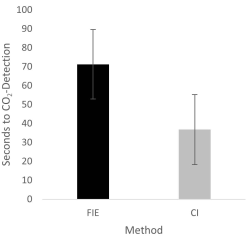
Figure 2: Time until CO2 detection in group comparison. For the group that was intubated using a flexible intubation endoscope, it took significantly longer until end-tidal CO2 could be detected, depicted as mean and standard deviation. Please click here to view a larger version of this figure.
to:
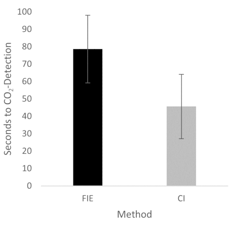
Figure 2: Time until CO2 detection in group comparison. For the group that was intubated using a flexible intubation endoscope, it took significantly longer until end-tidal CO2 could be detected, depicted as mean and standard deviation. n = 5 (for each group). Please click here to view a larger version of this figure.
In the Discussion, the fifth paragraph was updated from:
The increased duration had no clinical significance in this cohort. At no time was the termination criterion-a saturation of less than 93%-reached. This is shown in the results because a procedure change was unnecessary at any time. Prior adequate mask ventilation is a critical step to allow sufficient time for fiberoptic endotracheal tube placement to avoid rapid desaturation34. These results are consistent with previous studies comparing conventional intubation and endoscopically assisted intubations with inexperienced providers35.
to:
The increased duration had no clinical significance in this cohort. At no time was the termination criterion-a saturation of less than 93%-reached. This is shown in the results because a procedure change was unnecessary at any time. Prior adequate mask ventilation is a critical step to allow sufficient time for fiberoptic endotracheal tube placement to avoid rapid desaturation34. These results are consistent with previous studies comparing conventional intubation and endoscopically assisted intubations with inexperienced providers35. We attribute the prolonged duration of fiberoptic intubation to the fact that one must first reorient again after insertion, whereas with conventional intubation, one retains a view of the glottis. It is also important to avoid contact with the mucosa with the flexible intubation endoscope during advancement. This requires occasional corrective maneuvers. Last but not least, after successful placement, retraction of the relatively long endoscope is required, which increases the time to CO2 detection slightly.
