Summary

암세포의 내경동맥 주입에 의한 뇌전이 모델링

Published: August 02, 2022
doi:

Summary

뇌 전이는 암 환자의 심각한 이환율과 사망률의 원인입니다. 대부분의 뇌 전이 마우스 모델은 사망률 및 치료 개입 결과의 혼란 분석을 혼란시키는 전신 전이에 의해 복잡합니다. 여기에 제시된 것은 최소한의 전신 종양으로 일관된 두개 내 종양을 생성하는 암세포의 내 경동맥 주사를위한 프로토콜입니다.

Abstract

뇌 전이는 암 환자의 심각한 이환율과 사망률의 원인입니다. 복잡한 신경 미세 환경 및 기질 세포 상호 작용과 같은 전이성 질환의 중요한 측면은 시험관 내 분석으로 완전히 복제 할 수 없습니다. 따라서 동물 모델은 치료 적 개입의 효과를 조사하고 이해하는 데 중요합니다. 그러나 대부분의 뇌종양 이종 이식 방법은 시간 프레임과 종양 부담 측면에서 지속적으로 뇌 전이를 일으키지 않습니다. 암세포의 심장 내 주사에 의해 생성 된 뇌 전이 모델은 의도하지 않은 두개 외 종양 부담을 초래할 수 있으며 비 뇌 전이성 이환율 및 사망률을 초래할 수 있습니다. 암세포의 두개내 주사는 두개외 종양 형성을 제한할 수 있지만, 주사된 세포가 주사 부위에서 종종 단일 종양 덩어리를 형성하고, 높은 렙토메닌질 침범, 바늘 침투 중 뇌 혈관 구조 손상과 같은 몇 가지 주의 사항이 있습니다. 이 프로토콜은 내 경동맥 주사에 의해 생성 된 뇌 전이의 마우스 모델을 설명합니다. 이 방법은 다른 장기의 침범없이 일관되게 두개 내 종양을 생성하여 뇌 전이 치료제를 평가할 수 있습니다.

Introduction

뇌 전이는 매우 나쁜 예후와 관련된 널리 퍼진 악성 종양입니다 1,2. 뇌 전이 환자의 표준 치료는 환자의 일반적인 건강 상태, 두개외 질환 부담, 뇌 종양의 수와 위치에 따라 신경외과,전뇌 방사선 요법 및/또는 정위 방사선 수술로 구성된 다중 모드입니다3,4. 최대 3개의 두개내 병변이 있는 환자는 외과적 절제 또는 정위 방사선 수술을 받을 수 있으며, 다발성 병변이 있는 환자는 수술 관련 감염 및 부종의 위험을 피하기 위해 전뇌 방사선 요법을권장합니다5. 그러나 전뇌 방사선 치료는 방사능에 민감한 뇌 구조에 손상을 주어 삶의 질을 저하시킬 수 있습니다6.

전신 요법은 다발성 병변 환자를 치료하기 위한 비침습적 대안적이고 논리적인 접근법이다7. 그러나 혈류를 통한 세포 독성 약물의 수동 전달은 안전하지 않은 독성의 위험 없이는 뇌에서 치료 수준을 달성 할 수 없기 때문에 전신 요법이 효능이 좋지 않다는 오랜 개념으로 인해 덜 고려됩니다8. 이 패러다임은 최근 미국 식품의약국(FDA)이 승인한 전신 요법(전이성 HER2+ 유방암 뇌 전이에 대해 트라스투주맙 및 카페시타빈을 사용한 투카티닙)9,10,11,12 및 뇌 전이 환자에 대한 전신 요법 옵션 고려를 포함하는 치료 지침의 업데이트로 변화하기 시작했습니다13,14.

이러한 맥락에서, 분자 표적 요법, 면역 요법 및 표적 나노-약물 운반체와 같은 대체 약물 전달 시스템 분야의 개발은 잠재적으로 뇌 전이 치료15,16,17,18의 도전을 극복할 수 있다. 또한 뇌종양 장벽의 투과화를 통해 약물 전달을 개선하기위한 화학적 및 기계적 접근법도 조사되고 있습니다19,20. 이러한 접근법을 목적에 맞게 연구하고 최적화하려면 뇌 전이의 복잡한 생리학을 반영할 뿐만 아니라 두개내 약물 반응의 객관적인 분석을 허용하는 전임상 모델을 사용하는 것이 중요합니다.

대체로, 생체 내에서 뇌 전이를 모델링하기 위한 현재의 접근법은 마우스에서 암세포의 심장내(좌심실), 정맥내(일반적으로 꼬리 정맥), 두개내 또는 경동맥내(총경동맥) 주사를 포함합니다 21,22,23,24,25,26,27 . 종양 생착 전략 외에도, 종양 억제 유전자의 제거 또는 종양 유전자의 활성화에 의해 종양 형성이 촉발되는 유전자 조작 마우스 모델은 종양 모델링에 유용하다. 그러나 유전자 조작 마우스 모델은 소수에 불과하여 이차 종양을 생성하는 것으로 보고되었으며 뇌 전이를 안정적으로 생성하는 모델은 훨씬 적습니다28,29,30.

심장 내 (좌심실) 및 정맥 주사 (보통 꼬리 정맥) 주사와 같은 생착 방법은 암의 전신 전파를 모방합니다. 이러한 모델은 일반적으로 순환기 ‘첫 번째 통과’31 동안 대부분의 종양 세포를 가두는 모세 혈관에 따라 여러 장기 (예 : 뇌, 폐, 간, 신장, 비장)에 병변을 생성합니다. 그러나 뇌 생착의 일관되지 않은 속도는 원하는 통계적 검정력에 대한 샘플 크기를 달성하기 위해 더 많은 동물을 필요로합니다. 이러한 심장 내 및 정맥 주사 방법을 통해 결국 뇌에 확립되는 종양 세포의 수는 다양합니다. 따라서 뇌 전이 종양 부담은 동물마다 다를 수 있으며 진행의 차이로 인해 실험 일정을 표준화하고 결과를 해석하는 것이 어려울 수 있습니다. 두개외 종양 부담은 비뇌 전이 사망률로 이어질 수 있으며, 이러한 모델은 두개내 효능을 평가하는 데 적합하지 않습니다. 뇌-트로픽 세포주는 두개외 확립을 감소시키기 위해 인공 클론 선택 과정을 사용하여 확립되었지만, 복용률은 일관되지 않았고, 클론 선택 과정은 인간 종양에서 정상적으로 발견되는 이질성을 감소시킬 수 있다(32).

두개내 및 경동맥 내 주사와 같은 뇌 특이적 생착 방법은 보다 일관되고 효율적인 뇌 전이 모델링을 가능하게 합니다. 두개내 방법(33)에서, 암세포는 전형적으로 전두대뇌 피질 내로 주입되며, 이는 낮은 전신 침범으로 빠르고 재현가능한 종양 성장을 생성한다. 이 절차는 낮은 사망률로 잘 견디지만33, 주의할 점은 뇌에 (국소화된) 세포 덩어리를 빠르게 도입하고 초기 뇌 전이 병인을 모델링하지 않는 비교적 조잡한 접근 방식이라는 것입니다. 바늘은 뇌 조직 혈관을 손상시켜 국소 염증 5,34를 유발합니다. 경험에 비추어 볼 때, 바늘을 제거하는 동안 종양 세포 주사가 역류하는 경향이 있으며, 이는 렙토메닌 침범으로 이어집니다. 대안적으로, 경동맥 내 방법은 뇌 미세 혈관계를 갖는 세포를 총 경동맥으로 전달하여 순환, 혈관 외 유출 및 식민지화24에서의 생존을 모델링한다. 다른사람들과 일치 25,이 방법에 대한 우리의 경험은 외부 경동맥을 통해 이러한 조직의 모세 혈관으로 암세포를 의도하지 않게 전달하여 안면 종양을 유발할 수 있음을 발견했습니다 (미공개 데이터). 일반적인 경동맥 주사 전에 먼저 외부 경동맥을 결찰함으로써 안면 종양을 예방할 수 있습니다 (그림 1). 기사의 나머지 부분에서는이 방법을 ‘내 경동맥 주사’라고합니다. 경험을 통해 내경동맥 주사 방법은 전신적 사건이 거의 없는 뇌 전이를 지속적으로 생성하며 다양한 원발성 암(예: 흑색종, 유방암 및 폐암)의 뇌 전이 모델을 생성하는 데 성공했습니다(그림 1). 단점은 기술적으로 까다롭고 시간이 많이 걸리고 침습적이며 세포 수와 모니터링 일정을 신중하게 최적화해야 한다는 것입니다. 요약하면, 두개내 및 내경동맥 주사 방법 모두는 뇌종양 관련 생존 이점에 대한 치료 효과를 평가하기에 적합한 마우스 모델을 생성한다.

이 프로토콜은 내 경동맥 주사 방법을 설명하여 전신 침범이 거의없는 뇌 전이의 마우스 모델을 생성하므로 약물 분포 및 실험 치료제의 효능에 대한 전임상 평가에 적합합니다.

Figure 1
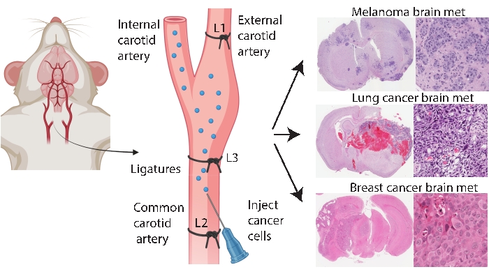
그림 1: 뇌 전이를 위한 내경동맥 주입 프로토콜의 개략적인 표현. 외부 경동맥 결찰을 통한 내 경동맥 주사는 다양한 원발성 암으로부터 뇌 전이 모델을 신뢰성있게 생성 할 수있다. 이 프로토콜에서는 3 개의 합자가 경동맥에 배치됩니다 (그림에서 L1-L3으로 주석이 달림). 이 그림의 더 큰 버전을 보려면 여기를 클릭하십시오.

Protocol

모든 연구는 퀸즐랜드 대학교 동물 윤리위원회 (UQCCR / 186 / 19)의 지침과 과학 목적을위한 동물 관리 및 사용에 대한 호주 코드에 따라 수행되었습니다. 1. 주사용 암세포의 제조 참고: 이 연구에서는 인간 유방암 세포주인 BT-474(BT474)를 사용했습니다. BT474는 10% 소 태아 혈청 및 1% 인슐린이 보충된 RPMI 1640 배지를 포함하는 완전 성장 배지에서 배양…

Representative Results

외부 경동맥 결찰 유무에 관계없이 일반적인 경동맥 주사 비교암세포를 먼저 외부 경동맥24를 결찰하지 않고 총 경동맥을 통해 주사했을 때, 이식 된 마우스의 77.8 %에서 안면 종양이 발견되었습니다 (n = 7/9 동물). 안면 종양의 예는 보충 그림 3에 설명되어 있습니다. 이 프로토콜에 설명 된 방법은 총 경동맥 전에 외부 경동맥을 결찰하여 …

Discussion

뇌 전이는 암세포가 원발성 부위에서 뇌로 퍼지는 복잡한 과정입니다. 이 다단계 과정의 특정 단계를 반영하는 다양한 동물 모델을 사용할 수 있으며 전임상 전이 연구41,42를 설계하는 데 생리적, 실제적 고려 사항이 있습니다. 뇌 전이 치료를위한 나노 의학의 사용을 조사하는 대부분의 발표 된 연구는 심장 내43,44 및 두개 내 45,46,47,48…

Divulgations

The authors have nothing to disclose.

Acknowledgements

이 연구는 호주 국립 보건 및 의학 연구위원회 (NHMRC), 보조금 번호 APP1162560의 자금 지원을 받았습니다. ML은 UQ 대학원 연구 장학금으로 자금을 지원받았습니다. 축산과 동물의 생체 내 이미징을 도와 주신 모든 분들께 감사드립니다. 이 연구를 위해 지르코늄 분취량을 기증해 주신 Royal Brisbane and Women’s Hospital에 감사드립니다.

Materials

100µm cell strainer Corning CLS431752
30G Microlance needle BD 23748
31G Ultra-Fine II insulin syringe BD 326103
Angled forceps Proscitech T67A-SS Fine pointed, angled without serrations, 18mm tip, length 128 mm
Animal heat mat
Antibiotic and antimycotic ThermoFisher Scientific 15240062
Autoclave bags
BT-474 (HTB-20) breast cancer cell line ATCC HTB-20
Buprenorphine (TEMGESIC)
Countess cell counter ThermoFisher Scientific C10227
Diet-76A ClearH2O 72-07-5022
Dissection microscope
Ear puncher
Electric clippers
Fine angled forceps Proscitech DEF11063-07 Angled 45°, Tip smooth, Tip width: 0.4 mm, Tip dimension: 0.4 x 0.3 mm, length 9cm
Fine tubing for cannula, Tubing OD (in) 1/32, Tubing ID (in) 1/100in Cole Parmer EW-06419-00
Foetal bovine serum ThermoFisher Scientific 26140079
Hank's Balanced Salt Solution without calcium and magnesium ThermoFisher Scientific 14170120
Hydrogel ClearH2O 70-01-5022
Isoflurane
Kimwipes Low lint disposable wipers Kimberly Clark- Kimwipes Z188964
Mashed mouse chow
Meloxicam (METACAM)
Nose cone Fashioned out of a microfuge tube
PAA ocular lubricant (Carbomer 2mg/g)  Bausch and lomb
Povidone-iodine solution Betadine 2505692
PPE (glove, mask, gown, hairnet)
Retractors Kent Scientific SURGI-5001
RPMI 1640 Media ThermoFisher Scientific 11875093
Silk suture 13mm 5-0, P3, 45cm Ethicon JJ-640G
Sterile normal saline ThermoFisher Scientific TM4469
Sticky tape
Surgical board A chopping board wrapped with autoclavable bag.
Surgical scissors Proscitech T104 Tip Dimensions (LxD): 38x7mm, Length 115mm
Suture forcep/ Curved Brophy forceps Proscitech T113C Curved, Rounded narrow 2 mm tip, with serrations, length 165 mm
Suture needle holder (Olsen Hegar needle holder) Proscitech TC1322-180 length 190 mm, ratchet clamp
Syringe driver with foot pedal/ UMP3 Ultra micro pump World Precision Instruments UMP3-3
T75 tissue culture flask ThermoFisher Scientific 156499
Thread
Trigene II surface disinfectant Ceva
Trypan Blue and Cell Counting Chamber Slides ThermoFisher Scientific C10228
TrypLE Express dissociating medium ThermoFisher Scientific 12605010

References

  1. Nayak, L., Lee, E. Q., Wen, P. Y. Epidemiology of brain metastases. Current Oncology Reports. 14 (1), 48-54 (2012).
  2. . Australian Institute of Health and Welfare. Cancer in Australia. , (2017).
  3. Maher, E. A., Mietz, J., Arteaga, C. L., DePinho, R. A., Mohla, S. Brain metastasis: opportunities in basic and translational research. Recherche en cancérologie. 69 (15), 6015-6020 (2009).
  4. Lin, N. U. Breast cancer brain metastases: new directions in systemic therapy. Ecancermedicalscience. 7, (2013).
  5. Zimmer, A. S., Van Swearingen, A. E. D., Anders, C. K. HER2-positive breast cancer brain metastasis: A new and exciting landscape. Cancer Reports. 5 (4), (2020).
  6. Brown, P. D., et al. Postoperative stereotactic radiosurgery compared with whole brain radiotherapy for resected metastatic brain disease (NCCTG N107C/CEC·3): a multicentre, randomised, controlled, phase 3 trial. Lancet Oncology. 18 (8), 1049-1060 (2017).
  7. Murrell, J., Board, R. The use of systemic therapies for the treatment of brain metastases in metastatic melanoma: Opportunities and unanswered questions. Cancer Treatment Reviews. 39 (8), 833-838 (2013).
  8. Stemmler, H. J., et al. Ratio of trastuzumab levels in serum and cerebrospinal fluid is altered in HER2-positive breast cancer patients with brain metastases and impairment of blood-brain barrier. Anticancer Drugs. 18 (1), 23-28 (2007).
  9. Venur, V. A., Leone, J. P. Targeted therapies for brain metastases from breast cancer. International Journal of Molecular Sciences. 17 (9), 1543 (2016).
  10. Murthy, R., et al. Tucatinib with capecitabine and trastuzumab in advanced HER2-positive metastatic breast cancer with and without brain metastases: a non-randomised, open-label, phase 1b study. The Lancet Oncology. 19 (7), 880-888 (2018).
  11. Murthy, R. K., et al. trastuzumab, and capecitabine for HER2-positive metastatic breast cancer. New England Journal of Medicine. 382 (7), 597-609 (2019).
  12. Shah, M., et al. FDA approval summary: Tucatinib for the treatment of patients with advanced or metastatic HER2-positive breast cancer. Clinical Cancer Research. 27 (5), 1220-1226 (2021).
  13. Vogelbaum, M. A., et al. Treatment for brain metastases: ASCO-SNO-ASTRO guideline. Journal of Clinical Oncology. 40 (5), 492-516 (2021).
  14. Ramakrishna, N., et al. Management of advanced human epidermal growth factor receptor 2-positive breast cancer and brain metastases: ASCO guideline update. Journal of Clinical Oncology. 10, (2022).
  15. Li, J., et al. A multifunctional polymeric nanotheranostic system delivers doxorubicin and imaging agents across the blood-brain barrier targeting brain metastases of breast cancer. ACS Nano. 8 (10), 9925-9940 (2014).
  16. Mittapalli, R. K., et al. Paclitaxel-hyaluronic nanoconjugates prolong overall survival in a preclinical brain metastases of breast cancer model. Molecular Cancer Therapeutics. 12 (11), 2389-2399 (2013).
  17. Hamilton, A. M., et al. Nanoparticles coated with the tumor-penetrating peptide iRGD reduce experimental breast cancer metastasis in the brain. Journal of Molecular Medicine. 93 (9), 991-1001 (2015).
  18. Patil, R., et al. MRI virtual biopsy and treatment of brain metastatic tumors with targeted nanobioconjugates: nanoclinic in the brain. ACS Nano. 9 (5), 5594-5608 (2015).
  19. Brighi, C., et al. MR-guided focused ultrasound increases antibody delivery to non-enhancing high-grade glioma. Neuro-Oncology Advances. 2 (1), (2020).
  20. Inamura, T., Black, K. L. Bradykinin selectively opens blood-tumor barrier in experimental brain tumors. Journal of Cerebral Blood Flow & Metabolism. 14 (5), 862-870 (1994).
  21. Priego, N., et al. Abstract 2746: Stat3 labels a subpopulation of reactive astrocytes required for brain metastasis. Recherche en cancérologie. 79, 2746 (2019).
  22. Wyatt, E. A., Davis, M. E. Method of establishing breast cancer brain metastases affects brain uptake and efficacy of targeted, therapeutic nanoparticles. Bioengineering & Translational Medicine. 4 (1), 30-37 (2018).
  23. Nakayama, J., et al. The in vivo selection method in breast cancer metastasis. International Journal of Molecular Sciences. 22 (4), 1886 (2021).
  24. Zhang, C., Lowery, F. J., Yu, D. Intracarotid cancer cell injection to produce mouse models of brain metastasis. Journal of Visualized Experiments. 120, 55085 (2017).
  25. Liu, Z., et al. Improving orthotopic mouse models of patient-derived breast cancer brain metastases by a modified intracarotid injection method. Scientific Reports. 9 (1), 622 (2019).
  26. Bos, P. D., et al. Genes that mediate breast cancer metastasis to the brain. Nature. 459, 1005-1009 (2009).
  27. Hu, X., Villodre, E. S., Woodward, W. A., Debeb, B. G. Modeling brain metastasis via tail-vein injection of inflammatory breast cancer cells. Journal of Visualized Experiments. 168, (2021).
  28. Cho, J. H., et al. AKT1 activation promotes development of melanoma metastases. Cell Reports. 13 (5), 898-905 (2015).
  29. Meuwissen, R., et al. Induction of small cell lung cancer by somatic inactivation of both Trp53 and Rb1 in a conditional mouse model. Cancer Cell. 4 (3), 181-189 (2003).
  30. Kato, M., et al. Transgenic mouse model for skin malignant melanoma. Oncogene. 17 (14), 1885-1888 (1998).
  31. Khanna, C., Hunter, K. Modeling metastasis in vivo. Carcinogenesis. 26 (3), 513-523 (2005).
  32. Sulaiman, A., Wang, L. Bridging the divide: preclinical research discrepancies between triple-negative breast cancer cell lines and patient tumors. Oncotarget. 8 (68), 113269-113281 (2017).
  33. Pierce, A. M., Keating, A. K. Creating anatomically accurate and reproducible intracranial xenografts of human brain tumors. Journal of Visualized Experiments. 91, 52017 (2014).
  34. Geisler, J. A., et al. Modeling brain metastases through intracranial injection and magnetic resonance imaging. Journal of Visualized Experiments. 160, (2020).
  35. Reid, Y., Storts, D., Riss, T., Minor, L., et al. . in Assay Guidance Manual. eds Markossian, S. et al.) Eli Lilly & Company and the National Center for Advancing Translational Sciences. , (2004).
  36. Janowicz, P. W., et al. Understanding nanomedicine treatment in an aggressive spontaneous brain cancer model at the stage of early blood brain barrier disruption. Biomaterials. , 283 (2022).
  37. Houston, Z. H., et al. Understanding the Uptake of Nanomedicines at Different Stages of Brain Cancer Using a Modular Nanocarrier Platform and Precision Bispecific Antibodies. ACS Cent Sci. 6 (5), 727-738 (2020).
  38. Matsumura, Y., Maeda, H. A new concept for macromolecular therapeutics in cancer chemotherapy: mechanism of tumoritropic accumulation of proteins and the antitumor agent smancs. Recherche en cancérologie. 46, 6387-6392 (1986).
  39. Clemons, T. D., et al. Distinction between active and passive targeting of nanoparticles dictate their overall therapeutic efficacy. Langmuir. 34 (50), 15343-15349 (2018).
  40. Wu, J., et al. Transcardiac perfusion of the mouse for brain tissue dissection and fixation. Bio-Protocol. 11 (5), (2021).
  41. Masmudi-Martín, M., et al. Brain metastasis models: What should we aim to achieve better treatments. Advanced Drug Delivery Reviews. 169 (20), 79-99 (2021).
  42. Carney, C. P., et al. Harnessing nanomedicine for enhanced immunotherapy for breast cancer brain metastases. Drug Delivery and Translational Research. 11 (6), 2344-2370 (2021).
  43. Hamilton, A. M., et al. Nanoparticles coated with the tumor-penetrating peptide iRGD reduce experimental breast cancer metastasis in the brain. Journal of Molecular Medicine. 93 (9), 991-1001 (2015).
  44. Bao, Y., et al. Synergistic chemotherapy for breast cancer and breast cancer brain metastases via paclitaxel-loaded oleanolic acid nanoparticles. Molecular Pharmaceutics. 17 (4), 1343-1351 (2020).
  45. Kotb, S., et al. Gadolinium-based nanoparticles and radiation therapy for multiple brain melanoma metastases: Proof of concept before phase I trial. Theranostics. 6 (3), 418-427 (2016).
  46. Zhang, T., et al. Multitargeted nanoparticles deliver synergistic drugs across the blood-brain barrier to brain metastases of triple negative breast cancer cells and tumor-associated macrophages. Advanced Healthcare Materials. 8 (18), 1900543 (2019).
  47. He, C., et al. Blood-brain barrier-penetrating amphiphilic polymer nanoparticles deliver docetaxel for the treatment of brain metastases of triple negative breast cancer. Journal of Controlled Release. 246, 98-109 (2017).
  48. Wang, X., et al. Enhanced anti-brain metastasis from non-small cell lung cancer of osimertinib and doxorubicin co-delivery targeted nanocarrier. International Journal of Nanomedicine. 15, 5491-5501 (2020).
  49. Gries, M., et al. Multiscale selectivity and in vivo biodistribution of NRP-1-targeted theranostic AGuIX nanoparticles for PDT of glioblastoma. International Journal of Nanomedicine. 15, 8739-8758 (2020).
check_url/fr/64216?article_type=t

Play Video

Citer Cet Article
Lim, M., Fletcher, N., McCart Reed, A., Flint, M., Thurecht, K., Saunus, J., Lakhani, S. R. Modeling Brain Metastasis by Internal Carotid Artery Injection of Cancer Cells. J. Vis. Exp. (186), e64216, doi:10.3791/64216 (2022).

View Video