Summary

מיקרוסקופ כוח אטומי ננו-הזחה מבוססת Cantilever: מדידות תכונות מכניות בקנה מידה ננומטרי באוויר ובנוזל

Published: December 02, 2022
doi:

Summary

כימות שטח המגע והכוח המופעל על ידי קצה גשושית כוח אטומי (AFM) על משטח הדגימה מאפשר קביעת תכונות מכניות ננומטריות. נדונות שיטות עבודה מומלצות ליישום ננו-הזחה מבוססת AFM באוויר או בנוזל על דגימות רכות וקשות למדידת מודולוס אלסטי או תכונות ננו-מכניות אחרות.

Abstract

מיקרוסקופ כוח אטומי (AFM) מודד באופן יסודי את האינטראקציה בין קצה גשושית AFM ננומטרי לבין משטח הדגימה. אם ניתן לכמת את הכוח המופעל על ידי קצה הגשושית ואת אזור המגע שלו עם הדגימה, ניתן לקבוע את התכונות המכניות הננומטריות (למשל, אלסטי או מודולוס יאנג) של המשטח הנבדק. כאן מובא הליך מפורט לביצוע ניסויי ננו-הזחה כמותיים מבוססי AFM cantilever, עם דוגמאות מייצגות לאופן שבו ניתן ליישם את הטכניקה כדי לקבוע את המודולים האלסטיים של מגוון רחב של סוגי דגימות, החל מ-kPa ועד GPa. אלה כוללים תאי גזע מזנכימליים חיים (MSCs) וגרעינים בחיץ פיזיולוגי, חתכי אורן לובולי מיובשים משובצים בשרף, ופצלי Bakken בהרכב משתנה.

בנוסף, ננו-הזחה מבוססת AFM משמשת לבדיקת חוזק הקרע (כלומר, כוח פריצת דרך) של דו-שכבות פוספוליפידים. שיקולים מעשיים חשובים כגון בחירת שיטה ופיתוח, בחירה וכיול של גשושיות, זיהוי אזור עניין, הטרוגניות מדגם, גודל תכונה ויחס גובה-רוחב, שחיקת קצוות, חספוס פני השטח וסטטיסטיקות ניתוח נתונים ומדידה נדונים כדי לסייע ביישום נכון של הטכניקה. לבסוף, מודגמת לוקליזציה משותפת של מפות ננומכניות שמקורן ב-AFM עם טכניקות של מיקרוסקופ אלקטרונים המספקות מידע נוסף לגבי הרכב היסודות.

Introduction

הבנת התכונות המכניות של חומרים היא אחת המשימות הבסיסיות והחיוניות ביותר בהנדסה. לניתוח תכונות חומר בתפזורת, קיימות שיטות רבות לאפיון התכונות המכניות של מערכות חומרים, כולל בדיקות מתיחה1, בדיקות דחיסה2 ובדיקות כיפוף שלוש או ארבע נקודות (גמישות)3. בעוד בדיקות מיקרוסקליות אלה יכולות לספק מידע רב ערך לגבי תכונות חומר בתפזורת, הן נערכות בדרך כלל לכישלון, ולכן הן הרסניות. בנוסף, הם חסרים את הרזולוציה המרחבית הדרושה כדי לחקור במדויק את התכונות המיקרו והננומטריות של מערכות חומרים רבות המעניינות כיום, כגון שכבות דקות, חומרים ביולוגיים וננו-מרוכבים. כדי להתחיל לטפל בחלק מהבעיות בבדיקות מכניות בקנה מידה גדול, בעיקר בטבעו ההרסני, אומצו בדיקות מיקרו-קשיות ממינרלוגיה. קשיות היא מדד לעמידות של חומר לעיוות פלסטי בתנאים מסוימים. באופן כללי, בדיקות מיקרו-קשיות משתמשות בבדיקה קשיחה, העשויה בדרך כלל מפלדה קשוחה או יהלום, כדי להיכנס לחומר. לאחר מכן ניתן להשתמש בעומק הכניסה ו/או באזור המתקבל כדי לקבוע את הקשיות. פותחו מספר שיטות, ביניהן ויקרס4, קנופ5 וקשיות ברינל6 ; כל אחד מהם מספק מדד לקשיות החומר המיקרוסקולרי, אך בתנאים שונים ובהגדרות שונות, וככזה מייצר רק נתונים שניתן להשוות לבדיקות המבוצעות באותם תנאים.

ננו-הזחה מכשורית פותחה כדי לשפר את הערכים היחסיים המתקבלים בשיטות בדיקת המיקרו-קשיות השונות, לשפר את הרזולוציה המרחבית האפשרית לניתוח תכונות מכניות, ולאפשר ניתוח של שכבות דקות. חשוב לציין, על ידי שימוש בשיטה שפותחה לראשונה על ידי אוליבר ופאר7, ניתן לקבוע את המודולוס האלסטי או של יאנג, E, של חומר לדוגמה באמצעות ננו-הזחה מכשורית. יתר על כן, על ידי שימוש בגשושית ננו-אינדנטרית פירמידלית תלת-צדדית של ברקוביץ’ (שפונקציית שטח החוד האידיאלית שלה תואמת לזו של הגשושית הפירמידלית הארבע-צדדית של ויקרס)8, ניתן לבצע השוואה ישירה בין מדידות קשיות ננומטריות למדידות מיקרוסקליות מסורתיות יותר. עם הגידול בפופולריות של AFM, ננו-הזחה מבוססת AFM החלה לקבל תשומת לב גם כן, במיוחד למדידת התכונות המכניות של חומרים רכים יותר. כתוצאה מכך, כפי שמתואר באופן סכמטי באיור 1, שתי השיטות הנפוצות ביותר כיום כדי לחקור ולכמת תכונות מכניות ננומטריות הן ננו-הזחה מכשירית (איור 1A) וננו-הזחה מבוססת AFM (איור 1B)9, שהאחרונה שבהן היא מוקד העבודה הזו.

Figure 1
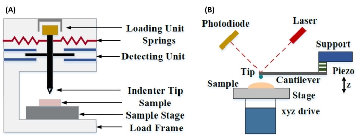
איור 1: השוואה בין מערכות ננו-הזחה מבוססות מכשור ו-AFM. דיאגרמות סכמטיות המתארות מערכות אופייניות לביצוע (A) ננו-הזחה מכשורית ו-(B) ננו-הזחה מבוססת AFM. נתון זה שונה מ Qian et al.51. קיצור: AFM = מיקרוסקופ כוח אטומי. אנא לחץ כאן כדי להציג גרסה גדולה יותר של איור זה.

הן ננו-הזחה מכשורית והן ננו-הזחה מבוססת AFM משתמשות בגשושית נוקשה כדי לעוות משטח דגימה מעניין ולנטר את הכוח והתזוזה המתקבלים כתוצאה מכך כפונקציה של זמן. בדרך כלל, פרופיל התזוזה הרצוי (כלומר, כוח) או (Z-piezo) מוגדר על ידי המשתמש באמצעות ממשק התוכנה ונשלט ישירות על ידי המכשיר, בעוד הפרמטר השני נמדד. התכונה המכנית המתקבלת לרוב מניסויי ננו-הזחה היא המודולוס האלסטי (E), המכונה גם מודולוס יאנג, שיש לו יחידות לחץ. המודולוס האלסטי של חומר הוא תכונה בסיסית הקשורה לנוקשות הקשר והוא מוגדר כיחס בין מתח מתיחה או דחיסה (σ, הכוח המופעל ליחידת שטח) למאמץ צירי (ε, העיוות היחסי לאורך ציר ההזחה) במהלך עיוות אלסטי (כלומר, הפיך או זמני) לפני תחילת העיוות הפלסטי (משוואה [1]):

Equation 1(1)

יש לציין כי מכיוון שחומרים רבים (בעיקר רקמות ביולוגיות) הם למעשה ויסקו-אלסטיים, במציאות, המודולוס (הדינמי או המורכב) מורכב הן מרכיבים אלסטיים (אחסון, בפאזה) והן צמיגים (אובדן, מחוץ לפאזה). בפועל, מה שנמדד בניסוי ננו-הזחה הוא המודולוס המופחת, E *, הקשור למודולוס המדגם האמיתי של עניין, E, כפי שמוצג במשוואה (2):
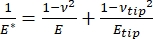
Equation 2(2)

כאשר קצה E וקצה ν הם המודולוס האלסטי והיחס של פואסון, בהתאמה, של קצה הננו-אינדנטר, ו-ν הוא היחס המשוער של פואסון לדגימה. יחס פואסון הוא היחס השלילי בין המתח הרוחבי למתח הצירי, ולכן מציין את מידת ההתארכות הרוחבית של דגימה כאשר היא נתונה למאמץ צירית (למשל, במהלך העמסת ננו-הזחה), כפי שמוצג במשוואה (3):
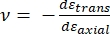
Equation 3(3)

ההמרה ממודולוס מופחת למודולוס ממשי נחוצה מכיוון ש-א) חלק מהמתח הצירי המוקנה על ידי קצה האינדנטר עשוי להיות מומר לזן רוחבי (כלומר, הדגימה עלולה להתעוות באמצעות התרחבות או כיווץ בניצב לכיוון ההעמסה), וב) קצה האינדנדר אינו קשה עד אינסוף, ולכן פעולת הזחת הדגימה גורמת לעיוות מסוים (קטן) של הקצה. שימו לב שבמקרה שבו קצה E >> E (כלומר, קצה האינדנטר קשה הרבה יותר מהדגימה, מה שנכון לעתים קרובות בעת שימוש בבדיקת יהלום), הקשר בין מודולוס הדגימה המוקטן לבין מודולוס הדגימה בפועל מפשט מאוד ל- E ≈ E*(1 v2). בעוד שננו-הזחה מכשורית עדיפה במונחים של אפיון כוח מדויק וטווח דינמי, ננו-הזחה מבוססת AFM מהירה יותר, מספקת בסדרי גודל כוח ורגישות תזוזה גדולים יותר, מאפשרת הדמיה ברזולוציה גבוהה יותר ואיתור כניסה משופר, ויכולה לחקור בו זמנית תכונות מגנטיות וחשמליות ננומטריות9. בפרט, ננו-הזחה מבוססת AFM עדיפה לכימות תכונות מכניות בקנה מידה ננומטרי של חומרים רכים (למשל, פולימרים, ג’לים, דו-שכבות ליפידים ותאים או חומרים ביולוגיים אחרים), שכבות דקות במיוחד (תת-מיקרומטר) (שבהן אפקטים של המצע יכולים להיכנס לפעולה בהתאם לעומק הכניסה)10,11, וחומרים דו-ממדיים מרחפים (דו-ממדיים) 12,13,14 כגון גרפן 15,16, נציץ17, בורון ניטריד משושה (H-BN)18, או דיכלקוגנידים של מתכת מעבר (TMDCs; למשל, MoS2)19. זאת בשל רגישות הכוח המעולה (sub-nN) והתזוזה (sub-nm), החשובה לקביעה מדויקת של נקודת המגע הראשונית ולהישאר בתוך אזור העיוות האלסטי.

בננו-הזחה מבוססת AFM, תזוזה של גשושית AFM לכיוון משטח הדגימה מופעלת על-ידי אלמנט פיאזואלקטרי מכויל (איור 1B), כאשר הכנף הגמיש מתכופף בסופו של דבר בשל כוח ההתנגדות הנחווה במגע עם משטח הדגימה. כיפוף או סטייה זו של המגן מנוטרת בדרך כלל על ידי החזרת לייזר מהחלק האחורי של הפתח לתוך פוטו-גלאי (גלאי רגיש מיקום [PSD]). יחד עם הידע על קשיחות הקנטיליבר (ב-nN/nm) ורגישות הסטייה (ב-nm/V), ניתן להמיר את הסטייה הנמדדת הזו (ב-V) לכוח (ב-nN) המופעל על הדגימה. לאחר מגע, ההבדל בין תנועת Z-piezo לבין הטיית הקנטליבר מניב את עומק הזחת הדגימה. בשילוב עם הידע של פונקציית אזור הקצה, זה מאפשר חישוב של אזור המגע של קצה הדגימה. לאחר מכן ניתן להתאים את השיפוע של חלקי המגע של עקומות מרחק הכוח או תזוזת הכוח (F-D) המתקבלות באמצעות מודל מכניקת מגע מתאים (ראה פרק ניתוח הנתונים בדיון) כדי לקבוע את התכונות הננו-מכניות של הדגימה. בעוד שלננו-הזחה מבוססת AFM יש כמה יתרונות ברורים על פני ננו-הזחה מכשורית כמתואר לעיל, היא גם מציגה מספר אתגרי יישום מעשיים, כגון כיול, שחיקת קצה וניתוח נתונים, שיידונו כאן. חיסרון פוטנציאלי נוסף של ננו-הזחה מבוססת AFM הוא ההנחה של גמישות ליניארית, מכיוון שרדיוס המגע ועומק ההזחה צריכים להיות קטנים בהרבה מרדיוס הכניסה, דבר שיכול להיות קשה להשגה בעבודה עם גשושיות AFM ננומטריות ו / או דגימות המציגות חספוס פני שטח משמעותי.

באופן מסורתי, ננו-הזחה הוגבלה למיקומים בודדים או לניסויי הזחת רשת קטנים, שבהם נבחר מיקום רצוי (כלומר, אזור עניין [ROI]) וכניסה מבוקרת יחידה, כניסות מרובות במיקום יחיד המופרדות על ידי זמן המתנה מסוים, ו/או רשת גסה של כניסות מבוצעות בקצב בסדר גודל של הרץ. עם זאת, ההתקדמות האחרונה ב- AFM מאפשרת רכישה בו זמנית של תכונות מכניות וטופוגרפיה באמצעות שימוש במצבי הדמיה מבוססי עקומת כוח במהירות גבוהה (המכונים בשמות מסחריים שונים בהתאם ליצרן המערכת), שבהם עקומות הכוח מתבצעות בקצב kHz תחת בקרת עומס, כאשר כוח הדגימה המרבי מנוצל כנקודת ההדמיה. פותחו גם שיטות point-and-shoot, המאפשרות רכישה של תמונת טופוגרפיה AFM ואחריה ננו-הזחה סלקטיבית בנקודות עניין בתוך התמונה, מה שמאפשר שליטה מרחבית ננומטרית על מיקום ננו-הזחה. למרות שזה לא המוקד העיקרי של עבודה זו, דוגמאות ספציפיות שנבחרו ליישומים הן של הדמיה מבוססת עקומת כוח והן של ננו-הזחה מבוססת קנטיליבר של point-and-shoot מוצגות בתוצאות המייצגות, וניתן להשתמש בהן בשילוב עם הפרוטוקול המתואר להלן אם זמין בפלטפורמת AFM הספציפית שבה נעשה שימוש. באופן ספציפי, עבודה זו מתווה פרוטוקול כללי ליישום מעשי של ננו-הזחה מבוססת AFM על כל מערכת AFM בעלת יכולת ומספקת ארבע דוגמאות מקרה שימוש (שתיים באוויר, שתיים בנוזל) של הטכניקה, כולל תוצאות מייצגות ודיון מעמיק בניואנסים, באתגרים ובשיקולים החשובים ליישום מוצלח של הטכניקה.

Protocol

הערה: בשל המגוון הרחב של AFM הזמינים מסחרית ומגוון סוגי הדגימות והיישומים הקיימים עבור ננו-הזחה מבוססת קנטילבר, הפרוטוקול הבא מתוכנן במכוון להיות כללי יחסית באופיו, תוך התמקדות בצעדים המשותפים הדרושים לכל ניסויי ננו-הזחה מבוססי קנטילבר ללא קשר למכשיר או ליצרן. מסיבה זו, המחברים מניחים שלקו…

Representative Results

עקומות תזוזת כוחאיור 7 מראה עקומות F-D מייצגות, כמעט אידיאליות, שהתקבלו מניסויי ננו-הזחה שבוצעו באוויר על דגימות אורן לובולי משובץ שרף (איור 7A) ובנוזל (מלוחים חוצצי פוספט [PBS]) על גרעיני תאי גזע מזנכימליים (MSC) (איור 7B). השימוש בכל מודל …

Discussion

הכנת דוגמאות
עבור ננו-הזחה באוויר, שיטות הכנה נפוצות כוללות הקפאה (למשל, דגימות רקמות), השחזה ו/או ליטוש ולאחריהן אולטרה-מיקרוטומינג (למשל, דגימות ביולוגיות משובצות שרף), כרסום יונים או הכנת קרן יונים ממוקדת (למשל, מוליכים למחצה, נקבוביים או דגימות קשיות מעורבות שאינן ניתנות לליט?…

Divulgations

The authors have nothing to disclose.

Acknowledgements

כל ניסויי AFM בוצעו במעבדה למדעי פני השטח של אוניברסיטת בויסי סטייט (SSL). אפיון SEM בוצע במרכז בויסי סטייט לאפיון חומרים (BSCMC). המחקר שדווח בפרסום זה בנוגע לחומרי הזנה לדלק ביולוגי נתמך בחלקו על ידי משרד האנרגיה האמריקאי, המשרד להתייעלות אנרגטית ואנרגיה מתחדשת, המשרד לטכנולוגיות ביו-אנרגיה כחלק מקונסורציום ממשק המרת חומרי הזנה (FCIC), ותחת חוזה משרד התפעול של DOE איידהו DE-AC07-051ID14517. מחקרים במכניקת התא נתמכו על ידי המכונים הלאומיים לבריאות (ארה”ב) תחת מענקים AG059923, AR075803 ו-P20GM109095, ועל ידי מענקי הקרן הלאומית למדע (ארה”ב) 1929188 ו-2025505. עבודת המודל של מערכות דו-שכבתיות שומנים נתמכה על ידי המכונים הלאומיים לבריאות (ארה”ב) תחת מענק R01 EY030067. המחברים מודים לד”ר אלטון גראוגנארד על הפקת התמונה המרוכבת שמוצגת באיור 11.

Materials

Atomic force microscope Bruker Dimension Icon Uses Nanoscope control software, including PeakForce Quantitative Nanomechanical Mapping (PF-QNM), FastForce Volume (FFV), and Point-and-Shoot Ramping experimental workspaces
AtomicJ American Institute of Physics https://doi.org/10.1063/1.4881683 Flexible, powerful, free open source Java-based force curve analysis software package. Supports numerous contact mechanic models, such as Hertz, Sneddon DMT, JKR, Maugis, and cone or pyramid (including blunt and truncated). Also includes a variety of initial contact point estimation methods to choose from. Supports batch processing of data and subsequent statistical analysis (e.g., averages, standard deviations, histograms, goodness of fit, etc.). Literature citation is: P. Hermanowicz, M. Sarna, K. Burda, and H. GabryEquation 1, “AtomicJ: An open source software for analysis of force curves” Rev. Sci. Instrum. 85: 063703 (2014), https://doi.org/10.1063/1.4881683
Buffer solution (PBS) Fisher Chemical (NaCl), Sigma Aldrich (KCl), Fisher BioReagents (Na2HPO4 and KH2PO4) S271 (>99% purity NaCl), P9541 (>99% purity KCl), BP332(>99% purity Na2HPO4), BP362 (>99% purity KH2PO4) Phosphate buffered saline (PBS) was prepared in the laboratory as an aqueous solution consisting of 137 mM NaCl, 2.7 mM KCl, 10 mM Na2HPO4, and 1.8 mM KH2PO4 dissolved in ultrapure water. Reagents were measured out using an analytical balance, and glassware was cleaned with soap and water followed by autoclaving immediately prior to use.
Chloroform
Diamond tip AFM probe Bruker PDNISP Pre-mounted factory-calibrated cube corner diamond (E = 1140 GPa) tip AFM probe (nominal R = 40 nm) with a stainless steel cantilever (nominal k = 225 N/m, f0 = 50 kHz). Spring constant is measured at the factory (k = 256 N/m for the probe, Serial #13435414, used here) and calibration data (including AFM images of indents showing probe geometry) is provided with the probe.
Diamond ultramicrotome blade Diatome Ultra 35° 2.1 mm width. Also used a standard glass blade for intial rough cut of sample surface before transitioning to diamond blade for final surface preparation
Epoxy Gorilla Glue 26853-31-6 Epoxy resin and hardner were mixed in a 1:1 ratio, a small drop was placed on a stainless steel sample puck (Ted Pella), and V1 grade muscovite mica (Ted Pella) was attached to create an atomically flat surface for preparation of phospholipid membranes.
Ethanol
LR white resin, medium grade (catalyzed) Electron Microscopy Sciences 14381 500 mL bottle, Lot #150629
Mesenchymal stem cells (MSCs) N/A N/A MSCs for nanomechanical studies were primary cells harvested from 8-10 week old male C57BL/6 mice as described in Goelzer, M. et al. "Lamin A/C Is Dispensable to Mechanical Repression of Adipogenesis" Int J Mol Sci 22: 6580 (2021) doi:10.3390/ijms22126580 and Peister, A. et al. "Adult stem cells from bone marrow (MSCs) isolated from different strains of inbred mice vary in surface epitopes, rates of proliferation, and differentiation potential" Blood 103: 1662-1668 (2004), doi:10.1182/blood-2003-09-3070.
Modulus standards Bruker PFQNM-SMPKIT-12M Used HOPG (E = 18 GPa) and PS (E = 2.7 GPa). Also contains 2x PDMS (Tack 0, E = 2.5 MPa; Tack 4, E = 3.5 MPa), PS-LDPE (E = 2.0/0.2 GPa), fused silica (E = 72.9 GPa), sapphire (E – 345 GPa), and tip characterization (titanium roughness) sample. All samples come pre-mounted on a 12 mm diameter steel disc (sample puck).
Muscovite mica Ted Pella 50-12 12 mm diameter, V1 grade muscovite mica
Nanscope Analysis Bruker Version 2.0 Free AFM image processing and analysis software package, but designed for, and proprietary/limited to Bruker AFMs; similar functionality is available from free, platform-independent AFM image processing and analysis software packages such as Gwyddion, WSxM, and others. Has built-in capabilities for force curve analysis, but AtomicJ is more flexible/full featured (e.g., more built-in contact mechanics models to choose from, statistical analysis of force curve fitting results, etc.) for force curve analysis and handles batch processing of force curves.
Phospholipids: POPC, Cholesterol (ovine) Avanti Polar Lipids POPC: CAS # 26853-31-6, Cholesterol: CAS # 57-88-5 POPC lipid dissolved in chloroform (25 mg/mL) was obtained from vendor and used without further purification. Cholesterol powder from the same vendor was dissolved in chloroform (20 mg/mL). 
Probe holder (fluid, lipid bilayers) Bruker MTFML-V2 Specific to the particular AFM used; MTFML-V2 is a glass probe holder for scanning in fluid on a MultiMode AFM.
Probe holder (fluid, MSCs) Bruker FastScan Bio Z-scanner Used with Dimension FastScan head (XY flexure scanners). Serial number MXYPOM5-1B154.
Probe holder (standard, ambient) Bruker DAFMCH Specific to the particular AFM used; DAFMCH is the standard contact and tapping mode probe holder for the Dimension Icon AFM, suitable for nanoindentation (PF-QNM, FFV, and point-and-shoot ramping)
Sample Puck Ted Pella 16218 Product number is for 15 mm diameter stainless steel sample puck. Also available in 6 mm, 10 mm, 12 mm, and 20 mm diameters at https://www.tedpella.com/AFM_html/AFM.aspx#anchor842459
Sapphire substrate Bruker PFQNM-SMPKIT-12M Extremely hard surface (E = 345 GPa) for measuring deflection sensitivity of probes (want all of the deflection to come from the probe, not the substrate). Part of the PF-QNM/modulus standards kit.
Scanning electron microscope Hitachi S-3400N-II Located at Boise State. Used to perform co-localized SEM/EDS on all samples except additively manufactured (AM) Ti-6Al-4V.
Silicon AFM probes (standard) NuNano Scout 350 Standard tapping mode silicon probe with reflective aluminum backside coating; k = 42 N/m (nominal), f0 = 350 kHz. Nominal R = 5 nm. Also available uncoated or with reflective gold backside coating. Probes with similar specifications are available from other manufacturers (e.g., Bruker TESPA-V2).
Silicon AFM probes (stiff) Bruker RTESPA-525, RTESPA-525-30  Rotated tip etched silicon probes with reflective aluminum backside coating; k = 200 N/m (nominal), f0 = 525 kHz. Nominal R = 8 nm for RTESPA-525, R = 30 nm for RTESPA-525-30. Spring constant of each RTESPA-525-30 is measured individually at the factory via laser Doppler vibrometry and supplied with the probe.
Silicon carbide grit paper (abrasive discs) Allied 50-10005 120 grit
Silicon nitride AFM probes (soft, large radius hemispherical tip) Bruker MLCT-SPH-5UM, MLCT-SPH-5UM-DC Also MLCT-SPH-1UM-DC. New product line of factory-calibrated (probe radius and spring constants of all cantilevers) large radius (R = 1 or 5 mm) hemispherical tip (at the end of a 23 mm long cylindrical shaft) probes. DC = drift compensation coating. 6 cantilevers/probe (A-F). Nominal spring constants: A, k = 0.07 N/m; B, k = 0.02 N/m; C, k = 0.01 N/m; D, k = 0.03 N/m; E, k = 0.1 N/m; F, k = 0.6 N/m.
Silicon nitride AFM probes (soft, medium sharp tip) Bruker DNP 4 cantilevers/probe (A-d). Nominal spring constants: A, k = 0.35 N/m; B, k = 0.12 N/m; C, k = 0.24 N/m; D, k = 0.06 N/m. Nominal radii of curvature, R = 10 nm.
Silicon nitride AFM probes (soft, sharp tip) Bruker ScanAsyst-Air Nominal values: resonance frequency, f0 = 70 kHz; spring constant, k = 0.4 N/m; radius of curvature, R = 2 nm. Designed for force curve based AFM imaging.
Superglue Henkel Loctite 495 Cyanoacrylate based instant adhesive. Lots of roughly equivalent products are readily available.
Syringe pump New Era Pump Systems NE1000US One channel syringe pump system with infusion and withdrawal capacity
Tip characterization standard Bruker PFQNM-SMPKIT-12M Titanium (Ti) roughness standard. Part of the PF-QNM/modulus standards kit.
Ultrahigh purity nitrogen (UHP N2), 99.999% Norco SPG TUHPNI – T T size compressed gas cylinder of ultrahigh purity (99.999%) nitrogen for drying samples
Ultramicrotome Leica EM UC6 Equipped with a glass blade (standard, for intial sample preparation) and a diamond blade (for final preparation)
Ultrapure water Thermo Fisher Barnstead Nanopure Model 7146 Model has been discontinued, but equivalent products are available. Produces ≥18.2 MΩ*cm ultrapure water with 1-5 ppb TOC (total organic content), per inline UV monitoring. Includes 0.2 µm particulate filter, ion exchange columns, and UV oxidation chamber.
Variable Speed Grinder Buehler EcoMet 3000 Used with silicon carbide grit papers during hand polishing.
Vibration isolation table (active) Herzan TS-140 Used with Bruker MultiMode AFM. Sits on a TMC 65-531 vibration isolation table. Bruker Dimension Icon AFM utilizes strictly passive vibration isolation (comes from manufacturer with custom acoustic hood, air table, and granite slab).
Vibration isolation table (passive) TMC 65-531 35" x 30" vibration isolation table with optional air damping (disabled). Used with Bruker MultiMode AFM. Herzan TS-140 "Table Stable" active vibration control table is located on top.

References

  1. Hart, E. W. Theory of the tensile test. Acta Metallurgica. 15 (2), 351-355 (1967).
  2. Fell, J. T., Newton, J. M. Determination of tablet strength by the diametral-compression test. Journal of Pharmaceutical Sciences. 59 (5), 688-691 (1970).
  3. Babiak, M., Gaff, M., Sikora, A., Hysek, &. #. 3. 5. 2. ;. Modulus of elasticity in three- and four-point bending of wood. Composite Structures. 204, 454-465 (2018).
  4. Song, S., Yovanovich, M. M. Relative contact pressure-Dependence on surface roughness and Vickers microhardness. Journal of Thermophysics and Heat Transfer. 2 (1), 43-47 (1988).
  5. Hays, C., Kendall, E. G. An analysis of Knoop microhardness. Metallography. 6 (4), 275-282 (1973).
  6. Hill, R., Storåkers, B., Zdunek, A. B. A theoretical study of the Brinell hardness test. Proceedings of the Royal Society of London. A. Mathematical and Physical Sciences. 423 (1865), 301-330 (1989).
  7. Oliver, W. C., Pharr, G. M. An improved technique for determining hardness and elastic modulus using load and displacement sensing indentation experiments. Journal of Materials Research. 7 (6), 1564-1583 (1992).
  8. Sakharova, N. A., Fernandes, J. V., Antunes, J. M., Oliveira, M. C. Comparison between Berkovich, Vickers and conical indentation tests: A three-dimensional numerical simulation study. International Journal of Solids and Structures. 46 (5), 1095-1104 (2009).
  9. Cohen, S. R., Kalfon-Cohen, E. Dynamic nanoindentation by instrumented nanoindentation and force microscopy: a comparative review. Beilstein Journal of Nanotechnology. 4 (1), 815-833 (2013).
  10. Saha, R., Nix, W. D. Effects of the substrate on the determination of thin film mechanical properties by nanoindentation. Acta Materialia. 50 (1), 23-38 (2002).
  11. Tsui, T. Y., Pharr, G. M. Substrate effects on nanoindentation mechanical property measurement of soft films on hard substrates. Journal of Materials Research. 14 (1), 292-301 (1999).
  12. Cao, G., Gao, H. Mechanical properties characterization of two-dimensional materials via nanoindentation experiments. Progress in Materials Science. 103, 558-595 (2019).
  13. Castellanos-Gomez, A., Singh, V., vander Zant, H. S. J., Steele, G. A. Mechanics of freely-suspended ultrathin layered materials. Annalen der Physik. 527 (1-2), 27-44 (2015).
  14. Cao, C., Sun, Y., Filleter, T. Characterizing mechanical behavior of atomically thin films: A review. Journal of Materials Research. 29 (3), 338-347 (2014).
  15. Lee, C., Wei, X., Kysar, J. W., Hone, J. Measurement of the elastic properties and intrinsic strength of monolayer graphene. Science. 321 (5887), 385-388 (2008).
  16. Elibol, K., et al. Visualising the strain distribution in suspended two-dimensional materials under local deformation. Scientific Reports. 6 (1), 28485 (2016).
  17. Castellanos-Gomez, A., et al. Mechanical properties of freely suspended atomically thin dielectric layers of mica. Nano Research. 5 (8), 550-557 (2012).
  18. Song, L., et al. Large scale growth and characterization of atomic hexagonal boron nitride layers. Nano Letters. 10 (8), 3209-3215 (2010).
  19. Castellanos-Gomez, A., et al. Elastic properties of freely suspended MoS2 nanosheets. Advanced Materials. 24 (6), 772-775 (2012).
  20. D’Costa, N. P., Hoh, J. H. Calibration of optical lever sensitivity for atomic force microscopy. Review of Scientific Instruments. 66 (10), 5096-5097 (1995).
  21. Wu, Y., et al. Evaluation of elastic modulus and hardness of crop stalks cell walls by nano-indentation. Bioresource Technology. 101 (8), 2867-2871 (2010).
  22. Barns, S., et al. Investigation of red blood cell mechanical properties using AFM indentation and coarse-grained particle method. BioMedical Engineering OnLine. 16 (1), 140 (2017).
  23. Hermanowicz, P., Sarna, M., Burda, K., Gabryś, H. AtomicJ: An open source software for analysis of force curves. Review of Scientific Instruments. 85 (6), 063703 (2014).
  24. Broitman, E. Indentation hardness measurements at macro-, micro-, and nanoscale: a critical overview. Tribology Letters. 65 (1), 23 (2016).
  25. Tiwari, A. . Nanomechanical Analysis of High Performance Materials. , (2015).
  26. Aggarwal, R. L., Ramdas, A. K. . Physical Properties of Diamond and Sapphire. , (2019).
  27. Boyd, E. J., Uttamchandani, D. Measurement of the anisotropy of Young’s modulus in single-crystal silicon. Journal of Microelectromechanical Systems. 21 (1), 243-249 (2012).
  28. Harding, J. W., Sneddon, I. N. The elastic stresses produced by the indentation of the plane surface of a semi-infinite elastic solid by a rigid punch. Mathematical Proceedings of the Cambridge Philosophical Society. 41 (1), 16-26 (2008).
  29. Lin, D. C., Dimitriadis, E. K., Horkay, F. Robust strategies for automated AFM force curve analysis-I. Non-adhesive indentation of soft, inhomogeneous materials. Journal of Biomechanical Engineering. 129 (3), 430-440 (2006).
  30. Lin, D. C., Dimitriadis, E. K., Horkay, F. Robust strategies for automated AFM force curve analysis-II: Adhesion-influenced indentation of soft, elastic materials. Journal of Biomechanical Engineering. 129 (6), 904-912 (2007).
  31. Haile, S., Palmer, M., Otey, A. Potential of loblolly pine: switchgrass alley cropping for provision of biofuel feedstock. Agroforestry Systems. 90 (5), 763-771 (2016).
  32. Lu, X., et al. Biomass logistics analysis for large scale biofuel production: Case study of loblolly pine and switchgrass. Bioresource Technology. 183, 1-9 (2015).
  33. Susaeta, A., Lal, P., Alavalapati, J., Mercer, E., Carter, D. Economics of intercropping loblolly pine and switchgrass for bioenergy markets in the southeastern United States. Agroforestry Systems. 86 (2), 287-298 (2012).
  34. Garcia, R. Nanomechanical mapping of soft materials with the atomic force microscope: methods, theory and applications. Chemical Society Reviews. 49 (16), 5850-5884 (2020).
  35. Derjaguin, B. V., Muller, V. M., Toporov, Y. P. Effect of contact deformations on the adhesion of particles. Journal of Colloid and Interface Science. 53 (2), 314-326 (1975).
  36. Ciesielski, P. N., et al. Engineering plant cell walls: tuning lignin monomer composition for deconstructable biofuel feedstocks or resilient biomaterials. Green Chemistry. 16 (5), 2627-2635 (2014).
  37. Liu, K., Ostadhassan, M., Zhou, J., Gentzis, T., Rezaee, R. Nanoscale pore structure characterization of the Bakken shale in the USA. Fuel. 209, 567-578 (2017).
  38. Maryon, O. O., et al. Co-localizing Kelvin probe force microscopy with other microscopies and spectroscopies: selected applications in corrosion characterization of alloys. JoVE. (184), e64102 (2022).
  39. Eliyahu, M., Emmanuel, S., Day-Stirrat, R. J., Macaulay, C. I. Mechanical properties of organic matter in shales mapped at the nanometer scale. Marine and Petroleum Geology. 59, 294-304 (2015).
  40. Li, C., et al. Nanomechanical characterization of organic matter in the Bakken formation by microscopy-based method. Marine and Petroleum Geology. 96, 128-138 (2018).
  41. Bouzid, T., et al. The LINC complex, mechanotransduction, and mesenchymal stem cell function and fate. Journal of Biological Engineering. 13 (1), 68 (2019).
  42. Dupont, S., et al. Role of YAP/TAZ in mechanotransduction. Nature. 474 (7350), 179-183 (2011).
  43. Wang, S., et al. CCM3 is a gatekeeper in focal adhesions regulating mechanotransduction and YAP/TAZ signalling. Nature Cell Biology. 23 (7), 758-770 (2021).
  44. Sen, B., et al. Mechanical strain inhibits adipogenesis in mesenchymal stem cells by stimulating a durable β-catenin signal. Endocrinology. 149 (12), 6065-6075 (2008).
  45. Sen, B., et al. mTORC2 regulates mechanically induced cytoskeletal reorganization and lineage selection in marrow-derived mesenchymal stem cells. Journal of Bone and Mineral Research. 29 (1), 78-89 (2014).
  46. Sen, B., et al. Mechanically induced nuclear shuttling of β-catenin requires co-transfer of actin. Stem Cells. 40 (4), 423-434 (2022).
  47. Newberg, J., et al. Isolated nuclei stiffen in response to low intensity vibration. Journal of Biomechanics. 111, 110012 (2020).
  48. Ding, Y., Xu, G. -. K., Wang, G. -. F. On the determination of elastic moduli of cells by AFM based indentation. Scientific Reports. 7 (1), 45575 (2017).
  49. Khadka, N. K., Timsina, R., Rowe, E., O’Dell, M., Mainali, L. Mechanical properties of the high cholesterol-containing membrane: An AFM study. Biochimica et Biophysica Acta. Biomembranes. 1863 (8), 183625 (2021).
  50. Castellana, E. T., Cremer, P. S. Solid supported lipid bilayers: From biophysical studies to sensor design. Surface Science Reports. 61 (10), 429-444 (2006).
  51. Qian, L., Zhao, H. Nanoindentation of soft biological materials. Micromachines. 9 (12), 654 (2018).
  52. Pittenger, B., Yablon, D. Improving the accuracy of nanomechanical measurements with force-curve-based AFM techniques. Bruker Application Notes. 149, (2017).
  53. Vorselen, D., Kooreman, E. S., Wuite, G. J. L., Roos, W. H. Controlled tip wear on high roughness surfaces yields gradual broadening and rounding of cantilever tips. Scientific Reports. 6 (1), 36972 (2016).
  54. Bhaskaran, H., et al. Ultralow nanoscale wear through atom-by-atom attrition in silicon-containing diamond-like carbon. Nature Nanotechnology. 5 (3), 181-185 (2010).
  55. Giannazzo, F., Schilirò, E., Greco, G., Roccaforte, F. Conductive atomic force microscopy of semiconducting transition metal dichalcogenides and heterostructures. Nanomaterials. 10 (4), 803 (2020).
  56. Melitz, W., Shen, J., Kummel, A. C., Lee, S. Kelvin probe force microscopy and its application. Surface Science Reports. 66 (1), 1-27 (2011).
  57. Kazakova, O., et al. Frontiers of magnetic force microscopy. Journal of Applied Physics. 125 (6), 060901 (2019).
  58. Kim, H. -. J., Yoo, S. -. S., Kim, D. -. E. Nano-scale wear: A review. International Journal of Precision Engineering and Manufacturing. 13 (9), 1709-1718 (2012).
  59. Heath, G. R., et al. Localization atomic force microscopy. Nature. 594 (7863), 385-390 (2021).
  60. Strahlendorff, T., Dai, G., Bergmann, D., Tutsch, R. Tip wear and tip breakage in high-speed atomic force microscopes. Ultramicroscopy. 201, 28-37 (2019).
  61. Lantz, M. A., et al. Wear-resistant nanoscale silicon carbide tips for scanning probe applications. Advanced Functional Materials. 22 (8), 1639-1645 (2012).
  62. Khurshudov, A. G., Kato, K., Koide, H. Wear of the AFM diamond tip sliding against silicon. Wear. 203, 22-27 (1997).
  63. Villarrubia, J. S. Algorithms for scanned probe microscope image simulation, surface reconstruction, and tip estimation. Journal of Research of the National Institute of Standards and Technology. 102 (4), 425 (1997).
  64. Kain, L., et al. Calibration of colloidal probes with atomic force microscopy for micromechanical assessment. Journal of the Mechanical Behavior of Biomedical Materials. 85, 225-236 (2018).
  65. Slattery, A. D., Blanch, A. J., Quinton, J. S., Gibson, C. T. Accurate measurement of Atomic Force Microscope cantilever deflection excluding tip-surface contact with application to force calibration. Ultramicroscopy. 131, 46-55 (2013).
  66. Dobrovinskaya, E. R., Lytvynov, L. A., Pishchik, V. . Sapphire: Material, Manufacturing, Applications. , (2009).
  67. te Riet, J., et al. Interlaboratory round robin on cantilever calibration for AFM force spectroscopy. Ultramicroscopy. 111 (12), 1659-1669 (2011).
  68. Pratt, J. R., Shaw, G. A., Kumanchik, L., Burnham, N. A. Quantitative assessment of sample stiffness and sliding friction from force curves in atomic force microscopy. Journal of Applied Physics. 107 (4), 044305 (2010).
  69. Slattery, A. D., Blanch, A. J., Quinton, J. S., Gibson, C. T. Calibration of atomic force microscope cantilevers using standard and inverted static methods assisted by FIB-milled spatial markers. Nanotechnology. 24 (1), 015710 (2012).
  70. Higgins, M. J., et al. Noninvasive determination of optical lever sensitivity in atomic force microscopy. Review of Scientific Instruments. 77 (1), 013701 (2006).
  71. Lévy, R., Maaloum, M. Measuring the spring constant of atomic force microscope cantilevers: thermal fluctuations and other methods. Nanotechnology. 13 (1), 33-37 (2001).
  72. Sikora, A. Quantitative normal force measurements by means of atomic force microscopy towards the accurate and easy spring constant determination. Nanoscience and Nanometrology. 2 (1), 8-29 (2016).
  73. Ohler, B. Cantilever spring constant calibration using laser Doppler vibrometry. Review of Scientific Instruments. 78 (6), 063701 (2007).
  74. Gates, R. S., Pratt, J. R. Accurate and precise calibration of AFM cantilever spring constants using laser Doppler vibrometry. Nanotechnology. 23 (37), 375702 (2012).
  75. Cleveland, J. P., Manne, S., Bocek, D., Hansma, P. K. A nondestructive method for determining the spring constant of cantilevers for scanning force microscopy. Review of Scientific Instruments. 64 (2), 403-405 (1993).
  76. Sader, J. E., Chon, J. W. M., Mulvaney, P. Calibration of rectangular atomic force microscope cantilevers. Review of Scientific Instruments. 70 (10), 3967-3969 (1999).
  77. Sader, J. E., et al. Spring constant calibration of atomic force microscope cantilevers of arbitrary shape. Review of Scientific Instruments. 83 (10), 103705 (2012).
  78. Sader, J. E. Frequency response of cantilever beams immersed in viscous fluids with applications to the atomic force microscope. Journal of Applied Physics. 84 (1), 64-76 (1998).
  79. Sader, J. E., Pacifico, J., Green, C. P., Mulvaney, P. General scaling law for stiffness measurement of small bodies with applications to the atomic force microscope. Journal of Applied Physics. 97 (12), 124903 (2005).
  80. Mendels, D. -. A., et al. Dynamic properties of AFM cantilevers and the calibration of their spring constants. Journal of Micromechanics and Microengineering. 16 (8), 1720-1733 (2006).
  81. Gao, S., Brand, U. In-situ nondestructive characterization of the normal spring constant of AFM cantilevers. Measurement Science and Technology. 25 (4), 044014 (2014).
  82. Gibson, C. T., Watson, G. S., Myhra, S. Determination of the spring constants of probes for force microscopy/spectroscopy. Nanotechnology. 7 (3), 259-262 (1996).
  83. Gates, R. S., Pratt, J. R. Prototype cantilevers for SI-traceable nanonewton force calibration. Measurement Science and Technology. 17 (10), 2852-2860 (2006).
  84. Neumeister, J. M., Ducker, W. A. Lateral, normal, and longitudinal spring constants of atomic force microscopy cantilevers. Review of Scientific Instruments. 65 (8), 2527-2531 (1994).
  85. Kim, M. S., Choi, I. M., Park, Y. K., Kang, D. I. Atomic force microscope probe calibration by use of a commercial precision balance. Measurement. 40 (7), 741-745 (2007).
  86. Kim, M. -. S., Choi, J. -. H., Park, Y. -. K., Kim, J. -. H. Atomic force microscope cantilever calibration device for quantified force metrology at micro- or nano-scale regime: the nano force calibrator (NFC). Metrologia. 43 (5), 389-395 (2006).
  87. Tian, Y., et al. A novel method and system for calibrating the spring constant of atomic force microscope cantilever based on electromagnetic actuation. Review of Scientific Instruments. 89 (12), 125119 (2018).
  88. Clifford, C. A., Seah, M. P. The determination of atomic force microscope cantilever spring constants via dimensional methods for nanomechanical analysis. Nanotechnology. 16 (9), 1666-1680 (2005).
  89. Chen, B. -. Y., Yeh, M. -. K., Tai, N. -. H. Accuracy of the spring constant of atomic force microscopy cantilevers by finite element method. Analytical Chemistry. 79 (4), 1333-1338 (2007).
  90. Mick, U., Eichhorn, V., Wortmann, T., Diederichs, C., Fatikow, S. Combined nanorobotic AFM/SEM system as novel toolbox for automated hybrid analysis and manipulation of nanoscale objects. 2010 IEEE International Conference on Robotics and Automation. , 4088-4093 (2010).
  91. Kim, M. -. S., Choi, J. -. H., Kim, J. -. H., Park, Y. -. K. Accurate determination of spring constant of atomic force microscope cantilevers and comparison with other methods. Measurement. 43 (4), 520 (2010).
  92. Zhang, G., Wei, Z., Ferrell, R. E. Elastic modulus and hardness of muscovite and rectorite determined by nanoindentation. Applied Clay Science. 43 (2), 271-281 (2009).
  93. Bobko, C. P., Ortega, J. A., Ulm, F. -. J. Comment on "Elastic modulus and hardness of muscovite and rectorite determined by nanoindentation by G. Zhang, Z. Wei and R.E. Ferrell. Applied Clay Science. 46 (4), 425-428 (2009).
  94. Zhang, G., Wei, Z., Ferrell, R. E. Reply to the Comment on "Elastic modulus and hardness of muscovite and rectorite determined by nanoindentation" by G. Zhang, Z. Wei and R. E. Ferrell. Applied Clay Science. 46 (4), 429-432 (2009).
  95. Jin, D. W., et al. Thermal stability and Young’s modulus of mechanically exfoliated flexible mica. Current Applied Physics. 18 (12), 1486-1491 (2018).
  96. Xiao, J., et al. Anisotropic friction behaviour of highly oriented pyrolytic graphite. Carbon. 65, 53-62 (2013).
  97. Hertz, H. Ueber die Berührung fester elastischer Körper. Journal für die reine und angewandte Mathematik. 1882 (92), 156-171 (1882).
  98. Johnson, K. L., Kendall, K., Roberts, A. D., Tabor, D. Surface energy and the contact of elastic solids. Proceedings of the Royal Society of London. A. Mathematical and Physical Sciences. 324 (1558), 301-313 (1971).
  99. Muller, V. M., Derjaguin, B. V., Toporov, Y. P. On two methods of calculation of the force of sticking of an elastic sphere to a rigid plane. Colloids and Surfaces. 7 (3), 251-259 (1983).
  100. Maugis, D. Adhesion of spheres: The JKR-DMT transition using a dugdale model. Journal of Colloid and Interface Science. 150 (1), 243-269 (1992).
  101. Muller, V. M., Yushchenko, V. S., Derjaguin, B. V. On the influence of molecular forces on the deformation of an elastic sphere and its sticking to a rigid plane. Journal of Colloid and Interface Science. 77 (1), 91-101 (1980).
  102. Muller, V. M., Yushchenko, V. S., Derjaguin, B. V. General theoretical consideration of the influence of surface forces on contact deformations and the reciprocal adhesion of elastic spherical particles. Journal of Colloid and Interface Science. 92 (1), 92-101 (1983).
  103. Johnson, K. L., Greenwood, J. A. An adhesion map for the contact of elastic spheres. Journal of Colloid and Interface Science. 192 (2), 326-333 (1997).
  104. Shi, X., Zhao, Y. -. P. Comparison of various adhesion contact theories and the influence of dimensionless load parameter. Journal of Adhesion Science and Technology. 18 (1), 55-68 (2004).
check_url/fr/64497?article_type=t

Play Video

Citer Cet Article
Enrriques, A. E., Howard, S., Timsina, R., Khadka, N. K., Hoover, A. N., Ray, A. E., Ding, L., Onwumelu, C., Nordeng, S., Mainali, L., Uzer, G., Davis, P. H. Atomic Force Microscopy Cantilever-Based Nanoindentation: Mechanical Property Measurements at the Nanoscale in Air and Fluid. J. Vis. Exp. (190), e64497, doi:10.3791/64497 (2022).

View Video