Here, we present a standard pipeline to obtain murine ATC tumors by spontaneous genetically engineered mouse models. Further, we present tumor dynamics and pathological information about the primary and metastasized lesions. This model will help researchers to understand tumorigenesis and facilitate drug discoveries.
Anaplastic thyroid cancer (ATC) is a rare but lethal malignancy with a dismal prognosis. There is an urgent need for more in-depth research on the carcinogenesis and development of ATC, as well as therapeutic methods, since standard treatments are essentially depleted in ATC patients. However, low prevalence has hampered thorough clinical studies and the collection of tissue samples, so little progress has been achieved in creating effective treatments. We used genetic engineering to create a conditionally inducible ATC murine model (mATC) in a C57BL/6 background. The ATC murine model was genotyped by TPO-cre/ERT2; BrafCA/wt; Trp53ex2-10/ex2-10 and induced by intraperitoneal injection with tamoxifen. With the murine model, we investigated the tumor dynamics (tumor size ranged from 12.4 mm2 to 32.5 mm2 after 4 months of induction), survival (the median survival period was 130 days), and metastasis (lung metastases occurred in 91.6% of mice) curves and pathological features (characterized by Cd8, Foxp3, F4/80, Cd206, Ki67, and Caspase-3 immunohistochemical staining). The results indicated that spontaneous mATC possesses highly similar tumor dynamics and immunological microenvironment to human ATC tumors. In conclusion, with high similarity in pathophysiological features and unified genotypes, the mATC model resolved the shortage of clinical ATC tissue and sample heterogeneity to some extent. Therefore, it would facilitate the mechanism and translational studies of ATC and provide an approach to investigate the treatment potential of small molecular drugs and immunotherapy agents for ATC.
Thyroid cancer is one of the most common endocrine malignancies1, originating from the thyroid epithelium. In recent years, the incidence of thyroid cancer has increased rapidly worldwide2. Thyroid cancer can be divided into distinct types according to the degree of tumor cell differentiation. On the basis of clinical behavior and histology, thyroid carcinomas are divided into well-differentiated carcinomas, including papillary thyroid carcinoma (PTC) and follicular thyroid carcinoma (FTC), poorly differentiated carcinoma (PDTC), and undifferentiated or anaplastic carcinoma of the thyroid (ATC)3. In contrast to PTC, which is a common type with mild behavior and better prognosis4, ATC is a rare and highly aggressive malignancy that accounts for 2% to 3% of all thyroid tumors5. Although ATC is rare, it is responsible for approximately 50% of thyroid cancer-related deaths, with dismal survival (6-8 months)6,7. Over 50% of ATC cases are diagnosed as lung metastasis8. In addition to the aggressive nature of ATC, limited effective treatment has been developed in the clinic. Therefore, ATC patients have a bleak prognosis9,10,11. This suggests that further in-depth studies are urgently needed on the molecular mechanisms underlying the development of ATC and treatment.
The tumorigenesis of ATC is a dynamic dedifferentiated process. The difficulty in collecting human tumor samples at each stage in clinical studies has hindered the understanding of the mechanism of development from well-differentiated to undifferentiated carcinomas. In contrast, the use of murine ATC models (mATC) favors the collection of mATC samples in the whole tumorigenesis course. Therefore, we can better understand the mechanisms of tumor formation by analyzing the dynamic dedifferentiated process. In addition, the heterogeneity of clinical ATC samples has also contributed to the difficulty in understanding the molecular mechanism. Nevertheless, mice shared the same genetic backgrounds and were maintained in similar living environments, ensuring each tumor's consistency. This facilitates exploring the generalized role of ATC development12,13,14. Additionally, mATC is an in situ tumor model that can restore the influence of the anatomic location and tissue-specific microenvironment. As such, compared with commonly used immunodeficient mice, mATC is a spontaneous murine model with an intact immune system and immune microenvironment.
Therefore, we constructed conditionally induced mATC with the C57BL/6 strain, which is a murine model capable of reproducing the pathological features of dedifferentiated thyroid carcinoma. Based on this model, we gave a brief overview of the molecular basis, construction ideas, pathological features, and applications of mATC. In addition, we observed and reported tumor growth, survival time, metastasis, and pathological features of mATC. We believe this will be an informatic overview to assist other researchers in using this model easier.
We constructed a conditional inducible mATC model, as first reported by McFadden15; initially, we constructed mice: TPO-cre/ERT2, Brafflox/wt, and Trp53flox/wt. Specifically, TPO-cre/ERT2 mice included the human thyroid peroxidase (TPO) promoter (a thyroid-specific promoter), driving the expression of a cre-ERT2 fusion gene (a cre recombinase fused to a human estrogen receptor ligand binding domain). Cre-ERT2 is usually confined to the cytoplasm and enters the nucleus only when exposed to tamoxifen, which induces cre to exert recombinant enzyme activity. When the mice are crossed with mice carrying loxP-flanked sequences, after tamoxifen-induction, cre-mediated recombination deletes the floxed sequences in the thyroid cells to achieve the purpose of knocking out or knocking in specific genes.
In addition, Brafflox/wt mice are a knock-in allele of human Braf based on the cre-loxP system. Brafflox/wt murine transcript is encoded by endogenous exons 1-14 and loxP-flanked human exons 15-18. After cre-mediated excision of the floxed regions, the mutant exon 15 (modified with a V600E amino acid substitution linked with constitutively active BrafV600E in human cancers) and the endogenous exons 16-18 are used to generate the transcripts. Furthermore, Trp53flox/wt mice are knockout alleles of human Trp53 and have loxP sites flanking exons 2-10 of Trp53. When crossed with mice with a cre recombinase, cre-mediated recombination deletes the floxed sequence to knock out Trp53. Then, TPO-cre/ERT2, Brafflox/w, and Trp53flox/wt mice were crossed to obtain TB (TPO-cre/ERT2; Brafflox/wt) mice and TBP (TPO-cre/ERT2; Brafflox/wt; Trp53flox/wt) mice, which could be used to generate PTC and ATC. After approximately 8 weeks, the mice were induced by an intraperitoneal (i.p.) administration of 150 mg/kg tamoxifen dissolved in corn oil for two administrations. Tumor growth could be monitored by high-frequency ultrasonography (the first time point of ultrasonography was recorded as Day 0). Initial ultrasonography was performed 40 days after tamoxifen introduction.
The animal procedures described here were performed with the approval of the Animal Ethics Committee of West China Hospital, Sichuan University, Chengdu, Sichuan, China.
1. Induction of TBP mice
2. Dissection and imaging of mouse thyroid tumors and metastatic tumors
3. HE staining of the primary tumor and lung
We induced mATC to investigate tumor growth, mouse survival time, and pathological characteristics. After induction, the mice were immediately sacrificed, and samples (thyroid, lung, and liver) were collected once one of the following conditions were found: 1) respiratory distress caused by tumor compression; 2) decreased appetite and abnormal vocalization; 3) unusually lethargy; and 4) body weight loss of over 20%. During the sampling process, we found that all mice (12/12) successfully formed tumors after induction. We recorded the mouse survival time, tumor features/size, and metastasized lesions.
Grossly, we observed the following: 1) the tumors were tender, and the size of the tumors on the left and right sides were inconsistent; 2) most mice (11/12) had lung metastases, but none had liver metastases. Specifically, the tumor size was monitored by animal high-frequency ultrasound and photoacoustic imaging systems (Vevo®3100) throughout the whole process17. Based on the ultrasound data, the tumor growth curve (Figure 1A) was plotted to observe the dynamic alteration of the tumor size. Furthermore, the tumors grew slowly at the early stage (average tumor size varied from 9.47 mm2 to 11.75 mm2 from Day 0 to Day 60), and became dramatically faster at the late stage (average tumor size varied from 11.75 mm2 to 23.95 mm2 (from Day 60 to Day 100). Most mice were sacrificed in the late stage. In short, mATC with a certain tumor latency period needs to be closely monitored after 60 days to prevent asphyxiation-related death.
On the other hand, the survival time of the mice was recorded, and a survival curve (Figure 1B) was plotted. The median survival of mATC was 130 days, ranging from 56-166 days. In addition, lung metastasis was found in most mATC (approximately 92%) (Figure 1C). We observed that only one mouse in this cohort did not show lung metastases on gross examination, and six mice had more than one lung metastatic lesion. No liver metastasis was found. In brief, these results were consistent with the biological behavior of ATC, which are prone to lung metastasis in clinics.
Furthermore, to better observe the dynamic process of mATC, we sacrificed the mice at two time points (1 month and 2 months after induction). We performed HE staining on the primary tumors and metastatic lung tissues of mATC (Figure 1D). In 1 month inducible tissue, we observed incomplete solidified features and the coexistence of follicular structures and malignant cells. The thyroid follicular structures disappeared, and the tumor solidified completely after a 2 month induction. HE staining of the primary tumor revealed that the tumor cells were morphologically diverse, with pleomorphic giant cells (indicated by a red arrow) and spindle-shaped cells (indicated by a yellow arrow). It also showed a wide variety in nuclear size, and many cells contained multiple nuclei. Clear metastatic foci (indicated by a circle) were seen in the lung. HE staining of metastatic lung tissues showed that the normal lung tissue was a reticular structure with clear alveolar structures and airspaces. Nevertheless, lung metastases showed a loss of normal reticular structure, air cavity thickening, and lung parenchyma.
Meanwhile, IHC staining was performed to further characterize (Cd8, Foxp3, F4/80, Cd206, Ki67, and Caspase-3) mATC tumors to quantify cell proliferation and apoptosis, and investigate the infiltration of lymphocytes, T-regular (Treg) cells, and myeloid cells (Figure 2A–D). Anti-Ki67 staining was highly positive, ranging from 86.9% to 95.07%, demonstrating a high degree of cell proliferation. Anti-activated caspase-3 antibody was used to test the rate of apoptosis, ranging from 5.2% to 51.9%. Specifically, mATC presented obvious CD8+ T cell infiltration, the ratio of which ranged from 0.47% to 10.55% (mean: 5.93%). This indicated that mATC was not an immune-desert tumor, which was consistent with ATC samples. Besides, Foxp3 staining defined Treg cells, which varied from 0.45% to 25.8%. In addition to lymphocytes, F4/80 and Cd206 were used to define macrophages and M2 macrophages, respectively. We found that myeloid cells extensively infiltrated tumors (F4/80 positive cell rate from 86.6% to 94.6%; Cd206 positive cell rate from 40.4% to 67.7%), which was consistent with the previous literature16. In brief, we found highly proliferative tumor cells, lymphocyte infiltration, and extensive infiltration of myeloid cells in mATC, which was consistent with clinical samples.
In conclusion, mATC samples showed homogeneity in tumor dynamics, metastasis, and pathological features, consistent with the clinical samples. Considering the tumor formation rate, mATC was a reliable murine model.
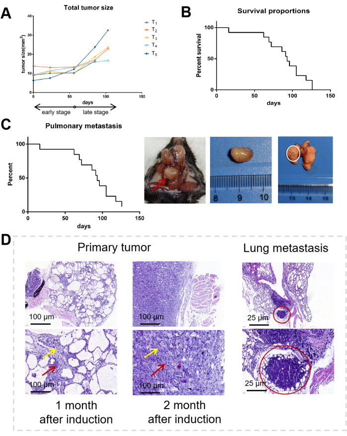
Figure 1: Tumor dynamics and pathological characteristics. (A) Tumor growth curve of mice (n = 5). Each line represents a mouse: the early stage ranges from Day 0 to Day 60, and the late stage ranges from Day 60 to Day 100. (B) Survival curves of mice (n = 12). The median survival of mATC was 130 days, ranging from 56 days to 166 days. (C) Lung metastasis curves of mice (n = 12). Representative dissection images of thyroid and lung metastasis. The red arrow indicates the thyroid tumor, and the white circle indicates metastasis in the lung. (D) HE staining of the primary tumor (1 month and 2 months after induction) and lung metastasis. The red arrow indicates the pleomorphic giant cell, the yellow arrow indicates the spindle-shaped cell, and the circled area indicates a metastasis in the lung. Please click here to view a larger version of this figure.
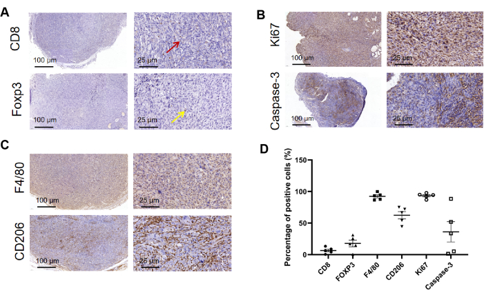
Figure 2: Brief description of immune cell infiltration in mATC. (A) Immunohistochemical staining of ATC murine primary tumors with antibodies (Cd8, Foxp3). The red arrow indicates a CD8 positive cell, the yellow arrow indicates a Foxp3 positive cell. (B) Immunohistochemical staining of ATC murine primary tumors with antibodies (Ki67, Caspase-3). (C) Immunohistochemical staining of ATC murine primary tumors with antibodies (F4/80, Cd206). (D) The quantification of IHC. Please click here to view a larger version of this figure.
Table 1: The list of primers and the PCR settings used in this study. Please click here to download this Table.
Critical steps within the protocol for thyroid tumor dissection
During dissection, the anatomical location of the thyroid gland needs to be correctly understood. The thyroid gland is a butterfly-shaped gland located on the dorsal side of the submandibular gland near the thyroid cartilage and the trachea. During the procedure, severing the blood arteries on both sides of the neck was carefully avoided.
Modification and troubleshooting of the mATC breed
ATC is a rare and highly aggressive malignancy. The clinical characteristics of mATC and ATC patients have certain similarities. Specifically, the major cause of death in mATC is asphyxiation, which is consistent with ATC patients. Therefore, during the experiment, the following should be noted: 1) gently grasp the mice and pay attention to their respiration during the operation; 2) closely monitor during the late stage to prevent sudden death and failure to obtain samples in time; 3) the thyroid and lung tissues are easily affected by the blood on the surface of the tissues for observation and photography, so the blood volume of mice can be reduced by eyeball enucleation.
Limitations of using mATC
The occurrence and development of human ATC is complex and changeable, but the genetic background of mATC is relatively simple, which can only simulate a part of human ATC. For example, some ATC tissues harbored TERT and BRAF mutations, the NOTCH2NL copy number variant18, instead of P53 mutations, which may have different tumorigenesis mechanisms and clinical pathological characteristics7,19,20. Additionally, ATC usually has a long time-course before diagnosis, but mATC obtains a diagnosis after an induction of 2 months. Therefore, there are inherent distinctions between mATC and human thyroid cancer, animal models cannot fully imitate all characteristics of human thyroid cancer, and many therapies that are restricted to humans cannot be evaluated on mATC. Furthermore, although mATC can be used to research the therapeutic effects of various small molecule medications, several biologics (e.g., antibody or antibody-related drugs) should be designed for mice specifically. In addition, the entire process of constructing conditional knockout mice and then obtaining the target genotype of mice by crossbreeding takes a long time and requires certain mouse culture conditions and costs21,22.
Significance of using mATC in thyroid cancer research
The heterogeneity of the human population and the rarity of human ATC hinder the exploration of potential mechanisms and therapeutic options for ATC. As a reliable mouse model, mATC facilitates the collection of ATC tissues and enriches the ATC tissue sample pool. mATC can also be used to study the effects of specific gene functions on ATC, study the cellular and molecular mechanisms of ATC development, understand the mechanisms of thyroid tumor progression and drug resistance, and ultimately improve the prognosis of ATC patients16.
Future applications of mATC
mATC can be used for research on various aspects of radiotherapy, chemotherapy, gene therapy, immunotherapy, and targeted therapy. mATC can also be used to explore the therapeutic effects and adverse effects of regimens, such as combinations between different drugs or with radiotherapy. We subsequently utilized mATC to investigate the therapeutic effects of radiotherapy combined with immunotherapy and small molecule inhibitor drugs on ATC. In addition, mATC can be used to investigate the effects of various delivery modalities or routes on the antitumor effects of medications to develop drug delivery systems with enhanced antitumor activity. In future clinical applications, we hope to minimize the recurrence rate and mortality rate of thyroid cancer patients and thus improve the survival rate of patients.
We expect that more ATC murine models will be developed in the future, as well as more in-depth analyses of ATC in different genetic backgrounds, such as PTEN and P13K23. These will more comprehensively reveal the molecular mechanism of ATC tumorigenesis and progression, predict the outcome of ATC treatment, and could provide patients with potential therapeutic targets24,25,26,27,28. We hope that through more experimental exploration, we can improve the modeling method of the mouse model and shorten the modeling time to make more contributions to basic research.
The authors have nothing to disclose.
This work was supported by the National Key Research Development Program of China (2021YFA1301203); the National Natural Science Foundation of China (82103031, 82103918, 81973408, 82272933); the Clinical Research Incubation Project, West China Hospital, Sichuan University (22HXFH019); the International Cooperation Project of Chengdu Municipal Science and Technology Bureau (2020-GH02-00017-HZ); Natural Science Foundation of Sichuan, 2022NSFSC1314; the "1.3.5 project for disciplines of excellence, West China Hospital, Sichuan University" (ZYJC18035, ZYJC18025, ZYYC20003, ZYJC18003); and Sichuan Science and Technology Program (2023YFS0098).
100x Citrate antigen retrieval solution (PH 6.0) | MXB | Cat# MVS-0101 | |
50x EDTA antigen retrieval solution(pH 9.5) | ZSGB-GIO | Cat# ZLI-9071 | |
Brafflox/wt mice | Collaboration with Institute of Life Science, eBond Pharmaceutical Technology Ltd, Chengdu, China | ||
Caspase-3 | Beyotime | Cat# AC033 | |
CD8 | Cell Signaling Technology | Cat# 98941; RRID:AB_2756376 | |
CD206 | Cell Signaling Technology | Cat# 24595; RRID:AB_2892682 | |
Chamber for anesthesia induction | RWDlifescience | ||
Enhanced DAB chromogenic kit | MXB | Cat# DAB-2031 | |
Eosin staining solution | ZSGB-GIO | Cat# ZLI-9613 | |
F4/80 | Abcam | Cat# 100790; RRID:AB_10675322 | |
Foxp3 | Cell Signaling Technology | Cat# 12653; RRID:AB_2797979 | |
Fully enclosed tissue dehydrator | Leica Biosystems | ASP300S | |
Hematoxylin staining solution | ZSGB-GIO | Cat# ZLI-9610 | |
HistoCore Arcadia fully automatic tissue embedding machine | Leica Biosystems | ||
Ki67 | Beyotime | Cat# AF1738 | |
Rotating Slicer | RWDlifescience | Minux S700 | |
SPlink detection kits (Biotin-Streptavidin HRP Detection Systems) | ZSGB-GIO | Cat# SP-9001 | |
TPO-cre/ERT2 mice | Collaboration with Institute of Life Science, eBond Pharmaceutical Technology Ltd, Chengdu, China | ||
Trp53flox/wt mice | Collaboration with Institute of Life Science, eBond Pharmaceutical Technology Ltd, Chengdu, China | ||
Ultrasonic cell crusher | Ningbo Xinyi Ultrasound Equipment Co., Ltd | JY92-IIN | |
Ultrasound gel | Keppler | KL-250 | |
Ultrasound system | VisualSonics | Vevo 3100 |