Surgical myotomy with a partial fundoplication may be used in selected patients as a definitive treatment for achalasia. This article provides a step-by-step description of a robotic myotomy and partial fundoplication in a 32-year-old patient with megaesophagus.
Laparoscopic Heller myotomy is currently considered the standard definitive treatment of achalasia. With the advancements in technology, robotic Heller myotomy has emerged as an alternative approach to traditional laparoscopy due to three-dimensional (3D) visualization, fine motor control, and improved ergonomics provided by the robot.
Although there is a lack of randomized controlled trials, robotic-assisted Heller myotomy seems to be associated with lower rates of intraoperative perforations compared to the laparoscopic approach. A robotic approach may also improve surgical outcomes by providing a more complete myotomy.
Here, we describe the detailed steps of robotic myotomy and partial fundoplication for achalasia.
Achalasia is a primary neurodegenerative esophageal motility disorder characterized by abnormal peristalsisand failure of the lower esophageal sphincter to relax1. Treatment of achalasia aims to reduce the resting pressure of the lower esophageal sphincter, thereby allowing esophageal emptying2. There are multiple options for treating achalasia, such as oral pharmacologic therapy, endoscopic pharmacologic therapy3, pneumatic dilatation4, peroral endoscopic myotomy (POEM)5, and surgical myotomy6.
Surgical myotomy, in which the muscle fibers of the lower esophageal sphincter are divided, has been described as one of the three definitive therapies for non-advanced achalasia, along with pneumatic dilatation and peroral endoscopic myotomy7,8. The addition of a fundoplication is performed as an anti-reflux procedure since the myotomy reduces the pressure of the lower esophageal sphincter, which can result in potential gastroesophageal reflux disease9,10.
Laparoscopic Heller myotomy has become the most common surgical procedure for treating achalasia due to decreased postoperative pain and reduced morbidity compared to other surgical approaches, such as thoracotomy, laparotomic, and thoracoscopic11,12. Robotic Heller myotomy has emerged as a minimally invasive alternative to laparoscopy for treating achalasia because of mechanical advantages provided by the robotic approach, such as magnified high-resolution three-dimensional visualization and minimized physiological tremor13,14,15.
This article presents a case of a 32-year-old patient with chronic dysphagia, regurgitation, and weight loss. The dysphagia was initially associated with solids, slowly progressing to liquids as well. The patient denied other clinical symptoms, such as pyrosis, epigastric pain, and postprandial fullness. An endoscopic evaluation was initially performed in order to exclude malignancy (Figure 1). The exam revealed dilatation and tortuosity of the esophagus, as well as retention of food, which was completely aspirated with the endoscope. Thickening of the mucosa was also identified, and no neoplastic lesions were detected. Narrow band imaging showed normal vascular and mucosal patterns. The gastroesophageal junction was located at the level of the diaphragmatic crus.
The investigation then proceeded with an esophageal manometry (Figure 2) and a barium esophagram (Figure 3). The manometry showed impaired gastroesophageal junction relaxation and esophagus with the absence of peristalsis. Barium esophagram findings were esophageal dilatation and delayed emptying of the barium. The diagnosis of achalasia was then established by the findings on the manometry and barium esophagram. The patient was considered eligible for robotic-assisted myotomy and partial fundoplication.
The aim of this article is to provide a step-by-step description of a robot-assisted Heller myotomy, performed at the University of São Paulo.
The recording of the surgical procedure and the use of its content for scientific and educational reasons were explained to the patient; he then signed a consent form, according to the Institution's human ethics committee. Written informed consent for the surgical and anesthetic procedures was also obtained.
NOTE: Patients with a confirmed diagnosis of achalasia by manometry and barium esophagram findings can be included in the protocol for robotic myotomy and partial fundoplication. A preoperative pre-anesthesia evaluation was performed, and patients with increased surgical risk were excluded. Patients who failed to meet the diagnostic criteria of achalasia and/or presented other esophageal motility disorders were excluded. Failure to sign both anesthetic and surgical forms also implied exclusion.
1. Operative setting and trocar placement
2. Dissection of the lower esophagus and division of the short gastric vessels
3. Heller myotomy
4. Creation of the partial fundoplication
Representative results are shown in Table 1. The operation time was 112 min with a measured blood loss of 20 mL. The postoperative course was uncomplicated. The post-operative care was carried out in a regular hospital room. There was no need for an intensive care unit since there were no complications. A liquid diet was started after day one of the surgery – the patient did not report dysphagia. The patient was discharged in good condition on postoperative day 2, on a liquid diet. Soft food was gradually introduced, after day 5. The patient did not develop any complications during the follow-up. A barium esophagram was performed 30 days after surgery, (Figure 5). The exam showed an adequate emptying of the barium, with no contrast retention and a normal aspect of the fundoplication.
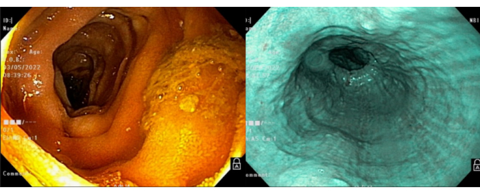
Figure 1: Endoscopy. The preoperative endoscopy showed no signs of malignancy or other diseases of the esophagus. Please click here to view a larger version of this figure.
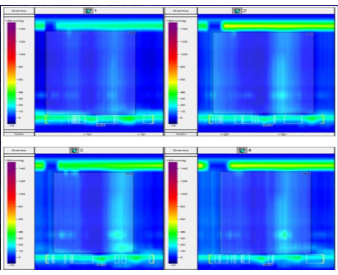
Figure 2: Manometry. This figure displays the results of a high-resolution esophageal manometry study in a patient with suspected esophageal motility disorder. The plot shows the pressure waves detected along the length of the esophagus during a series of swallows. In this case, there is a complete absence of peristaltic contractions and no detectable pressurization of the esophageal lumen. These findings are consistent with a diagnosis of achalasia type I. Please click here to view a larger version of this figure.
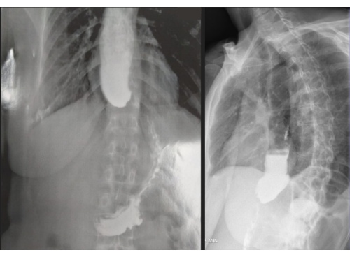
Figure 3: Barium esophagram. On the preoperative esophagogram, typical signs of achalasia were observed, including dilation of the distal esophagus, stasis of contrast material, and absence of air in the stomach. These findings are consistent with impaired esophageal emptying due to a failure of the lower esophageal sphincter to relax properly, which is characteristic of achalasia. The esophagus in this patient dilates up to 6 cm, indicating a grade III megaesophagus according to the Rezende-Mascarenhas classification. Please click here to view a larger version of this figure.
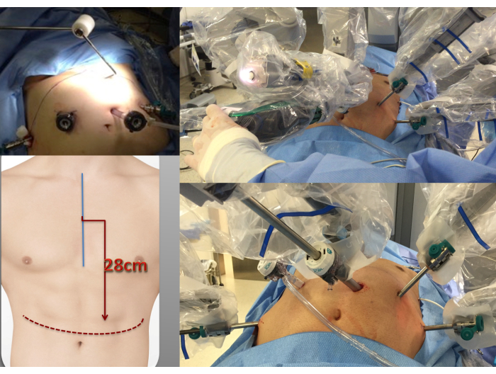
Figure 4: Trocar placement. The trocars are positioned in a linear arrangement directly above the umbilicus, with a distance of 28cm from the sternal angle. Please click here to view a larger version of this figure.
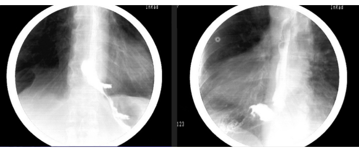
Figure 5: Postoperative barium esophagram. The postoperative esophagogram reveals improved contrast clearance from the esophagus to the stomach, along with a reduction in esophageal stasis. These findings suggest that the surgical intervention has been successful in improving esophageal motility and facilitating the emptying of ingested material into the stomach. Please click here to view a larger version of this figure.
Operation time | 112 minutes |
Blood loss | 20 ml |
Hospital discharge | 2 days |
Table 1: Postoperative outcomes.
This protocol describes robotic myotomy and partial fundoplication as a treatment for achalasia. The article highlights a Heller Pinotti fundoplication, which consists of a variation of the classic Dor fundoplication. This technique, presented in the article, demonstrates the performance of three rows of sutures, instead of the classic two sutures performed in the Dor fundoplication. The optimal type of fundoplication, including total, anterior, or posterior has been extensively studied in the literature, but there is still controversy surrounding the best approach for improving outcomes17. Modifications have been implemented to the technique over time, for example, the introduction of a posterior suture line to the classic Dor fundoplication, resulting in a postero-lateral-anterior fundoplication that provides enhanced reflux control compared to the anterior fundoplication alone. This technique, widely known as the Heller Pinotti procedure, has gained popularity in Brazil17.
Critical steps during the procedure include the proper placement of the robotic arms and trocars and careful dissection of the muscular layers of the esophagus. While laparoscopic Heller myotomy is currently considered the gold standard treatment for achalasia, robotic-assisted myotomy can also be a successful alternative in the hands of experienced surgeons.
Possible reasons for the low rate of robotic-assisted operations for Heller myotomy and other gastro-esophageal procedures have already been presented by several authors16 those include, mostly, a longer learning curve and cost. Although the robotic myotomy technique requires specialized equipment and may be more expensive than other minimally invasive procedures, it is expected that these limitations will become less significant over time as robotic technology becomes more widely available and costs decrease. It is the authors’ opinion that robotic assisted Heller myotomy should be considered as a feasible treatment option, in the field of minimally invasive treatments, for achalasia.
The authors have nothing to disclose.
None.
Da Vinci Surgical System | Intuitive Surgical | ||
Needle driver | Intuitive Surgical | ||
Bipolar forceps | Intuitive Surgical | ||
Bipolar Fenestrated Grasper | Intuitive Surgical | ||
Ultracision | Johnson & Johnson |