Here, we describe a protocol for developing a chick chorioallantoic membrane (CAM) xenografting model for human ovarian tissue and demonstrate the effectiveness of the technique, the graft revascularization time frame, and the tissue viability across a 6 day grafting period.
Ovarian tissue cryopreservation and transplantation is an effective strategy for preserving fertility but has one major drawback, namely massive follicle loss occurring shortly after reimplantation due to abnormal follicle activation and death. Rodents are benchmark models for investigating follicle activation, but the cost, time, and ethical considerations are becoming increasingly prohibitive, thus driving the development of alternatives. The chick chorioallantoic membrane (CAM) model is particularly attractive, being inexpensive and maintaining natural immunodeficiency up to day 17 postfertilization, making it ideal to study short-term xenografting of human ovarian tissue. The CAM is also highly vascularized and has been widely used as a model to explore angiogenesis. This gives it a remarkable advantage over in vitro models and allows the investigation of mechanisms affecting the early post-grafting follicle loss process. The protocol outlined herein aims to describe the development of a CAM xenografting model for human ovarian tissue, with specific insights into the effectiveness of the technique, the graft revascularization time frame, and the tissue viability across a 6 day grafting period.
The demand for fertility preservation for oncological and benign indications, as well as social reasons, has dramatically increased over recent decades. However, various treatments used to cure malignant and non-malignant diseases are highly toxic to the gonads and can result in iatrogenic premature ovarian insufficiency, ultimately leading to infertility1. Established techniques for fertility preservation include embryo cryopreservation, immature or mature oocyte vitrification, and ovarian tissue cryopreservation2,3,4. Ovarian tissue freezing is the only available option for preserving fertility in prepubertal girls or women who require immediate cancer therapy. The restoration of endocrine function following ovarian tissue transplantation occurs in over 95% of subjects, with live birth rates ranging from 18% to 42%5,6,7,8,9.
Although the transplantation of frozen-thawed ovarian tissue has proven successful, there is still room for improvement. Indeed, as ovarian cortical fragments are transplanted without vascular anastomosis, they experience a period of hypoxia during which graft revascularization takes place10,11,12. The vast majority of studies investigating human ovarian tissue transplantation have used a xenografting model, in which ovarian tissue is transplanted to immunodeficient mice. The complete revascularization of the xenografts takes around 10 days, with both the host and graft vessels contributing to the formation of functional vessels12,13,14. Around 50%-90% of the follicle reserve is lost during this hypoxic window before the completion of graft revascularization10,15,16. It has been strongly suggested that this massive follicle loss occurs through both direct follicle death, as demonstrated by a decrease in the absolute follicle numbers left after grafting, and the activation of primordial follicle growth, as indicated by changes in follicle proportions towards increased rates of growing follicles17,18.
Interestingly, previous research works using various animal ovarian tissues grafted to chick chorioallantoic membrane (CAM), which has a constitution mimicking the typical grafting site of the peritoneum, have reported the inhibition of spontaneous follicle activation, with the primordial follicle reserve staying intact for up to 10 days19,20,21,22. Our team previously demonstrated that the grafting of frozen-thawed human ovarian tissue to CAM constituted a reliable approach for investigating human ovarian tissue transplantation in its first ischemic stages23 and recently showed that this grafting method was able to counteract follicle activation24.
The CAM model is especially appealing not only because eggs are much cheaper than mice but also because of the highly vascularized nature of CAM, allowing scrutiny of the association between follicle activation and ovarian graft revascularization. The avian system is, indeed, one of the most common and versatile ways of studying angiogenesis25. Chick embryo development (ED) takes 21 days until hatching, and the CAM is formed within the first 4-5 days through the fusion of the allantois and chorion26. Notably, the chick embryo is a naturally immunodeficient host until day 17 of ED, so xenografting experiments can be performed without any risk of graft rejection27,28. Moreover, the CAM model approach does not raise any ethical or legal concerns in terms of European law29, making it an attractive alternative to other animal models. With regard to breeding conditions, chick embryos only need an incubator set at 37 °C with a relative air humidity of 40%-60%. These limited experimentation requirements significantly reduce the research costs compared to use of immunodeficient mice.
The protocol presented herein aims to describe the development of a CAM xenografting model for human ovarian tissue and provide specific insights into the effectiveness of the technique, the time frame of graft revascularization, and the tissue viability over a 6 day grafting period. This protocol could be of great interest for investigating the mechanisms behind early post-grafting follicle loss and studying the impact of several agents (growth factors, hormones, etc.) on this phenomenon.
The use of human tissue was approved by the Institutional Review Board of the Catholic University of Louvain. The patients gave their written informed consent for the use of their ovarian tissue for research purposes.
1. Ordering day 0 eggs that are highly likely to be embryonated
2. Preparing the eggs for incubation
3. Opening the eggshell on day 3 of ED
NOTE: A rectangular window is made in the eggshell on day 3 of ED.
4. Grafting frozen-thawed human ovarian tissue to the CAM
NOTE: Transplantation to the CAM should ideally be initiated between days 7-10 of ED.
5. Harvesting the grafts
NOTE: Xenografts should be harvested at the latest by day 17 of ED, since the immune system of the embryo becomes mature and competent from day 18.
Chick embryo survival rates
The embryo survival rate from windowing (day 3 of ED) to ovarian tissue grafting (day 7 of ED) was 79% (33/42). Since the percentage of embryonated day 0 eggs is unknown, supernumerary day 0 eggs from Lohman-selected white Leghorn chickens were ordered to ensure sufficient embryonated eggs would be available for grafting. A total of 23 viable day 7 eggs were used for grafting, one of which perished during the first 24 h, resulting in an overall embryo survival rate of 96% after transplantation (22/23).
Macroscopic aspect of the grafts
After 1 day of grafting, the grafts looked viable in 100% (10/10) of cases and were already at least partially adherent to the CAM, showing a wheel-spoke pattern of blood vessels needed for their vascularization (Figure 2A,B). After 6 days of grafting, all the implants were still adherent (Figure 2C), apart from two that did not attach to the CAM. They took on a necrotic appearance and were excluded from further analysis (Figure 3), resulting in a grafted tissue survival rate of 83% (10/12) after 6 days. All in all, the viability assessment of the human ovarian grafts revealed an overall tissue survival rate of 91% (20/22), irrespective of the day of grafting (Table 2).
Around day 3 post-transplantation, the grafts were found to be covered with a second layer of CAM, and they eventually became encapsulated, leading to even better graft vascularization (Figure 2C). Overall, 80% of the transplants had also penetrated the egg (Figure 2D), in some cases making it hard to retrieve them on day 6.
Microscopic assessment of the grafts
A total of 30 frozen-thawed human ovarian fragments obtained from five different patients were analyzed and fixed in 4% paraformaldehyde on grafting day 0, day 1, or day 6, embedded in paraffin, and serially cut into 5 µm sections for histological evaluation (Figure 4).
In order to assess the ovarian follicle survival rates, follicles were counted in 12 random hematoxylin and eosin-stained sections per time point and per patient. Only morphologically normal follicles with a visible oocyte were taken into consideration for analysis32. Healthy follicles were observed at all time points (Figure 4B–D). The follicle survival rates were calculated by determining the remaining follicle density (number of follicles/mm3) after grafting and normalizing it to the follicle density in non-grafted controls (considered 100%). The follicle densities tended to decrease after transplantation, but not enough to reach statistical significance (p > 0.8) (Table 2). Similarly, the follicle survival rates were maintained during the grafting period, standing at 95% ± 19% on day 1 and 83% ± 27% on day 6 (p > 0.5).
The revascularization process was further investigated by the detection of avian red blood cells in vessels from the xenografted ovarian tissue. Avian erythrocytes are easily discernible since they are nucleated (Figure 4A,C,D). Surprisingly, we were able to observe avian erythrocytes in ovarian vessels in 30% of implants after only 1 day of transplantation and in all the implants by day 6, exhibiting very rapid revascularization (Table 2).
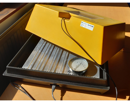
Figure 1: Egg incubator. Please click here to view a larger version of this figure.
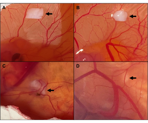
Figure 2: Macroscopic aspect of viable implants. (A) Grafting day 0. (B) Implants on day 1, with the CAM showing a wheel-spoke pattern of blood vessels towards the ovarian tissue. (C) Implants encapsulated by CAM on day 6, leading to even better graft vascularization. (D) Graft penetrating the egg. The arrows point to the grafted ovarian tissue. Please click here to view a larger version of this figure.
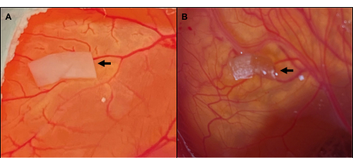
Figure 3: Macroscopic aspect of necrotic implants. (A) Healthy-looking ovarian tissue on day 0 of grafting. (B) Necrotic aspect of the implant 6 days later. The arrows point to the ovarian tissue. Please click here to view a larger version of this figure.
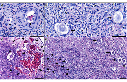
Figure 4: Microscopic aspect of implants. Hematoxylin and eosin-stained sections of implants grafted for (A,B) 1 day and for (C,D) 6 days. The arrowheads point to avian red blood cells perfusing the ovarian vessels after (A) 1 day and (C,D) 6 days of grafting. The arrows indicate healthy-looking primordial follicles in implants grafted for (B) 1 day and (C,D) 6 days. Scale bar: 50 µm. Please click here to view a larger version of this figure.
Steps | Baths | Time |
Second fixation | 1 Formalin 10% bath | 2 h |
Dehydration | 7 methanol baths | 7x 1 h |
Clarification | 3 toluene baths | 3x 1 h |
Impregnation | 3 liquid paraffin baths (60°C) | 15 min – 30 min – 30 min |
Table 1: Paraffin embedding steps.
Non-grafted | Grafting day 1 | Grafting day 6 | P-value | |
Macroscopic aspect of grafts | ||||
Tissue survival rate | – | 100% (10/10) | 83% (10/12) | / |
Microscopic aspect of grafts | ||||
Follicle density (n/mm3) | 848 ± 272 | 817 ± 370 | 684 ± 236 | p > 0.8 |
Follicle survival rate | 100% | 95% ± 19% | 83% ± 27% | p > 0.5 |
Avian red blood cells present in implants | 0% (0/10) | 30% (3/10) | 100% (10/10) | / |
Table 2: Results. Statistical analysis conducted using one-way ANOVA, followed by Sidak correction. Results are expressed as mean ± standard deviation.
The most challenging part of the protocol described here is making the small hole required to aspirate the albumen in order to detach the CAM from the eggshell prior to creating a window. Applying too much pressure can result in overpenetration or may even crack and destroy the egg, causing irrevocable damage to the CAM and its vasculature. To keep mistakes to a minimum during initial attempts to separate the CAM, it is strongly advised to practice making small holes in the eggshell of non-fertilized, grocery-bought eggs using a straight pin. Furthermore, given the natural variability of fertility across batches, along with the fact that not all embryos survive the procedure, obtaining supernumerary eggs from the egg supplier is recommended. In order to achieve optimal embryo survival rates and a well-developed CAM, the eggs need to be incubated in ideal conditions. In the case of low viability, different aspects may be to blame and need investigating. The egg supplier should be contacted, as the viability of embryos from the supplier may simply be lower at this point in time. Low egg viability could also be due to inaccurate or inappropriate incubator settings, which may compromise the CAM development. The use of an independent hygrometer and thermometer can ensure that the settings are accurate and stable, but detailed troubleshooting instructions should be provided by the manufacturer. Finally, it has also been suggested that checking transplanted eggs too often may result in temperature and humidity fluctuations, which could have deleterious effects on the grafted embryos33.
In the present protocol, the windowing of the eggshell was performed on day 3 of ED by aspirating around 2 mL of albumen from the embryo to detach the CAM from the shell, which, in turn, allowed the creation of a window without damaging the CAM. The embryo survival rate 4 days later was 79%, consistent with previous studies34,35. Other methods have been reported, with varying degrees of success. One alternative is to prise the CAM away from the shell by applying suction to the air sac, typically around day 7-10 of ED. This method does not require the egg to be punctured33,36. Studies reporting embryo survival rates using the latter technique are few and far between in the literature, with one team reporting up to 90% of embryo survival36. Another option to consider is shell-less chick embryo culture, also known as ex ovo culture. Removing the eggshell provides unrestricted access to the embryo, thus facilitating embryo manipulation and surgery and also allowing the use of high-resolution imaging techniques for live experiments, such as fluorescence microscopy and microcomputed topography37,38. In order to conduct ex ovo culture, the embryo, including its extraembryonic membranes, is transferred to a specific culture containment between day 2 and day 4 of ED. However, this technique is highly invasive, with embryos usually dying around day 12 and fewer than 18% of them surviving until day 1538,39.
During the xenografting experiments reported herein, human ovarian implants were grafted to CAMs that had been gently traumatized by applying a small strip of sterile ether-extracted lens paper to the surface of the epithelium and removing it immediately. Overall, the embryo survival rates were excellent and reached 96% during the grafting period, which is consistent with previous studies involving the transplantation of ovarian tissue fragments to CAMs23,40. Moreover, this approach has been shown to increase graft adhesion rates and enhance the subsequent establishment of neovascularization, with angiogenesis likely induced by the initial wound-healing process23. Indeed, xenografting rat ovarian tissue to murine wound-healing granulation tissue was found to improve graft vascularization41. Likewise, in the first live birth reported after orthotopic transplantation of cryopreserved human ovarian tissue, Donnez et al. created a peritoneal window and coagulated its margins 7 days before implantation to stimulate angiogenesis and neovascularization in the transplantation site42.
With regard to the histological assessment of grafted human ovarian tissue, the follicle survival rates were encouraging, with more than 80% of follicles remaining intact after 6 days of grafting. Interestingly, the follicle survival rates reported in the literature after the transplantation of human frozen-thawed ovarian tissue to the peritoneum of immunodeficient mice are only around 30%10,14,18,43,44, which is much lower than in the present study. Our findings of enhanced follicle survival after the grafting of human ovarian tissue to the CAM corroborate our recently published data, in which we demonstrate that CAM inhibits follicle activation and apoptosis24. Moreover, avian blood vessels were able to penetrate the implants, as demonstrated by the presence of avian erythrocytes after just 1 day of transplantation. All in all, these results demonstrate that the CAM approach is a suitable model for further scrutinizing the transplantation of human ovarian tissue, since it supports the survival of the tissue.
The major drawback of the CAM model is the limited period during which grafting is possible. Indeed, transplantation can be performed at the earliest on day 7 of ED, as soon as the CAM has developed sufficiently, until day 17, before the chick embryos acquire immunocompetence and hatch. Despite the short grafting duration, the CAM model is still a useful tool for studying human ovarian tissue transplantation in its first ischemic stages. One can also use this model to test the impact of proangiogenic factors, protective antioxidant agents, cytokines, growth- and reproduction-related hormones, and factors controlling follicle activation in grafted human ovarian tissue, all under experimental conditions that can be easily controlled25,45.
The authors have nothing to disclose.
The authors thank Mira Hryniuk, BA, for reviewing the English language of the article.
Agani hypodermic needle, 19 G | Terumo Europe | AN*1950R1 | 19 G needle to aspirate albumen |
Terumo syringe, 5 mL concentric Luer lock | Terumo Europe | SS*05LE1 | 5-mL sterile syringe |
Caseviewer v2.2 | 3DHISTECH | Image analysis software | |
Diethyl ether | Merck Chemicals | 603-022-00-4 | Sterile ether to traumatize the CAM |
Eosin Y aqueous solution 0.5% | Merck | 1098441000 | Staining solution |
Formaldehyde 4% aqueous solution buffered (Formalin 10%) | VWR | 97139010 | Formaldehyde used for tissue fixation |
Fridge | Liebherr | 7081260 | Fridge at 4 °C used for paraffin-embedding |
Heating plate | Schott | SLK2 | Hot plate used to dry the slides |
Incubator | Thermo Forma Scientific 3111 | 10365156 | Oven used for slide incubation |
Leica CLS 150 XE microscope cold light source | Leica | CLS 150 XE | Focal cold light source to candle the eggs |
Lens cleaning tissue, grade 541 | VWR | 111-5003 | Tissue to soak in sterile ether to traumatize the CAM |
Mayer's hematoxylin | MERCK | 1092491000 | Staining solution |
Methanol | VWR | 20847307 | Methanol |
Microtome | ThermoScientific-MICROM | HM325-2 | Microtome |
Pannoramic P250 Flash III | 3DHISTECH | / | Slide scanner at 20x magnification |
Paraformaldehyde | Merck | 1,04,00,51,000 | Paraffin-embedding solution |
Paraplast Plus R | Sigma | P3683-1KG | Paraffin |
Petri dish, 60×15 mm, sterile | Greiner | 628161 | Sterile petri dish |
Pin holder | Fine Science Tools | 26016-12 | Pin holder |
Polyhatch | Brinsea | CP01F | Egg incubator with automatic rotator |
Scroll saw blade, 132 mm | Sencys | / | Saw blade to create a window in the eggshell |
Stainless steel insert pins | Fine Science Tools | 26007-02 | Straight pin to make a hole in the eggshell |
Steril-Helios | Angelantoni Industrie | ST-00275400000 | Laminar flow hood |
Superfrost Plus bords rodés 90° | VWR | 631-9483 | Glass slides |
Tissue-Tek VIP 6Al | Sakura | 60320417-0711 VID6E3-1 | Automatic embedding device |
Titanium forceps | Fine Science Tools | 11602-16 | Forceps for eggshell removal and ovarian tissue manipulation |
Toluene, pa | VWR | 28701364 | Paraffin-embedding solution |