Complete resection of the uncinate process and mesopancreas is one of the most important and difficult processes in laparoscopic pancreatoduodenectomy (LPD). This article presents a method for managing the uncinate process in no-touch LPD using the median-anterior and left-posterior approaches to the superior mesenteric artery (SMA).
Laparoscopic pancreatoduodenectomy (LPD) is a demanding abdominal operation that necessitates meticulous surgical skills and teamwork. The management of the pancreatic uncinate process is one of the most important and difficult processes in LPD because of its deep anatomical location and difficult exposure. Complete resection of the uncinate process and mesopancreas has become the cornerstone of LPD. In particular, it is even more difficult to avoid positive surgical margins and incomplete lymph node dissection when the tumor is located in the uncinate process. No-touch LPD, which is an ideal oncological operation process fitting the “tumor-free” principle, has been reported by our group previously. This article introduces the management of the uncinate process in no-touch LPD. Based on the multi-angle arterial approach, in this protocol, the median-anterior and left-posterior approaches to the SMA are used to correctly deal with the important vascular structure, the inferior pancreaticoduodenal artery (IPDA), in order to ensure the safe and complete excision of the uncinate process and mesopancreas. For the achievement of the no-touch isolation technique in LPD, the pancreatic head and the blood supply to the duodenal region must be severed at the very early stage of the operation; after that, the tumor can be isolated intact, resection can be performed in situ, and finally, the tissue can be removed en bloc. This paper aims to show the distinctive ways to manage the uncinate process in no-touch LPD and investigate the viability and safety of this approach. Moreover, the technique may increase the R0 resection rate.
The pancreatic uncinate process is the protrusion from the bottom left of the pancreatic head that is situated behind the SMA and superior mesenteric vein (SMV)1. The management of the uncinate process is a challenging area in pancreatic surgery due to its deep anatomy and challenging exposure; therefore, especially for pancreatic cancer located in the uncinate process, surgery is prone to result in a positive surgical margin, incomplete lymph node dissection, and poor prognosis2. Therefore, improved surgical techniques and strategies are urgently required.
Most resection procedures of the pancreatic head and uncinate process are performed along the right side of the SMV and SMA3. Although this approach largely works for ampullary tumors, it has drawbacks for pancreatic head cancer, particularly for large tumors in the uncinate process2. During the procedure, it is frequently required to spin and pull the SMV and SMA in order to expose them. The IPDA cannot be dissected clearly with such approaches, which usually results in bleeding or insufficient cleaning of the area. In this work, the authors describe an approach that can support the early control of the IPDA, which results in better bleeding control, less blood loss, and better dissection of the uncinate process.
Meanwhile, conventional laparotomy or laparoscopy pancreatoduodenectomy requires a Kocher maneuver for the wide mobilization of the duodenum and pancreatic head4. However, with this maneuver, the tumor cells have the potential capacity to metastasize via the portal vein (PV), as the surgeon might squeeze the tumor while holding it during surgery4,5,6. The no-touch isolation technique is one of the popular concepts in pancreatoduodenectomy. Although it has not been proven by large clinical trials whether this surgical intervention could enhance the cancer-related prognosis of pancreatic cancer patients, the study of Hirota et al.7 reported the significance of no-touch technology in preventing cancer cell metastasis by using molecular markers (CEA mRNA) to detect cancer cells in the portal vein blood. In their study, the no-touch technique group had a lower spread rate of portal vein cancer cells and a higher 3 year survival rate than the conventional technique group. No-touch LPD has been reported by the team of the current authors previously as an ideal oncological operation process fitting the "tumor-free" principle8,9.
This article presents the management of the uncinate process in no-touch LPD. The median-anterior and left-posterior approaches to the SMA were performed to deal precisely with the IPDA. To achieve the no-touch isolation technique in LPD, the blood supply to the duodenum and pancreatic heads must be cut off at the very early stage of the operation, after which the tumor can be isolated intact, resected in situ, and finally, removed en bloc.
The purpose and advantages of this strategy are to ensure the safe and complete excision of the uncinate process and mesopancreas based on a multi-angle arterial approach10. This article aims to explore the efficacy and safety of this technique for the management of the uncinate process in no-touch LPD, which might improve the R0 resection rate11.
This study was approved by the Ethics Committee of the Second Affiliated Hospital of Guangzhou University of Chinese Medicine, and informed written consent was obtained from the patients involved in this study.
1. Preoperative work-up
2. Anesthetization steps
3. Installation
4. Surgery techniques
5. Postoperative management
A male patient aged 59 years old with symptoms including upper abdominal pain and jaundice was hospitalized in our department. He had no previous medical history and a relatively moderate normal body mass index (23.94 kg/m2). A contrast-enhanced CT scan was performed, and a tumor with a diameter of approximately 5.5 cm x 4.6 cm x 6.3 cm was found at the head and uncinate process of the pancreas (Figure 10). No evidence of distant metastasis was found, and percutaneous transhepatic cholangical drainage19 was performed before the operation. The total bilirubin declined from 201.4 µmol/mL to 36.0 µmol/mL.
Representative results are shown in Table 1. The operation lasted 314 min, and the total blood loss was 80 mL. The pancreatic fistula was not found, as the amylase level of the abdominal drainage fluid was normal throughout the postoperative period, and the drainage tubes were removed before postoperative day 8. No other significant morbidity occurred, and this patient was discharged on the 11th postoperative day. Histopathology confirmed a pancreatic intraductal papillary mucinous neoplasm with focal moderately differentiated invasive adenocarcinoma (Figure 11). The surgical margins were microscopically negative (R0), and none of the 18 lymph nodes were involved.
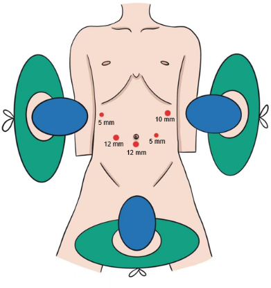
Figure 1: Distribution of the trocars. The five-port method was used. The patient was laid down in a supine position with his legs spread out. Please click here to view a larger version of this figure.
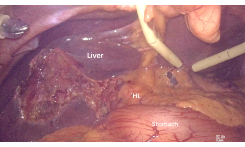
Figure 2: Removal of the gallbladder and suspending the liver to expose the hepatoduodenal ligament and hilum. Abbreviation: HL = hepatoduodenal ligament. Please click here to view a larger version of this figure.
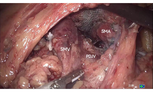
Figure 3: The PDJV between the SMV and the SMA is exposed in the inferior colonic region. Abbreviations: PDJV = proximal-dorsal jejunal vein; SMV = superior mesenteric vein; SMA = superior mesenteric artery. Please click here to view a larger version of this figure.
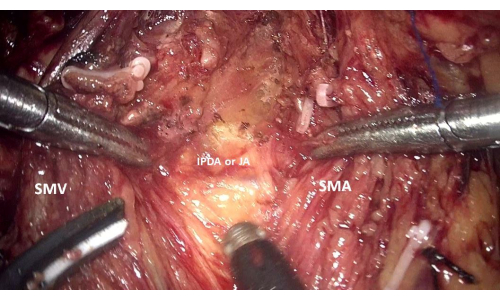
Figure 4: Exposure of the main branches of the IPDA and JA along the right side of the SMA through the median-anterior approach to the SMA. Abbreviations: IPDA = inferior pancreaticoduodenal artery; JA = jejunal artery; SMV = superior mesenteric vein; SMA = superior mesenteric artery. Please click here to view a larger version of this figure.
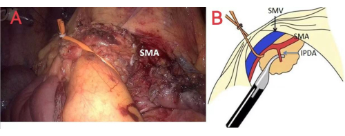
Figure 5: Method of hanging the SMA and SMV. (A) and (B) represent the method of hanging the SMA and SMV to establish a sufficient view of the left-posterior surface of the SMA. Abbreviations: IPDA = inferior pancreaticoduodenal artery; SMA = superior mesenteric artery; SMV = superior mesenteric vein. Please click here to view a larger version of this figure.
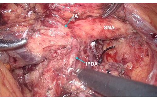
Figure 6: Dissection along the SMA circumferentially exposing the IPDA, which originates directly from the SMA. Abbreviations: IPDA = inferior pancreaticoduodenal artery; JA = jejunal artery; SMA = superior mesenteric artery. Please click here to view a larger version of this figure.
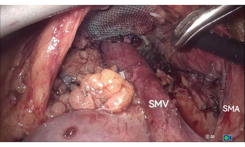
Figure 7: Separating the SMA and SMV from the uncinate process and mesopancreas. Using the median-anterior and left-posterior approaches to the SMA, the SMA and SMV are completely separated from the uncinate process and mesopancreas. Abbreviations: SMA = superior mesenteric artery; SMV = superior mesenteric vein. Please click here to view a larger version of this figure.
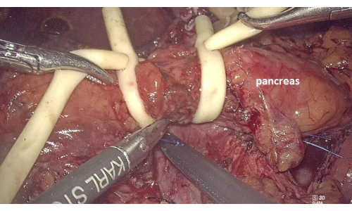
Figure 8: Placing two Fr14 catheters to block the pancreatic neck blood supply and reduce bleeding. Please click here to view a larger version of this figure.
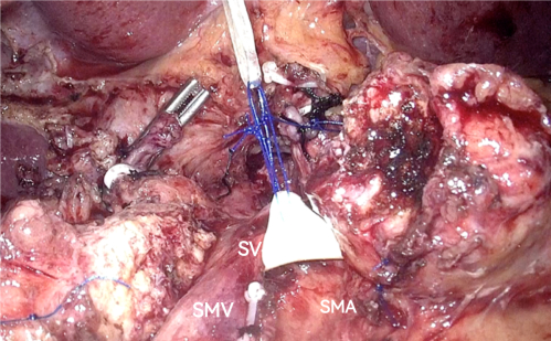
Figure 9: Suspending the SV to establish the exposure. Abbreviations: SV = splenic vein; SMA = superior mesenteric artery; SMV = superior mesenteric vein. Please click here to view a larger version of this figure.
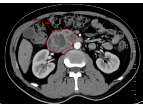
Figure 10: Image showing the tumor in the head and uncinate process of the pancreas. Please click here to view a larger version of this figure.
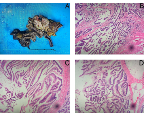
Figure 11: Histopathological study. Histopathology confirmed a pancreatic intraductal papillary mucinous neoplasm with focal moderately differentiated invasive adenocarcinoma. (A) Postoperative paraffin specimens in which the size of the stomach is 7 cm x 5 cm x 2.5 cm, the length of the small intestine is about 22 cm, the size of the pancreas is 6 cm x 4 cm x 3 cm, and there is a gray mass in the head of the pancreas, which is about 6 cm x 3 cm x 1.5 cm in size. (B) Atypical glands, suggesting a focally infiltrating adenocarcinoma. (C,D) An intraductal papillary mucinous tumor. Magnifications: B, 200x; C, 400x; D, 300x. Please click here to view a larger version of this figure.
Variable | Outcome |
Intraoperative | |
operative time, min | 314 |
Intraoperative blood loss, mL | 80 |
Postoperative | |
Postoperative pancreatic fistula | Not found |
Drain removal, postoperative day | 8 |
Postoperative hospital stay, days | 11 |
Pathological diagnosis | Pancreatic intraductal papillary mucinous neoplasm with focal moderately differentiated invasive adenocarcinoma |
Table 1: Representative results of the surgery.
With intestinal rotation, the ventral pancreas flips to the dorsal side and unites with the dorsal pancreas throughout the development of the human embryo, and the ventral pancreas also grows into the lower portion of the pancreatic head and the uncinate process1. The pancreatic uncinate process is described as the protrusion from the bottom left of the pancreatic head that is situated behind the SMA and the SMV1.
In the meantime, the mesopancreas rotates around the SMA into a spiral along with intestinal rotation20. As the IPDA and IPDV arise from the posterior area of the SMA and SMV, their lengths are also relatively shorter due to the rotation of the mesenteric vascular axis. These vessels may be inadvertently injured and lead to massive bleeding when dissecting the pancreatic uncinate process and mesopancreas from the SMA. Due to this issue, the management of the pancreatic uncinate process is one of the most important and difficult processes in LPD. Importantly, the complete resection of the uncinate process and mesopancreas has become a cornerstone of LPD.
This study provides a unique strategy for the management of the uncinate process in no-touch LPD, which might potentially develop into a reproducible, standardized, and effective oncological procedure for the safe and complete excision of the uncinate process and mesopancreas. The median-anterior and left-posterior approaches to the SMA are used to ligate and dissect the IPDA, IPDV, and uncinate process artery in situ. The blood supply to the pancreatic head and duodenal region must be severed at the very early stage of the operation so that the tumor can be isolated intact, and then SMA and SMV can be separated from the uncinate process and mesopancreas completely, and the specimen can be resected en bloc using the Kocher maneuver.
The pancreatic uncinate process is situated behind the SMA1. Preoperative high-resolution contrast-enhanced CT should be performed to classify the anatomical features of the interaction between them into one of two types: type I, when the uncinate process is located at the right side of the SMA (this type is common in ampullary tumors and some cases of pancreatic head tumor); and type II, when the uncinate process extends to the left side of the SMA (this type is more commonly seen when the tumor is located in the uncinate process of pancreas)21. It is relatively easy to dissociate the uncinate process and mesopancreas from the SMA in type I. However, the SMA, the SMV, and their branches are presented on the ventral side of the uncinate process and tumor in type II. This kind of uncinate process excision is challenging and calls for a multi-angle combined arterial approach and advanced surgical skills. In these situations, dissecting the 14th group of lymph nodes is challenging. Using the median-anterior and left-posterior approaches to the SMA is beneficial and necessary.
The goal and benefit of this method are the demonstration of the feasibility and safety of the management of the uncinate process in no-touch LPD. This complex procedure is recommended to be performed in relatively high-volume centers by experienced surgical teams. The learning curve is inevitably steep as this operation strategy requires effective cooperation among the surgical teams, comprehensive anatomical knowledge of the epigastrium, and a flexible response capability against unexpected variations. Furthermore, a limited number of cases are suitable for this procedure, and randomized clinical trials are difficult to design and implement; as a result, high-level evidence regarding the short-term and long-term outcomes of this strategy is difficult to establish.
The authors have nothing to disclose.
Our article #64904 was supported by the Research project of Traditional Chinese Medicine Bureau of Guangdong Province (ID:20222077).
3D Laparoscope | STORZ | TC200,TC302 | |
Cisatracurium Besylate Injection | Hengrui Pharma | H20183042 | |
Dual-source dual-energy CT | SOMATOM | Definition FLASH | |
Echelon Flex Powered Plus Articulating Endoscopic Linear Cutter and Endopath Echelon Endoscopic Linear Cutter Reloads with Gripping Surface Technology | Ethicon Endo-Surgery | ECR60G/GST60G | |
ENDOPATH XCEL Trocars | Johnson & Johnson | CB5LT/CB12LT | |
HARMONIC ACE Ultrasonic Surgical Devices | Ethicon Endo-Surgery | HAR36 | |
Laparoscopic bulldog clamps | Aesculap | FB367R,FB369R | |
Ligating Clips | Teleflex Medical | 5,44,22,05,44,23,05,44,000 | |
Nacrotrend anaesthesia monitoring system | Monitor Technik,Bad Bramsted | ||
PROLENE Polypropylene Nonabsorbable Suture/5/0 | Johnson & Johnson | W8556 | |
Propofol Injectable Emulsion | Aspen Pharma Trading Limited | H20171275 | |
Remifentanil Hydrochloride for injection | Humanwell Healthcare | H20030197 | |
Sevoflurane for Inhalation | Hengrui Pharma | H20070172 | |
Sufentanil Citrate Injecton | Humanwell Healthcare | H20054171 | |
Trocars | AOFO | FQ-D1/5.5mm 10.5mm | |
Ultrasonic Surgical & Electrosurgical Generator | Ethicon Endo-Surgery | GEN11CN |