RNA Fluorescence In Situ Hybridization for Long Non-Coding RNA Localization in Human Osteosarcoma Cells
Summary
The present protocol describes a method of RNA fluorescence in situ hybridization to localize the lncRNAs in human osteosarcoma cells.
Abstract
The important roles of long non-coding RNAs (lncRNAs) in cancer have been studied, such as regulating the proliferation, epithelial-mesenchymal transition (EMT), migration, infiltration, and autophagy of cancer cells. Localization detection of lncRNAs in cells can provide insight into their functions. By designing the lncRNA-specific antisense chain sequence followed by labeling with fluorescent dyes, RNA fluorescence in situ hybridization (FISH) can be applied to detect the cellular localization of lncRNAs. Together with the development of microscopy, the RNA FISH techniques now even allow for visualization of the poorly expressed lncRNAs. This method can not only detect the localization of lncRNAs alone, but also detect the colocalization of other RNAs, DNA, or proteins by using double-color or multicolor immunofluorescence. Here, we have included the detailed experimental operation procedure and precautions of RNA FISH by using lncRNA small nucleolar RNA host gene 6 (SNHG6) in human osteosarcoma cells (143B) as an example, to provide a reference for researchers who want to perform RNA FISH experiments, especially lncRNA FISH.
Introduction
Our understanding of the human genome has been greatly expanded by recent advances in whole-genome technology. About 93% of the human genome can be transcribed into RNAs, but only 2% of the RNAs can be translated into proteins; the remaining 98% of RNAs that have no protein translation function are called non-coding RNA (ncRNA)1. As a class of noncoding RNAs (ncRNAs), long ncRNAs (lncRNAs), containing over 200 nucleotides2, have attracted increasing attention due to their involvement in many physiological and pathological processes of the cells, such as differentiation, cycle control, apoptosis, migration, and invasion3,4,5. LncRNAs play their roles through various mechanisms, such as regulating chromatin structure and nuclear gene expression, controlling the mRNA splicing process, and posttranscriptional modification6. LncRNAs regulate the occurrence, development, and metastasis of malignancies at both the transcriptional and posttranscriptional levels. Transcriptional regulation is realized in the nucleus by affecting RNA transcription via binding to the chromosomal structures, while posttranscriptional regulation is realized in the cytoplasm by controlling the target genes via an endogenous competitive RNA (ceRNA) mechanism5, 7, 8. CeRNA has revealed a new mechanism of RNA interaction, namely that lncRNAs can act as a sponge to adsorb miRNAs and inhibit the miRNA-mediated degradation of related target genes9. Therefore, the information regarding the subcellular localization of lncRNAs, whether a specific lncRNA is located in the cytoplasm or nucleus, is important to help identify their biological functions.
At present, lncRNA localization is mainly detected by two methods, one is by nucleus/cytoplasm fraction isolation assay, and the other is by RNA FISH. In the former, RNAs in the cytoplasmic and the nuclear fractions are extracted respectively, and then PCR amplification is performed with specific lncRNA primers to detect the ratio of lncRNAs in the cytoplasm and nucleus. The advantage of this method is time efficiency, while the disadvantage is that the actual lncRNA localization is not directly reflected by the relative proportion of lncRNAs in the cytoplasm and nucleus. RNA FISH can detect lncRNA localization in cells through the design of lncRNA-specific antisense chain sequences followed by labeling with fluorescent dyes10. The RNA FISH methods have been improved with advances in probe techniques and detection methods, including fluorophore-labeled multiple oligo probe sets11, LNA probes12, and branched-DNA (bDNA) probes13. RNA FISH can not only detect the localization of lncRNA, but also detect the colocalization of other RNAs, DNA, or proteins by using double-color or multicolor immunofluorescence14.
In this work, we have included the detailed intracellular localization detection protocol of lncRNA small nucleolar RNA host gene 6 (SNHG6) in osteosarcoma cells (143B) by RNA FISH as an example. SNHG6 is a 600-730 nucleotide lncRNA in its mature spliced form and identified as a novel oncogene in diverse human cancers, including colorectal cancer, gastric cancer, ovarian clear cell carcinoma, osteosarcoma, and hepatocellular carcinoma15,16,17,18. Studies have confirmed the involvement of SNHG6 in biological behaviors of cancer cells, such as proliferation, EMT, and autophagy, and shown the cytoplasmic localization of SNHG6 where it may affect the target genes by binding (sponging) the miRNAs15,16,17. This detailed detection protocol of SNHG6 intracellular localization by RNA FISH is presented herein.
Protocol
See the Table of Materials for details of all materials, reagents, and instruments used in this protocol. Figure 1 shows the overall protocol for RNA FISH; Table 1 contains the composition of all solutions and Table 2 contains the primer sequences used in this protocol.
1. Probe preparation
- Identify and acquire the FASTA sequence of a target lncRNA of interest, for example, from GenBank (https://www.ncbi.nlm.nih.gov/genbank/). Following the indications on the website, design the ISH probes online19, and review the design algorithm's suggestions to list the probes to be ordered with Cy3 label.
- Incubate the hybridization buffer at 37 °C for 2 h in advance.
- Dissolve 4 optical density (OD) probe in 160 µL of diethylpyrocarbonate (DEPC)-treated ddH2O at the concentration of 1 mg/mL away from light.
NOTE: Optical density (OD) represents the measurement unit of DNA and RNA. Usually, 1 OD unit = 33 µg/mL DNA. - Prepare 200 µL of the probe mixture for each well (1.2 µL-10 µL of probe, 70 µL of hybridization buffer, make up the volume with DEPC-treated ddH2O to 200 µL), and set up a series of 50 µg/mL, 25 µg/mL, 12.5 µg/mL, and 6 µg/mL.
NOTE: The probe concentration needs to be explored experimentally in advance. A high probe concentration will lead to non-specific binding of lncRNAs, while a low probe concentration will lead to insensitive or failed detection of lncRNAs. - Denature the probe mix at 73 °C for 5 min.
NOTE: If the Cy3-labeled DNA probe is double-stranded, denature the probe to single-stranded DNA at 95 °C for 5 min, then quickly cool for 2 min on ice.
2. Cell preparation
- Seed 50,000 143B cells per well on sterile glass coverslips in a 12-well cell culture plate and incubate them for 24 h (37 °C, 5% CO2) in DMEM.
NOTE: The specific cell number seeded here varies with cell size, which is appropriate for the seeded cells to reach a confluence of 50% after being incubated for 24 h. The sterile glass coverslips are round to be suitable for the 12-well plate. - Remove the medium and wash for 2 x 5 min with 1x phosphate-buffered saline (PBS).
NOTE: Prepare 1x PBS with DEPC-treated ddH2O. PBS without DEPC-treatment cannot be used because it contains RNase. Perform all the following steps in RNase-free conditions. - Remove 1x PBS and add 200 µL of 100% ethanol in each well to fix for 15 min at room temperature.
- Remove the ethanol, add 200 µL of 0.1% Triton X-100 (in 1x PBS) in each well, and incubate for 15 min at room temperature.
NOTE: The time needs to be strictly controlled with Triton X-100 permeabilization and cannot be too long. - Remove 0.1% Triton X-100 and wash 2 x 5 min with 1x PBS.
NOTE: If the protocol has to be paused here, replace the PBS with 70% ethanol (dilution of 100% ethanol with RNase-free ddH2O) and store the sample at 4 °C for up to 3 months. - Remove 1x PBS, add 200 µL of 2x sodium saline citrate (SSC) buffer (dilution of 20x SSC with RNase-free ddH2O) into each well, and incubate for 30 min at 37 °C.
- Remove 2x SSC buffer, add 200 µL of 70% ethanol into each well, and incubate for 3 min at room temperature.
- Discard the 70% ethanol, add 200 µL of 85% ethanol (dilution of 100% ethanol with RNase-free ddH2O) into each well, and incubate for 3 min at room temperature.
- Discard the 85% ethanol, add 200 µL of 100% ethanol into each well, and incubate for 3 min at room temperature.
- Absorb and discard 100% ethanol; let the wells dry.
3. Fluorescence in situ hybridization (FISH)
- Add 200 µL of probe mixture (denatured as in step 1.5) to each well and incubate at 37 °C overnight (16-18 h).
NOTE: There is a strong positive correlation between hybridization temperature and probe concentration; therefore, to optimize the background, both the hybridization temperature and the probe concentration should be reduced. - The next day, take out samples from 37 °C and discard the probe mixture. Add 200 µL of 0.4x SSC/0.3% Tween-20 buffer to each well (preheated at 65 °C) and wash for 2 min at room temperature.
- Remove the 0.4x SSC/0.3% Tween-20 buffer, add 200 µL of 2x SSC/0.1% Tween-20 buffer to each well, and wash for 2 min at room temperature.
- Remove 2x SSC/0.1% Tween-20 buffer, add 200 µL of 4',6-diamidino-2-phenylindole (DAPI) staining solution (1 µg/mL), and stain for 20 min away from light.
- Discard the DAPI dye solution and wash with 1x PBS for 2 min at room temperature.
- Add 50 µL of mounting medium containing gum onto the slide and place the glass coverslip on the slide to fix.
NOTE: Be sure to place the coverslip on the slide with the cell side down. - Observe under a fluorescence microscope.
NOTE: Use a fluorescence microscope or a laser confocal microscope; the latter produces a higher sensitivity and clarity imaging.
Representative Results
Representative images of SNHG6 FISH in human osteosarcoma cells are shown (Figure 2). The negative control is treated with the negative Ctrl probe; positive control is treated with U6 probe 20. SNHG6 probe and U6 probe are labeled with Cy3, which emits red fluorescence. DAPI is a dye that stains the DNA, which emits blue fluorescence. This result shows that SNHG6 is mainly localized in the cytoplasm, and this information can provide an important direction for further study of SNHG6.
If a very high concentration of probe is used, a background color can be seen under the fluorescence microscope. Figure 3 shows the representative images when a high concentration of SNHG6 probe is used in RNA FISH. The white arrows represent nonspecific staining.
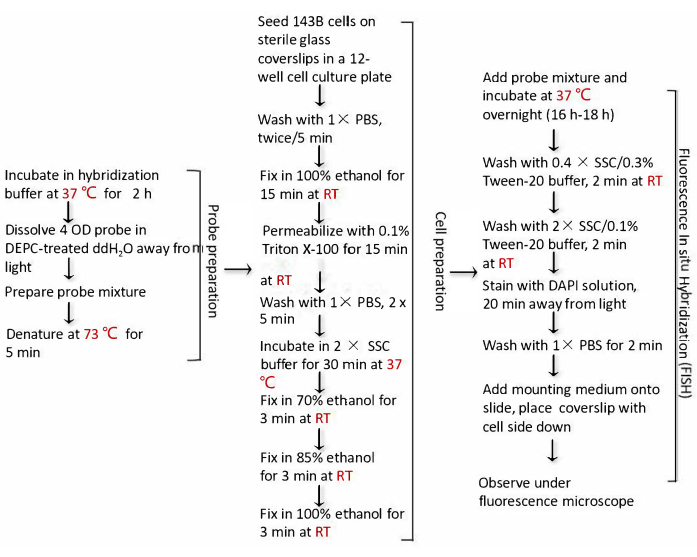
Figure 1: Flow chart of the RNA FISH protocol for lncRNA. This chart shows the key protocol of the RNA FISH experiment process. Abbreviations: FISH = fluorescence in situ hybridization; lncRNA = long noncoding RNA. Please click here to view a larger version of this figure.
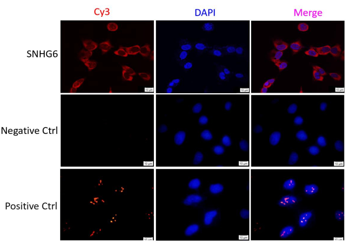
Figure 2: The localization of LncRNA (SNHG6) in human osteosarcoma cells (143B). Negative control is treated with a negative control probe. Positive control is treated with a U6 probe. SNHG6 and U6 probes are labeled with Cy3, which emits red fluorescence. DAPI is a dye that stains the DNA and emits blue fluorescence. Red and blue merge to make pink. The images are taken using a fluorescence microscope. Scale bar = 10 µm. Abbreviations: LncRNA = long non-coding RNA; DAPI = 4',6-diamidino-2-phenylindole. Please click here to view a larger version of this figure.
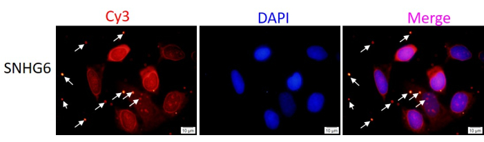
Figure 3: The representative images of high concentration of SNHG6 probe in RNA FISH. SNHG6 probes are labeled with Cy3, which emits red fluorescence. DAPI is a dye that stains the DNA and emits blue fluorescence. Red and blue merge to make pink. The images are taken using a fluorescence microscope. Scale bar = 10 µm. Please click here to view a larger version of this figure.
Table 1: The composition of solutions used in the RNA FISH hybridization experiment. All dilutions should be done with sterile, RNase-free water. All added water is DEPC-treated ddH2O. Abbreviations: FISH = fluorescence in situ hybridization; DEPC = diethylpyrocarbonate. Please click here to download this Table.
Table 2: All probe sequences used in this experiment. Please click here to download this Table.
Discussion
This RNA FISH protocol can not only detect the localization of lncRNAs in cells, but also detect the colocalization of other RNAs, DNA, or proteins in cells, which can also be used to detect the location of lncRNAs in paraffin-embedded tissues. However, the specific protocol in such cases is different because the paraffin-embedded tissues need to be dewaxed21. This experimental procedure can be applied in 48- or 96-well plates, but 384-well plates are too small to be used here.
Several critical steps of this method need to be noted, including the RNase-free environment, probe concentration, hybridization temperature, and the setting of negative and positive controls. First, the RNase-free condition is crucial because the RNase breaks down the bonds between nucleotides and leads to the degradation of lncRNAs in general. Second, the commonly recommended probe concentration ranges from 5 to 50 µg/mL. A high probe concentration will lead to non-specific binding of lncRNAs, while a low probe concentration will lead to insensitive or failed detection of lncRNAs19. If a very high concentration of probe is used, a background color can be seen under the fluorescence microscope even in the presence of a scrambled probe (a negative control) only; therefore, the same concentrations of negative controls are usually used to set up the baseline (same as the internal control) during the preliminary experiments exploring the appropriate probe concentration to avoid any non-specific binding. The highest probe concentration without background color in the negative control group, presenting the strongest signaling without non-specific binding, was used in the following experiments. Third,it is recommended to design at least two or three probes for optimization with a specific lncRNA. At the same time, two or more probes can also be mixed to improve the detection sensitivity of target lncRNAs. Fourth, the normal hybridization temperature of the lncRNA probe is 37 °C. During the hybridization process, the hybridization temperature should be kept constant and uniform. The range of hybridization temperature between different probes is 34 to 38 °C. The hybridization temperature is strongly positively correlated with the probe concentration; therefore, to optimize the background, the probe concentration should be reduced. Lastly, it is also critical to set negative and positive controls. The group without probe is used as a negative control to obtain background signals. A U6 probe or a ribosomal RNA probe is used as a positive control to rule out the possibility of false positive results.
This approach has several advantages. The fluorescence color of lncRNA probes can be changed. Green fluorescence is emitted by fluorescein isothiocyanate (FITC), fluorescein (FAM), and Alexa Fluor 488, while red fluorescence is emitted by Cy3 and Alexa Fluor 555. FITC is used for typical green and Cy3 for typical red. In addition, the images can be obtained by using either a fluorescence microscope or a laser confocal microscope. The latter’s images are more sensitive and clearer than those acquired by using a fluorescence microscope.
The limitation of the RNA FISH method is that this is a qualitative method; therefore, the results are not quantifiable. Due to the change in hybridization time and the influence of subjective factors during fluorescence microscope observation, lncRNA expression cannot be accurately quantified in cells. The expression levels of lncRNAs in different cellular fractions can only be qualitatively evaluated. However, with improvement in the technique, a single-molecule fluorescent in situ hybridization (smFISH) method22 has been developed to quantify the RNA expression level; more smFISH detection may be reported in the future.
The RNA FISH technique has a wide range of applications. The intracellular localization detection of lncRNAs will benefit the study of the biological mechanisms of lncRNAs. At the same time, this technique can also be used to detect the localization of other non-coding RNAs in cells, including circRNAs, miRNAs, and tRNAs.
Divulgations
The authors have nothing to disclose.
Acknowledgements
This work is supported by grants from (1) the National Key R&D Program of China (2020YFE0201600); (2) the National Nature Science Foundation (81973877 and 82174408); (3) Shanghai Collaborative Innovation Center of Industrial Transformation of Hospital TCM Preparation; (4) Research Projects within Budget of Shanghai University of Traditional Chinese Medicine (2021LK047).
Materials
| Automatic cell counter | Shanghai Simo Biological Technology Co., Ltd | IC1000 | Counting cells |
| Cell culture plate-12 | Shanghai YueNian Biotechnology Co., Ltd | 3513,corning | Place the coverslips in the plate |
| Cell line (143B) | Cell Bank of Chinese Academy of Sciences | CRL-8303 | osteosarcoma cancer cell line |
| Centrifuge tube (15 mL, 50 mL) | Shanghai YueNian Biotechnology Co., Ltd | 430790, Corning | Centrifuge the cells |
| Coverslips | Shanghai YueNian Biotechnology Co., Ltd | abs7026 | The cells are seeded on the coverslips |
| Cy3 label-SNHG6 DNA probe | Shanghai GenePharma Co.,Ltd | A10005 | Detect SNHG6 location |
| DMEM media | Shanghai YueNian Biotechnology Co., Ltd | LM-E1141 | Cell culture medium |
| Dry Bath Incubator | Haimen Kylin-Bell Lab Instruments Co.,Ltd. | DKT200-2 | Incubation at different high temperatures |
| Ethanol 100% | Sinopharm Chemical ReagentCo., Ltd | 10009218 | dehydration |
| Fluorescence microscope | Shanghai Waihai Biotechnology Co., LTD | Olympus BX43 equipped with a camera of Olympus U-TV0.5XC-3(SN:5J01719),olympus | Observation and positioning |
| Incubator | Shanghai Yiheng Scientific Instrument Co., LTD | DHP-9051 | The samples were incubated at 37 °C. |
| Mounting Medium | Sangon Biotech (Shanghai) Co., Ltd. | E675004 | Attach the coverslips to the slide |
| Shaker | Haimen Kylin-Bell Lab Instruments Co.,Ltd. | TS-8S | Washing sample |
| Slide | Shanghai YueNian Biotechnology Co., Ltd | 188105 | The coverslips is placed on the slide |
| Triton X-100 | Sangon Biotech (Shanghai) Co., Ltd. | A600198 | Permeable membrane and nuclear membrane |
| Trypsin (0.25%) | Shanghai YueNian Biotechnology Co., Ltd | 25200056, Gibco | trypsin treatment of cells |
| Tween-20 | Sangon Biotech (Shanghai) Co., Ltd. | A600560 | detergent |
References
- Djebali, S., et al. Landscape of transcription in human cells. Nature. 489 (7414), 101-108 (2012).
- Li, C. H., Chen, Y. Insight into the role of long noncoding RNA in cancer development and progression. International Review of Cell and Molecular Biology. 326, 33-65 (2016).
- Pan, R., et al. lncRNA FBXL19-AS1 regulates osteosarcoma cell proliferation, migration and invasion by sponging miR-346. OncoTargets and Therapy. 11, 8409-8420 (2018).
- Jia, D., Niu, Y., Li, D., Liu, Z. lncRNA C2dat1 promotes cell proliferation, migration, and invasion by targeting miR-34a-5p in osteosarcoma cells. Oncology Research. 26 (5), 753-764 (2018).
- Liu, Y., Wang, D., Ji, Q., Yan, J. LncRNA MATN1-AS1 for prediction of prognosis in osteosarcoma patients and its cellular function. Molecular Biotechnology. 64 (1), 66-74 (2022).
- Luo, M. L. Methods to study long noncoding RNA biology in cancer. Advances in Experimental Medicine and Biology. 927, 69-107 (2016).
- Zhao, A., et al. lncRNA TUSC7 inhibits osteosarcoma progression through the miR-181a/RASSF6 axis. International Journal of Molecular Medicine. 47 (2), 583-594 (2021).
- Tong, C. J., et al. LncRNA RUSC1-AS1 promotes osteosarcoma progression through regulating the miR-340-5p and PI3K/AKT pathway. Aging. 13 (16), 20116-20130 (2021).
- Qi, X., et al. ceRNA in cancer: possible functions and clinical implications. Journal of Medical Genetics. 52 (10), 710-718 (2015).
- Tripathi, V., Fei, J., Ha, T., Prasanth, K. V. RNA fluorescence in situ hybridization in cultured mammalian cells. Methods in Molecular Biology. 1206, 123-136 (2015).
- Femino, A. M., Fay, F. S., Fogarty, K., Singer, R. H. Visualization of single RNA transcripts in situ. Science. 280 (5363), 585-590 (1998).
- Thomsen, R., Nielsen, P. S., Jensen, T. H. Dramatically improved RNA in situ hybridization signals using LNA-modified probes. RNA. 11 (11), 1745-1748 (2005).
- Player, A. N., Shen, L. P., Kenny, D., Antao, V. P., Kolberg, J. A. Single-copy gene detection using branched DNA (bDNA) in situ hybridization. The Journal of Histochemistry and Cytochemistry. 49 (5), 603-612 (2001).
- Hazra, R., Spector, D. L. Simultaneous visualization of RNA transcripts and proteins in whole-mount mouse preimplantation embryos using single-molecule fluorescence in situ hybridization and immunofluorescence microscopy. Frontiers in Cell and Developmental Biology. 10, 986261 (2022).
- Xu, M., et al. lncRNA SNHG6 regulates EZH2 expression by sponging miR-26a/b and miR-214 in colorectal cancer. Journal of Hematology & Oncology. 12 (1), 3 (2019).
- Yan, K., Tian, J., Shi, W., Xia, H., Zhu, Y. LncRNA SNHG6 is associated with poor prognosis of gastric cancer and promotes cell proliferation and EMT through epigenetically silencing p27 and sponging miR-101-3p. Cellular Physiology and Biochemistry: International Journal of Experimental Cellular Physiology, Biochemistry, and Pharmacology. 42 (3), 999-1012 (2017).
- Zhu, X., Yang, G., Xu, J., Zhang, C. Silencing of SNHG6 induced cell autophagy by targeting miR-26a-5p/ULK1 signaling pathway in human osteosarcoma. Cancer Cell International. 19, 82 (2019).
- Birgani, M. T., et al. Long non-coding RNA SNHG6 as a potential biomarker for hepatocellular carcinoma. Pathology Oncology Research: POR. 24 (2), 329-337 (2018).
- Nielsen, B. S., et al. Detection of lncRNA by LNA-based in situ hybridization in paraffin-embedded cancer cell spheroids. Methods in Molecular Biology. 2348, 123-137 (2021).
- Li, Y., et al. Long noncoding RNA SNHG6 regulates p21 expression via activation of the JNK pathway and regulation of EZH2 in gastric cancer cells. Life Sciences. 208, 295-304 (2018).
- Traylor-Knowles, N. In situ hybridization techniques for paraffin-embedded adult coral samples. Journal of Visualized Experiments: JoVE. (138), e57853 (2018).
- Wang, S. Single molecule RNA FISH (smFISH) in whole-mount mouse embryonic organs. Current Protocols in Cell Biology. 83 (1), 79 (2019).
