Here, we established in vivo osteo-organoids triggered by bone morphogenetic protein-2-loaded gelatin scaffolds to harvest therapeutic hematopoietic stem/progenitor cells for the reconstruction of a damaged hematopoietic and immune system. Overall, this approach can provide a promising cell source for cell therapies.
Hematopoietic stem cell transplantation (HSCT) requires a sufficient number of therapeutic hematopoietic stem/progenitor cells (HSPCs). To identify an adequate source of HSPCs, we developed an in vivo osteo-organoid by implanting scaffolds loaded with recombinant human bone morphogenetic protein-2 (rhBMP-2) into an internal muscle pouch near the femur in mice. After 12 weeks of implantation, we retrieved the in vivo osteo-organoids and conducted flow cytometry analysis on HPSCs, revealing a significant presence of HSPC subsets within the in vivo osteo-organoids.
We then established a sublethal model of hematopoietic/immune system injury in mice through radiation and performed hematopoietic stem cell transplantation (HSCT) by injecting the extracted osteo-organoid-derived cells into the peripheral blood of radiated mice. The effect of hematopoietic recovery was evaluated through hematological, peripheral blood chimerism, and solid organ chimerism analyses. The results confirmed that in vivo osteo-organoid-derived cells can rapidly and efficiently reconstruct damaged peripheral and solid immune organs in irradiated mice. This approach holds potential as an alternative source of HSPCs for HSCT, offering benefits to a larger number of patients.
Hematopoietic stem cell transplantation stands as the conventional therapy for a variety of hematological malignancies, as well as numerous inherited and autoimmune disorders1,2,3,4. Nevertheless, the restricted quantity and origin of hematopoietic stem/progenitor cells (HSPCs) have emerged as a substantial impediment to the clinical implementation of hematopoietic stem cell transplantation (HSCT)5,6.
Large-scale in vitro cell expansion is a commonly employed method for harvesting therapeutic cells5,7. Various studies have developed conditions that stimulate HSPCs to self-renew in vitro, typically through the use of a combination of self-renewal agonists (such as cytokines and growth factors) and serum albumin, resulting in the ex vivo expansion of HSPCs8. However, it is important to note that current methods are still time-consuming and challenging to maintain the self-renewal capacity of expanded HSPCs9.
In contrast to the aforementioned method, harvesting cells in vivo presents a new and innovative strategy. This approach involves the establishment of an in vivo osteo-organoid that mimics the native bone marrow structure10,11. To achieve this, we form in vivo osteo-organoids using bone morphogenetic protein-2 (BMP-2)-loaded gelatin scaffolds to obtain abundant and high-quality autologous cell cocktails, including HSPCs. By therapeutically applying these osteo-organoids, we have successfully treated irradiation damage and demonstrated that HSPCs derived from the osteo-organoids can rapidly and stably reconstitute the experimentally impaired immune system.
Male and female C57BL/6 mice, aged 8-10 weeks, were included in the study. All mice were housed in the animal facility of East China University of Science and Technology. All the experimental procedures were approved by the Institutional Animal Care and Use Committees of East China University of Science and Technology (ECUST-21010).
1. Fabrication of bioactive scaffold
2. Surgical implantation of bioactive scaffolds
NOTE: All surgical instruments are sterilized for use.
3. Characterization of in vivo osteo-organoids 12 weeks after implantation
NOTE: The bioactive scaffolds will develop in vivo to form osteo-organoids after implantation.
4. Mouse irradiation model
NOTE: C57BL/6 mice were irradiated with X-rays using an X-ray irradiator.
5. Cell therapy process
NOTE: All procedures should be done in sterile conditions.
6. Evaluation of treatment effects
As per the protocol, we have created a bioactive scaffold by dripping BMP-2 into a degradable gelatin sponge under sterile conditions. The scaffold was then implanted into the lower limb muscles of mice to establish in vivo osteo-organoids. After an incubation period of 12 weeks, we conducted macroscopic photography, histological analysis, and flow cytometry analysis on the osteo-organoids (Figure 1A). The gelatin sponge was cut into cubes with dimensions of 5 mm x 5 mm x 5 mm, and after absorbing 30 µL of BMP-2 stock solution (1.0 mg/mL), a bioactive scaffold was obtained. The bioactive scaffold retained its original shape even after freeze-drying (Figure 1B). Scanning electron microscopy (SEM) images depict a porous structure within the bioactive scaffold that promotes cell adhesion and proliferation (Figure 1C). The interconnections among various pores could enhance blood vessel sprouting and invasion, facilitating nutrient transport to support cell growth and aiding in osteo-organoid formation. After 12 weeks of induction in vivo, the implanted osteo-organoids exhibited a deep red color (Figure 1D). H&E-stained image showed that the bioactive scaffold induced the formation of a natural bone marrow-like structure composed of osteocytes and bone marrow cells, further confirming the formation of in vivo osteo-organoids (Figure 1E).
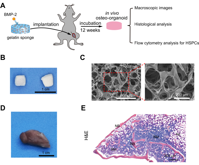
Figure 1: Formation and characterization of in vivo osteo-organoids. (A) Schematic diagram for harvesting and evaluating in vivo osteo-organoids induced by bioactive scaffolds. Freeze-dried gelatin scaffolds loaded with BMP-2 were surgically implanted into the lower limb muscles of mice. The in vivo osteo-organoids were explanted after 12 weeks of incubation for subsequent analysis. (B) Macroscopic images and (C) scanning electron microscope images of the bioactive scaffold. Figure 1A–C are reproduced from Dai et al.11. (D) Macroscopic image and (E) H&E-stained sections of the osteo-organoid 12 weeks after implantation. Figure 1D,E are reproduced from Dai et al.10. n = 4 biological replicates. Bone marrow and new bone are shown. Scale bars = 200 µm (C, right; E), 500 µm (C, left), 1 cm (B,D). Abbreviations: BMP-2 = bone morphogenetic protein-2; H&E = hematoxylin and eosin; BM = bone marrow; NB = new bone. Please click here to view a larger version of this figure.
To assess the long-term therapeutic cell-generating potential of osteo-organoids, we analyzed single-cell suspensions from the osteo-organoids using flow cytometry for quantitative analysis of HSPCs (Supplemental Figure S1). We analyzed the types of HSPC subsets 12 weeks after implantation (Figure 2A). Hematopoietic stem cells (HSCs; Lin−c-kit+Sca-1+CD48−CD150+) and their differentiated hematopoietic progenitor cells (HPCs), including multipotent progenitors (MPPs; Lin−c-kit+Sca-1+CD48−CD150−), common lymphoid progenitors (CLPs; Lin−c-kit–Sca-1–Flt3+IL7Rα+), common myeloid progenitors (CMPs; Lin−c-kit+Sca-1−CD34+FCγR−), granulocyte-monocyte progenitors (GMPs; Lin−c-kit+Sca-1−CD34+FCγR+), and megakaryocyte erythroid progenitors (MEPs; Lin−c-kit+Sca-1−CD34−FCγR−) were all present in the osteo-organoids. We then conducted further quantitative analysis of the cell numbers in different cell populations within the osteo-organoids (Figure 2B). After 12 weeks of implantation, the numbers of HSCs, MMPs, CLPs, CMPs, GMPs, and MEPs in the osteo-organoids were approximately 530, 860, 2750, 80,000, 43,000, and 55,000, respectively. In conclusion, these results supported that osteo-organoids contained an abundant supply of HSPCs for cell therapy.
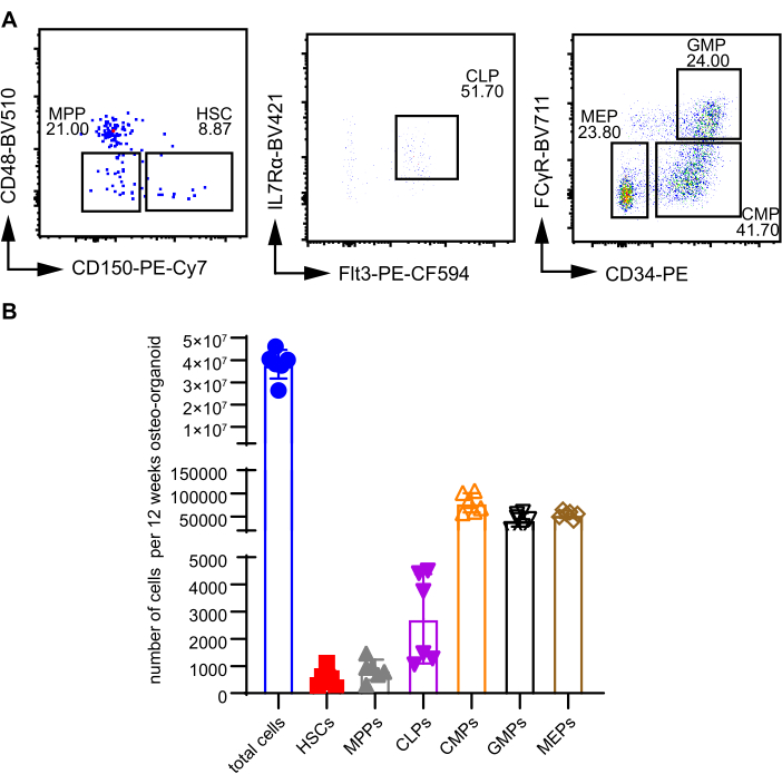
Figure 2: In vivo osteo-organoids contain abundant HSPCs. (A) Representative flow cytometric plots of HSPCs in the osteo-organoids 12 weeks after implantation. (B) Absolute numbers of total cells, Hematopoietic stem cells (Lin−c-kit+Sca-1+CD48−CD150+) and differentiated hematopoietic progenitors: multipotent progenitors (Lin−c-kit+Sca-1+CD48−CD150−), common lymphoid progenitors (Lin−c-kit–Sca-1–Flt3+IL7Rα+), common myeloid progenitors (Lin−c-kit+Sca-1−CD34+FCγR−), granulocyte-monocyte progenitors (Lin−c-kit+Sca-1−CD34+FCγR+), and megakaryocyte erythroid progenitors (Lin−c-kit+Sca-1−CD34−FCγR−). Figure 2A,B was modified from Dai et al.10. n = 6 biological replicates. Data are presented as means ± SD. Statistical differences among groups are identified by one-way ANOVA, followed by Tukey's multiple comparison tests. *P < 0.05, **P < 0.01, and ***P < 0.001. Abbreviations: HSPCs = hematopoietic stem/progenitor cells; HSCs = Hematopoietic stem cells; MPPs = multipotent progenitors; CLPs = common lymphoid progenitors; CMPs = common myeloid progenitors; GMPs = granulocyte-monocyte progenitors; MEPs = megakaryocyte erythroid progenitors; PE = phycoerythrin. Please click here to view a larger version of this figure.
HSCT has proven to be an effective cell therapy for treating radiation-induced immune system injury12,13,14. We transplanted cells derived from in vivo osteo-organoids (incubated for 12 weeks) into sublethally irradiated mice and collected samples from different parts of the mice at indicated time points for subsequent analysis (Figure 3A). Hematological analysis showed that, compared to the native BM-treated group, the WBC count in the 5 Gy osteo-organoid-treated group significantly increased at the 4th week post transplantation, suggesting an enhanced WBC recovery capability of the osteo-organoid-derived cells (Figure 3B). In the second week after transplantation, the RBC count of both the 5 Gy osteo-organoid-treated group and the 5 Gy native BM-treated group returned to normal levels (Figure 3C). At 2 weeks post transplantation, the PLT count was significantly increased in the 5 Gy osteo-organoid-treated group and the 5 Gy native BM-treated group compared to the 5 Gy PBS-treated group (Figure 3D). In conclusion, these results confirmed that cells derived from osteo-organoids exerted a more robust capacity to enhance hematopoietic recovery compared to native BM cells.
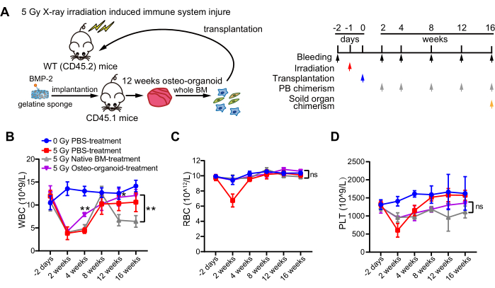
Figure 3: Cell therapy from osteo-organoids for the treatment of sublethally irradiated mice. (A) Experimental scheme of HSCT. The osteo-organoids were generated by implanting bioactive scaffolds into the femur muscle pocket of mice (donors, CD45.1) for 12 weeks. The osteo-organoids were collected and digested to obtain a whole bone marrow cell suspension, which was then transplanted to WT mice (recipients, CD45.2) with immune system injury induced by 5 GY X-ray. peripheral blood was collected at 2, 4, 8, 12, and 16 weeks after transplantation for subsequent analysis of peripheral blood chimerism. Solid organ was collected at 16 weeks after transplantation for subsequent analysis of solid organ chimerism. (B–D) peripheral blood cells from the four groups were analyzed at indicated time points. WT mice in the 0 Gy PBS-treated group did not receive irradiation and received PBS. WT mice in the 5 Gy PBS-treated, 5 Gy native BM-treated, and 5 Gy osteo-organoid-treated groups received 5 Gy irradiation 1 day prior to transplantation and were transplanted with PBS, native BM cells, or osteo-organoid-derived cells, respectively. n = 4 to 5 biological replicates. Figure 3A–D was modified from Dai et al.10. Data are presented as means ± SD. (B–D) Significant differences among groups are identified by two-way ANOVA followed by Bonferroni's post hoc test. **P < 0.01. Abbreviations: HSCT = Hematopoietic stem cell transplantation; BMP-2 = bone morphogenetic protein-2; WT = wild type; PB = peripheral blood; PBS = phosphate-buffered saline; BM = bone marrow; WBC = white blood cell; RBC = red blood cell; PLT = platelet. Please click here to view a larger version of this figure.
To assess the contribution of transplanted cells to the peripheral immune system reconstitution in irradiated mice, we performed quantitative analysis of CD3+ T cells, B220+ B cells, and CD11b+ myeloid cells using flow cytometry. This allowed us to evaluate the chimerism of osteo-organoid-derived and native BM-derived cells in the peripheral blood of the recipients. (Figure 4A and Supplemental Figure S2). We collected peripheral blood samples at 2, 4, 8, 12, and 16 weeks after cell therapy in different groups (Figure 3A). We observed sustained high levels of chimerism in the peripheral blood, with donor-derived CD3+ T cells, B220+ B cells, and CD11b+ myeloid cells consistently exceeding 80% in both the 5 Gy osteo-organoid treated groups and the 5 Gy native BM treated groups. (Figure 4B and Figure 4E,F). Consistent with hematological analysis, the osteo-organoid-derived cells exhibited enhanced regenerative capacity in the T cell subset chimerism in comparison to the native BM-derived cells (Figure 4C,D). The CD3+ T cell subset from the donor source achieved stable chimerism at 8 weeks post transplantation, while the B220+ B cell and CD11b+ myeloid cell subsets from the donor source achieved stable chimerism as early as 2 weeks after transplantation (Figure 4B–F). These data substantiated the role of osteo-organoid-derived cells from the donor in accelerating the recovery of T cell subsets in peripheral blood compared to the native BM-derived cells from the donor.
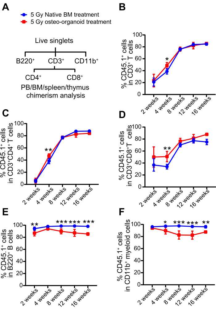
Figure 4: Reconstitution of the peripheral immune system in sublethally irradiated mice by osteo-organoid-derived cells. (A) The flow cytometric gating strategy for the chimerism analysis of recipient mice peripheral blood, BM, spleen, and thymus. (B–F) peripheral blood chimerism from the 5 Gy native BM-treated and 5 Gy osteo-organoid-treated groups was analyzed by the proportion of T cells, B cells, and myeloid cells at the indicated time points. Figure 4A–F was reproduced from Dai et al.10. n = 4 to 5 biological replicates. Data are presented as means ± SD. (B–F) Significant differences among groups were identified by two-way ANOVA followed by Bonferroni's post hoc test. *P < 0.05, **P < 0.01, and ***P < 0.001. Abbreviations: PB = peripheral blood; BM = bone marrow. Please click here to view a larger version of this figure.
To assess the regenerative capacity of transplanted cells in the irradiated mice's solid immune organs, we utilized the same flow cytometry gating strategy to analyze the chimerism of both native BM-derived and osteo-organoid-derived cells in the recipients' BM, spleen, and thymus (Figure 4A). At 16 weeks after cell therapy, the chimerism of T cells derived from donor osteo-organoid cells in recipients' BM was found to be increased, while the chimerism of B cells and myeloid cells was observed to be reduced, as compared to the chimerism of native BM cells from the donor (Figure 5A). Donor cells from both the 5 Gy osteo-organoid-treated group and the 5 Gy native BM-treated group exhibited pronounced chimerism in the spleen and thymus of the recipients, characterized by high levels of T cells, B cells, and myeloid cells (typically exceeding 80%) (Figure 5B,C). These findings provided evidence that osteo-organoid-derived and native BM-derived cells efficiently reconstituted the damaged immune system in mice exposed to irradiation.
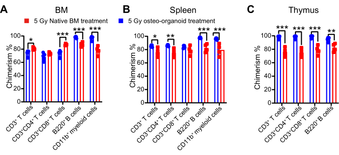
Figure 5: Reconstitution of the solid immune organ in sublethally irradiated mice by osteo-organoid-derived cells. (A–C) BM, spleen, and thymus chimerism from the 5 Gy native BM-treated and 5 Gy osteo-organoid-treated groups were analyzed by the chimerism rates of T cells, B cells, and myeloid cells at 16 weeks after transplantation. Figure 5A–C were reproduced from Dai et al.10. n = 4 to 5 biological replicates. Data are presented as means ± SD. Statistical differences among groups are identified by two-way ANOVA followed by Bonferroni's post hoc test. *P < 0.05, **P < 0.01, and ***P < 0.001. Abbreviation: BM = bone marrow. Please click here to view a larger version of this figure.
Supplemental Figure S1: Gating strategy used to identify HSPCs in the osteo-organoid. Single-cell suspensions from the osteo-organoid were subjected to flow cytometry for quantitative analysis of HSPCs. This figure was reproduced from Dai et al.10. Please click here to download this File.
Supplemental Figure S2: Gating strategy used to identify donor derived CD45.1+ cells in PB, BM, spleen, and thymus of CD45.2 recipients. Single-cell suspensions from PB, BM, spleen, and thymus of the CD45.2 recipients were analyzed via flow cytometry for the quantitative analysis of donor chimera. This figure was reproduced from Dai et al.10. Abbreviations: PB = peripheral blood; BM = bone marrow. Please click here to download this File.
In this protocol, we present an approach to establish in vivo osteo-organoids with bone marrow-like structures by implanting gelatin sponge scaffolds loaded with BMP-2. We demonstrate that these in vivo osteo-organoids can stably produce therapeutic HSPCs over a long period of time (more than 12 weeks). Compared to existing in vitro expansion or in vivo incubation methods that load cells, this protocol can obtain cell cocktails with diverse cell types, including HSPCs and various immune cells, within 3 weeks10. Animal experiments have also shown that transplantation of in vivo osteo-organoid-derived cells can accelerate hematopoietic recovery in irradiated mice. Given that gelatin sponges loaded with BMP-2 can retain their osteogenic activity for over 12 months after lyophilization when stored at -20 °C and that the material does not necessitate the incorporation of any cells, this variety of gelatin sponge infused with BMP-2 has the potential to be fashioned into an off-the-shelf product for immediate utilization.
There are two pivotal stages in the protocol that require special attention. First, during the material preparation process, it is crucial to maintain sterility to avoid bacterial contamination, which could result in bone formation failure and the inability to attain in vivo osteo-organoids. Second, after lyophilization, it is imperative to store the material at a low temperature to preserve the activity of BMP-2 protein. Additionally, the process of obtaining single-cell suspensions for transplantation must be carried out in a gentle and prompt manner to ensure cell viability. If the cell suspension is placed on ice, we recommend completing the transplantation within 4 h.
Although various biocompatible materials, such as hydroxyapatite, decalcified bone matrix, and matrix gel, have been reported in the literature as scaffold materials for loading BMP-2 to establish in vivo organoids15, we chose gelatin sponges as the scaffold material due to their availability and clinical translational needs. In addition, we chose to lyophilize the gelatin sponge after loading it with BMP-2 solution. This approach can increase the convenience of use and control the release ability of BMP-2 to a certain extent, reducing the dosage of BMP-2 used. From this perspective, this will significantly reduce the side effects of current clinical products that mainly consist of BMP-2 solution. In the cell transplantation process, in vivo organoid-derived cells can be transplanted after simple filtration without lysis of red blood cells, further simplifying the experimental process. In addition to routine peripheral blood chimerism analysis, we also systematically evaluated the engraftment levels of in vivo osteo-organoid derived cells in multiple solid organs of recipient mice, including bone marrow, spleen, and thymus. This approach will provide a more comprehensive evaluation of the therapeutic potential of in vivo osteo-organoid-derived cells in terms of hematopoietic function.
Although in vivo organoid derived cells can accelerate hematopoietic recovery in mice with bone marrow injuries, the number of HSPCs obtained is still lower than that obtained from single femurs. How to modify the material system to obtain a higher quantity and stronger functional HSPCs is an area that needs further exploration. We recently found that sulfated polysaccharides (SCS), especially sulfated chitooligosaccharides (SCOS), can regulate the function of HSPCs in in vivo osteo-organoids11. This result also suggests that the complexation of BMP-2 with more functional molecules should be a key research direction in the future.
Animal experiments have shown that functional HSPCs can be obtained through the in vivo osteo-organoid approach. However, in large animals such as miniature pigs or macaques, it is still unclear how high doses of BMP-2 can induce functional HSPCs, which is an important step for clinical translation and requires further research.
In summary, the in vivo osteo-organoid approach proposed in this protocol can efficiently and stably produce therapeutic HSPCs in vivo and provide a stable cell source for hematopoietic stem cell transplantation. It is worth exploring more HSPC enhancement factors to benefit more patients. In the following clinical application, we are investigating the use of immediate family members of patients as biogenerators to establish the in vivo osteo-organoid for therapeutic cell provision. This approach has the potential to minimize the risk of immunological rejection and eliminate potential ethical issues.
The authors have nothing to disclose.
This research was supported by the Basic Science Center Program (No. T2288102), the Key Program of the National Natural Science Foundation of China (No. 32230059), the National Natural Science Foundation of China (No. 32301123), the Foundation of Frontiers Science Center for Materiobiology and Dynamic Chemistry (No. JKVD1211002), the Wego Project of Chinese Academy of Sciences (No. (2020) 005), the Project of National Facility for Translational Medicine (Shanghai) (No. TMSK-2021-134), and the China Postdoctoral Science Foundation (No. 2022M721147).
AF700-anti-CD11b (M1/70) | eBioscience | 56-0112-82 | Store at 4 °C. Dilute 1:200 for staining. |
AF700-anti-Sca-1 (D7) | BioLegend | 108141 | Store at 4 °C. Dilute 1:200 for staining. |
APC-anti-CD3e (145-2C11) | Tonbo | 20-0031-U100 | Store at 4 °C. Dilute 1:200 for staining. |
bio-anti-CD34 (RAM34) | eBioscience | 13-0341-82 | Store at 4 °C. Dilute 1:200 for staining. |
BV421-anti-CD127 (IL-7Rα) | BioLegend | 135023 | Store at 4 °C. Dilute 1:200 for staining. |
BV510-anti-CD48 (HM48-1) | BioLegend | 103443 | Store at 4 °C. Dilute 1:200 for staining. |
BV711-anti-CD16/32 (93) | BioLegend | 101337 | Store at 4 °C. Dilute 1:200 for staining. |
Capillary tube | Shanghai Huake Labware Co. | DC616297403604-100mm/0.5mm | |
Cell strainer | CORNING | 352340 | |
Ethanol | GENERAL-REAGENT | 01158566 | |
Ethylenediamine tetraacetic acid (EDTA) solution | Servicebio | G1105 | |
Ethylenediaminetetraacetic acid dipotassium salt dihydrate (EDTA-K2) | Solarbio | E8651 | |
FITC-anti-CD45.2 (104) | BioLegend | 109806 | Store at 4 °C. Dilute 1:200 for staining. |
Flowjo | Becton, Dickinson & Company | A flow cytometry. | |
Gelatin sponge | Jiangxi Xiangen Co. | Use under sterile conditions. | |
HBSS without Ca2+ and Mg2+ | Gibco | 14170112 | HBSS without Ca2+ and Mg2+ can prevent cell aggregation. |
Hematology analyzer | Sysmex | pocH-100i Diff | |
iodine swabs | Xiangtan Mulan Biological Technology Co., Ltd. | 01011 | To prevent the infection after operation. |
Isoflurane | RWD | R510-22-10 | To avoid adverse effects of anesthesia waste gases on the environment and laboratory personnel, a gas recovery system should be used in conjunction. |
Kraft paper | absorbent paper | ||
LIVE/DEAD Fixable Near IR Dead Cell Staining Kit(used in 3.4.5) | Thermo Fisher Scientific | L34962 | A live/dead staining kit. Store at -20 °C. Dissolve in 50 μL of DMSO for working solution. |
lubricating vet ointment | Pfizer | To prevent dryness and counteract the ocular irritations caused by isoflurane. | |
Neutral balsam | Solarbio | G8590 | |
nylon filter | Shanghai Shangshai Wire Mesh Manufacturing Co., Ltd. | Used for cell filtration. | |
Paraffin liquid | Macklin | P815706 | |
Paraformaldehyde (PFA) solution | Servicebio | G1101 | Immersion fixation is used for routine animal tissues. The volume of fixative used is generally 10-20 times the tissue volume, and fixation at room temperature for 24 hours is sufficient. |
PE-anti-CD45.1 (A20) | BioLegend | 110708 | Store at 4 °C. Dilute 1:200 for staining. |
PE-CF594-anti-CD135 (A2F10.1) | BD Biosciences | 562537 | Store at 4 °C. Dilute 1:200 for staining. |
PE-Cy5-anti-c-kit (2B8) | BD Biosciences | 105809 | Store at 4 °C. Dilute 1:200 for staining. |
PE-Cy7-anti-B220 (RA3-6B2) | BioLegend | 103222 | Store at 4 °C. Dilute 1:200 for staining. |
PE-Cy7-anti-CD150 (TC15-12F12.2) | BioLegend | 115914 | Store at 4 °C. Dilute 1:200 for staining. |
PE-Dazzle594-anti-CD4 (GK1.5) | BioLegend | 100456 | Store at 4 °C. Dilute 1:200 for staining. |
Pentobarbital sodium salt | Sigma-Aldrich | 57-33-0 | Prepare for use at a concentration of 1% (w/v). |
PerCp-Cy5.5-anti-CD8a (53-6.7) | BioLegend | 100734 | Store at 4 °C. Dilute 1:200 for staining. |
PerCp-Cy5.5-anti-lineage cocktail | BD Biosciences | 561317 | Store at 4 °C. Dilute 1:10 for staining. |
Red blood cell lysis buffer | Beyotime | C3702 | Store at 4 °C. Use in clean bench. |
rhBMP-2 | Shanghai Rebone Biomaterials Co. | The concentration of rhBMP-2 in the stock solution is 1.0 mg/mL. | |
Staining buffer | BioLegend | 420201 | Store at 4 °C. |
Xylene | GENERAL-REAGENT | 01018114 | |
Zombie UV Fixable Viability Kit (used in 6.2.5) | BioLegend | 423108 | A live/dead staining kit. For reconstitution, bring the kit to room temperature; add 100 µL of DMSO to one vial of Zombie UV dye until fully dissolved. |
.