The experimental protocol was approved by the Institutional Animal Care and Use Committee of Jianghan University and was conducted in accordance with Experimental Animals Ethical Guidelines issued by the Center for Disease Control of China. Adult male C57BL/6J mice, 10 weeks old, weighted 24-26 g, were used in this protocol. All mice were housed under a 12-h light/dark cycle controlled environment with food and water ad libitum.
1. Preoperative preparation
NOTE: The key instruments and equipment required for this protocol are shown in Figure 1.
2. Distal MCAO model
3. Sham Operation
4. Behavioral tests
NOTE: Prior to the dMCAO, the mice underwent behavioral training twice daily for 3 days. On 3rd day post-dMCAO, move the mice to the behavioral testing room for a 2 h environmental adaptation before testing.
5. Perfusion and sample preparation
6. Identification of infarction volume
7. Histopathological and immunofluorescent staining analysis
The key instruments used to perform the dMCAO are the microsurgical instruments set, the isoflurane vaporizer, and the monopolar microsurgical electrocoagulation generator shown in Figure 1. The experimental procedure of this study is illustrated in Figure 2. In brief, a small bone window craniotomy was employed to expose the distal MCA, which was subsequently coagulated to induce permanent focal cerebral ischemia in C57BL/6 mice. Furthermore, the ischemic insults and stroke outcomes were assessed through TTC staining, histological examinations, and behavioral evaluations at 3 days post dMCAO. During the surgical procedure, only 1 mouse died from surgical bleeding. Furthermore, all the remaining mice survived during the 3-day observation period after surgery.
Macroscopic observation revealed that the dMCAO generated visible hyperemia and edema in the cortex (Figure 3A, bottom). No discernible macroscopic alterations were observed in the sham-operated group (Figure 3A, top). Additionally, dMCAO-induced cortical infarction was also verified using TTC staining (Figure 3B). The infarct volume was 16.6% ± 0.8% 3 days after the surgery in the dMCAO group, demonstrating the stability and repeatability of this cerebral ischemia model (Figure 3C).
Several behavioral tests were conducted to evaluate the neurological deficits on 3rd day post dMCAO. As shown in Figure 4A, the fore limb grip strength exhibited a significant reduction in the dMCAO group compared to the sham-operated group (70.8 g ± 4.2 g vs. 114.0 g ± 6.2 g, P < 0.001). The dMCAO group mice exhibited prolonged latencies in both turning around and descending to the ground during the pole test, as compared to the sham-operated group (15.8 s ± 1.7 s vs. 8.7 s ± 1.3 s, P < 0.01, 45.1 s ± 3.3 s vs. 29.2 s ± 2.1 s, P < 0.001) (Figure 4B,C). For the adhesive removal test, a significant increase in the removal time was observed in the dMCAO group compared to the sham operation group (20.5 s ± 2.5 s vs. 7.8 s ± 1.1 s, P < 0.001) (Figure 4D). The statistical analysis of the cylinder test data demonstrated a significant reduction in contralateral forepaw usage rates within the dMCAO group compared to the sham operation group (35.2% ± 2.6% vs. 48.7% ± 2.2%, P < 0.001) (Figure 4E).
Black squares in Figure 5A illustrate the analysis region for immunofluorescent staining. The H&E staining results revealed the disordered arrangement of neuron cells in the peri-infarct area of the dMCAO group, characterized by prominent pyknosis, vacuolization, and nuclear hyperstaining (Figure 5B, bottom). However, the sham-operation group did not exhibit any discernible alterations in neuronal morphology (Figure 5B, top). Figure 5C illustrates that dMCAO resulted in a significant reduction in the density of NeuN-positive cells within the peri-infarct area (319.6 ± 19.0 vs. 765.0 ± 26.0, P < 0.001). Figure 5D,E illustrates that the densities of microglia (665.8 ± 30.6 vs. 207.4 ± 16.2, P < 0.001) and astrocytes (305.2 ± 17.2 vs. 17.2 ± 2.1, P<0.001) increased greatly in dMCAO group compared with the sham-operation group. These findings offer compelling evidence supporting the presence of neuronal loss and the excessive activation of microglia and astrocytes on the 3rd day post-dMCAO.
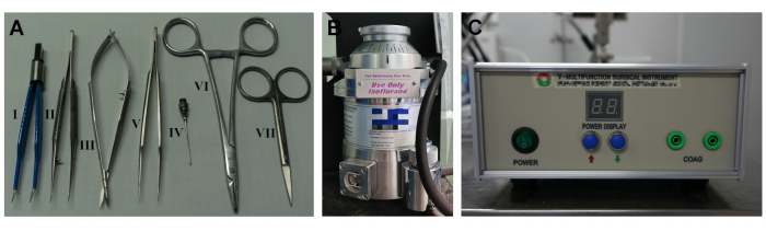
Figure 1: Key instruments used to establish the dMCAO model. (A) Essential surgical instruments. The Roman numerals I-VII refer to electric coagulation forceps, curved micro forceps, micro scissors, straightmicro forceps, retractors, needle holders, and surgical scissors. (B) Isoflurane vaporizer. (C) Electro-surgical generator. Please click here to view a larger version of this figure.
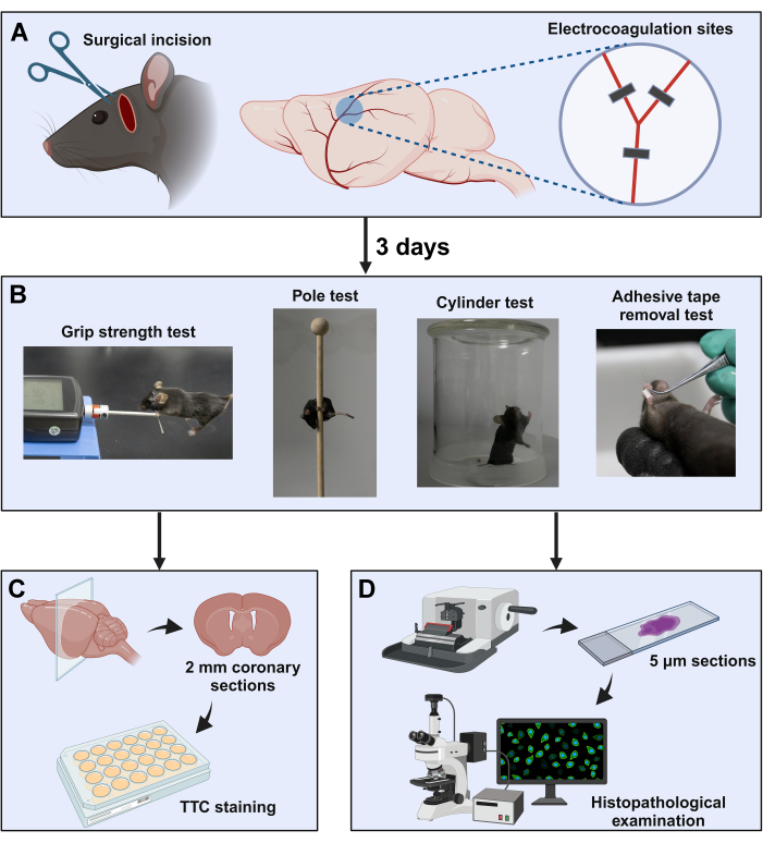
Figure 2: Schematic diagram of the experimental procedure. (A) The surgical window is located between the orbit and the ear canal. The distal MCA was exposed by subtemporal craniotomy, followed by coagulation at three sites near the bifurcation (indicated with black squares). (B) Neurological behavior evaluation paradigms were used in this study. After completion of the neurological behavior tests, the brain samples were collected for (C) TTC staining and (D) histopathology examinations. Please click here to view a larger version of this figure.
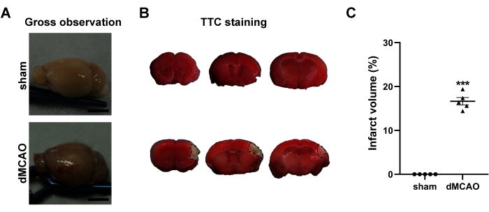
Figure 3: Macroscopic evaluation of the brains at 3rd day post dMCAO. (A) Gross observation of the brains from the sham operation and dMCAO groups. (B) Representative TTC-stained brain coronary sections of sham operation group (top) and dMCAO group (bottom). The black line demarcates the non-infarcted (unstained) cortex of the ipsilateral brain. (C) Quantification of the infarct volume. Scale bar = 5 mm. Data are presented as mean ± SEM. N = 5, ***P < 0.001 compared with the sham group, paired t-test. Please click here to view a larger version of this figure.
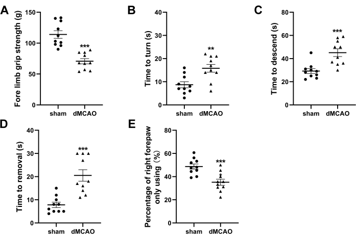
Figure 4: Neurological behavior tests at 3rd day post-dMCAO. (A) Quantification ofthefore limb grip strength. (B,C) Quantification of the time latencies to turn around and descend to the ground in the pole test. (D) Quantification of time required to remove the adhesive tape in the adhesive removal test. (E) Quantification of cylinder test presented as the usage ratio of the right forepaw. Data are presented as mean ± SEM, N = 10, **P < 0.01, ***P < 0.001 compared with the sham group, paired t-test. Please click here to view a larger version of this figure.
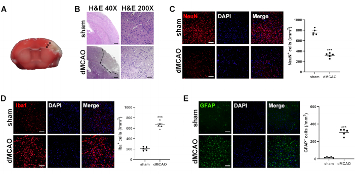
Figure 5: Histological analysis of brains at 3rd day post dMCAO. (A) Immunofluorescence measurement from the cortex of the selected three fields in the ischemic penumbra. (B) Representative images of H&E-stained brain sections from the sham operation and dMCAO groups. Dotted lines surround unstained parts, indicating an infarct lesion. (H&E 40x, Scale bar = 200 µm; H&E 200x, Scale bar = 50 µm). Representative images of immunofluorescence staining and quantitative analysis for (C) NeuN (a marker for neuron), (D) Iba-1 (a marker for microglia), and (E) GFAP(a marker for astrocyte). Scale bar = 50 µm. Data are presented as mean ± SEM, N = 5, ***P < 0.001 compared with the sham group, paired t-test. Please click here to view a larger version of this figure.
2,3,5-Triphenyltetrazolium Chloride (TTC) |
Sigma-Aldrich | 108380 | Dye for TTC staining |
24-well culture plate | Corning (USA) | CLS3527 | Vessel for TTC staining |
4% paraformaldehyde | Wuhan Servicebio Technology Co., Ltd. |
G1101 | Tissue fixation |
5% bovine serum albumin | Wuhan BOSTER Bio Co., Ltd. | AR004 | Non-specific antigen blocking |
5-0 Polyglycolic acid suture | Jinhuan Medical Co., Ltd | KCR531 | Material for surgery |
Anesthesia machine | Midmark Corporation | VMR | Anesthetized animal |
Antifade mounting medium | Beyotime Biotech | P0131 | Seal for IF staining |
Automation-tissue-dehydrating machine |
Leica Biosystems (Germany) | TP1020 | Dehydrate tissue |
Depilatory cream | Veet (France) | 20220328 | Material for surgery |
Diclofenac sodium gel | Wuhan Ma Yinglong Pharmaceutical Co., Ltd. |
H10950214 | Analgesia for animal |
Drill tip (0.8 mm) | Rwd Life Science Co., Ltd. | Equipment for surgery | |
Eosin staining solution | Wuhan Servicebio Technology Co., Ltd. |
G1001 | Dye for H&E staining |
Eye ointment | Guangzhou Pharmaceutical Co., Ltd | H44023098 | Material for surgery |
Fluorescence microscope | Olympus (Japan) | BX51 | Image acquisition |
GFAP Mouse monoclonal antibody | Cell Signaling Technology Inc. (Danvers, MA, USA) |
3670 | Primary antibody for IF staining |
Goat anti-mouse Alexa 488-conjugated IgG |
Cell Signaling Technology Inc. (Danvers, MA, USA) |
4408 | Second antibody for IF staining |
Goat anti-rabbit Alexa 594-conjugated IgG |
Cell Signaling Technology Inc. (Danvers, MA, USA) |
8889 | Second antibody for IF staining |
Grip strength meter | Shanghai Xinruan Information Technology Co., Ltd. | XR501 | Equipment for behavioral test |
Hematoxylin staining solution | Wuhan Servicebio Technology Co., Ltd. |
G1004 | Dye for H&E staining |
Iba1 Rabbit monoclonal antibody | Abcam | ab178846 | Primary antibody for IF staining |
Isoflurane | Rwd Life Science Co., Ltd. | R510-22-10 | Anesthetized animal |
Laser doppler blood flow meter | Moor Instruments (UK) | moorVMS | Blood flow monitoring |
Meloxicam | Boehringer-Ingelheim | J20160020 | Analgesia for animal |
Microdrill | Rwd Life Science Co., Ltd. | 78001 | Equipment for surgery |
Microsurgical instruments set | Rwd Life Science Co., Ltd. | SP0009-R | Equipment for surgery |
Microtome | Thermo Fisher Scientific (USA) | HM325 | Tissue section production |
Microtome blade | Leica Biosystems (Germany) | 819 | Tissue section production |
Monopolar electrocoagulation generator | Spring Scenery Medical Instrument Co., Ltd. |
CZ0001 | Equipment for surgery |
Mupirocin ointment | Tianjin Smith Kline & French Laboratories Ltd. |
H10930064 | Anti-infection for animal |
NeuN Rabbit monoclonal antibody | Cell Signaling Technology Inc. (Danvers, MA, USA) |
24307 | Primary antibody for IF staining |
Neutral balsam | Absin Bioscience | abs9177 | Seal for H&E staining |
Paraffin embedding center | Thermo Fisher Scientific (USA) | EC 350 | Produce paraffin blocks |
Pentobarbital sodium | Sigma-Aldrich | P3761 | Euthanized animal |
Phosphate buffered saline | Shanghai Beyotime Biotech Co., Ltd | C0221A | Rinsing for tissue section |
Shaver | Shenzhen Codos Electrical Appliances Co.,Ltd. |
CP-9200 | Equipment for surgery |
Sodium citrate solution | Shanghai Beyotime Biotech Co., Ltd. | P0083 | Antigen retrieval for IF staining |
Ischemic stroke remains the predominant cause of mortality and functional impairment among the adult populations globally. Only a minority of ischemic stroke patients are eligible to receive intravascular thrombolysis or mechanical thrombectomy therapy within the optimal time window. Among those stroke survivors, around two-thirds suffer neurological dysfunctions over an extended period. Establishing a stable and repeatable experimental ischemic stroke model is extremely significant for further investigating the pathophysiological mechanisms and developing effective therapeutic strategies for ischemic stroke. The middle cerebral artery (MCA) represents the predominant location of ischemic stroke in humans, with the MCA occlusion serving as the frequently employed model of focal cerebral ischemia. In this protocol, we describe the methodology of establishing the distal MCA occlusion (dMCAO) model through transcranial electrocoagulation in C57BL/6 mice. Since the occlusion site is located at the cortical branch of MCA, this model generates a moderate infarcted lesion restricted to the cortex. Neurological behavioral and histopathological characterization have demonstrated visible motor dysfunction, neuron degeneration, and pronounced activation of microglia and astrocytes in this model. Thus, this dMCAO mouse model provides a valuable tool for investigating the ischemiastroke and worth of popularization.