Exonucleases play critical roles in ensuring genome stability. Loss of WRN exonuclease function results in premature aging. Studying substrates and other requirements of the nuclease in vitro can help elucidate its role in vivo. Here we demonstrate a rapid and reproducible fluorescence-based assay to measure its nuclease activity.
WRN exonuclease is involved in resolving DNA damage that occurs either during DNA replication or following exposure to endogenous or exogenous genotoxins. It is likely to play a role in preventing accumulation of recombinogenic intermediates that would otherwise accumulate at transiently stalled replication forks, consistent with a hyper-recombinant phenotype of cells lacking WRN. In humans, the exonuclease domain comprises an N-terminal portion of a much larger protein that also possesses helicase activity, together with additional sites important for DNA and protein interaction. By contrast, in Drosophila, the exonuclease activity of WRN (DmWRNexo) is encoded by a distinct genetic locus from the presumptive helicase, allowing biochemical (and genetic) dissection of the role of the exonuclease activity in genome stability mechanisms. Here, we demonstrate a fluorescent method to determine WRN exonuclease activity using purified recombinant DmWRNexo and end-labeled fluorescent oligonucleotides. This system allows greater reproducibility than radioactive assays as the substrate oligonucleotides remain stable for months, and provides a safer and relatively rapid method for detailed analysis of nuclease activity, permitting determination of nuclease polarity, processivity, and substrate preferences.
Nucleases serve a vital role in cells in removing damaged DNA, resolving nonduplex structures such as Holliday junctions and providing proof-reading capacity during DNA replication, both intrinsic within DNA polymerases and extrinsic to them1. Nucleases can act either by sequentially degrading DNA from free ends (exonucleases) or by cleaving internal phosphodiester bonds within a longer DNA molecule (endonucleases). Loss of nuclease activity can result in highly specific genome instability phenotypes. While mutation of the RecQ helicase family member BLM result in excessively high rates of sister chromatid exchange and globally elevated cancer rates (reviewed byPayne and Hickson2), mutation of the highly related WRN protein leads to premature aging3; the major significant difference between these two family members is the presence of a 3'-5' exonuclease domain with in WRN4. Evidence of a critical role of the WRN exonuclease in maintaining genome stability has accumulated from analysis of genotypes in WS patients5, together with point mutation and deletion studies in human cells, backed by crystallographic studies of the isolated exonuclease domain6. However, cooperation and cross talk between WRN's exonuclease activity and its central helicase activity7 makes it difficult to tease apart the functionality of each and their relative contributions to genome stability. In plants and lower metazoan animals, WRN exonuclease activity is present on a single polypeptide lacking helicase activity8-10 (reviewed in Cox and Boubriak11); it has been demonstrated biochemically in Arabidopsis that this exonuclease acts coordinately with the cognate WRN helicase, effectively reconstituting the combined enzyme activities observed in vertebrate WRN9. We have studied WRN exonuclease in Drosophila since the excellent genetic tools allow analysis of the impact of exonuclease mutation (without impacting on the presumptive cognate helicase) at the whole organism level and through development10,12. Moreover, we have cloned, expressed, and purified recombinant Drosophila WRN exonuclease (DmWRNexo) allowing full biochemical analysis of its enzyme properties13,14.
Nuclease analysis in vitro has traditionally been conducted using radiolabeled oligonucleotides, assessing degradation by looking for laddering of products on acrylamide gels4,8,15. While sensitive, such assays are not quantitatively reproducible day-to-day because of radioactive decay of the labeled substrates. Additionally, handling and disposal of radioactive reagents pose significant environmental and health issues; sourcing of radiolabel is also becoming increasingly problematic. An alternative recent method assesses the amount of the final degradation product by mass spectrometry16. However, it is time consuming (taking several days), requires specialized equipment, and the readout is the amount of end product (single nucleotide) so is not suitable for sensitive measurement of aspects such as enzyme processivity or for determining whether some nucleotides, sequences, or modifications lead to nuclease pausing or halt. To overcome these problems, we have adapted the traditional gel-based assays for use with fluorescent oligonucleotide substrates, generating stably labeled substrates that can be used reproducibly over long time periods and thus allow direct comparison of nuclease activities under different conditions.
1. Preparation of Substrate Oligonucleotides
2. Preparation of Acrylamide Gels
3. Preparation of His-tagged Recombinant DmWRNexo Protein
4. Nuclease Assay
5. Gel Analysis and Imaging
Performing in vitro analysis of exonuclease activity requires a number of preparatory steps in addition to the actual analysis. An overview of the procedures is shown in Figure 1.
Prior to conducting fluorescence-based exonuclease assays, it is critical to optimize detection of the fluorescently labeled oligonucleotide substrate following separation on urea-acrylamide gels using a suitable fluorescence imaging system. Filter choice is extremely important as this can have a marked impact on the sensitivity of the overall assay – our oligonucleotide substrate is fluorescein-labeled so requires a filter that permits excitation at 470 nm and emission at 520 nm. Using this filter, it is possible to increase the ability to detect low concentrations of substrate (or product) by adjusting both the sensitivity and resolution of the imager (Figures 2A and 2B). Note there is some tolerance in terms of filter bandwidth, as a suboptimal filter choice (ex 520/em 580 nm) still allows partial detection of the substrate, though with much lower sensitivity (Figure 2C). We suggest using a combination of labeled oligonucleotide concentration and imager settings that allow robust detection of substrate (Figure 2B and dark grey bars on right panel of Figure 2) so that degradation products, which are present at lower amounts in each band as the substrate is sequentially fragmented, can be reliably detected.
A successful exonuclease assay will cause degradation of the substrate sequentially towards the fluorophore (according to the polarity of the exonuclease being tested, this can be either a 5' or 3' end label on one strand of a duplex), resulting in a characteristic ladder-like pattern of DNA on the gel representative of exonuclease degradation (e.g. a time course of degradation is shown in Figure 3). Single stranded oligonucleotides can also be used but note that nucleases may show a length requirement for cleavage of ss DNA15,17 so false negatives may result from using too short an oligonucleotide. The kinetics of the enzyme activity can be approximated by performing densitometry of the gel images of a time course experiment and quantifying amount of nondegraded substrate remaining compared with amount present in smaller fragments (i.e. products of degradation – see Figure 2 of Mason et al.14 for an example). One simple method is to divide the gel equally into four vertical sections then obtain densitometry readings of each quarter. This avoids issues with minor variation in mobility of the substrate across the gel, though of course does not permit sensitive measurements such as cleavage of one or a few nucleotides from the substrate. It is important to normalize densitometry measurements against a region of the gel lacking any DNA (e.g. leave at least one lane empty), and to calculate degradation against an oligonucleotide-only control that is loaded for every experiment (see Mason et al.14).
Once optimized for detection, the assay can be used to detect differential activities both qualitatively and quantitatively. For example, the presence or absence of activity can be determined for nuclease mutants compared with wild type protein (Figure 4), while different substrates may also be tested for their ability to be degraded by the nuclease under investigation (Figure 4). If processivity measurements are required, it is possible to add excess unlabeled oligonucleotide at a defined time point and determine cleavage activity post-addition. A highly processive enzyme will continue to cleave the labeled substrate, while a poorly processive enzyme will show greatly diminished capacity to cleave the labeled substrate as it dissociates from substrate and reassociates with the unlabeled oligonucleotide that is in molar excess (e.g. see Mason et al.14 Figure 2).
Similarly, the intensity and migration of bands of the ladder will provide data on the activity and processivity of a particular exonuclease, either intrinsically or under differential conditions such as cation availability or temperature. As described previously14, DmWRNexo is partially processive but does not readily cleave the 5' fluorescein-labeled 50 nucleotide template to completion under the conditions used here. Single nucleotide bands are thus rarely seen with this enzyme and substrate. However, the assay can readily detect single nucleotide cleavage products, as observed when DmWRNexo (a 3'-5' exonuclease) acts on a 3' fluorescein-labeled substrate (see Figure 3 of Mason et al.14). Indeed, determination of directionality of exonuclease activity is achieved by analysis using substrates with backbones labeled either at the 5' or 3' end – a 3' exonuclease will immediately cleave the fluorophore from a 3'-labeled strand, rendering the subsequent laddering activity invisible to analysis, and resulting in only a single very-high mobility species apparent on the gel (see Figure 3 of Mason et al.14). Endonucleases that cut internally will show products at specific mobilities rather than a ladder. Similarly, altered bases that are not susceptible to nuclease cleavage will result in strong pause or stop sites that can be detected as more intense bands representing cessation of cleavage at those modified bases (e.g. See Figure 7 of Mason et al.14).
Nonoptimal results include smearing i.e. nonspecific degradation. Other issues to be aware of are gel problems that cause poor separation e.g. where the gel has not set evenly (Figure 5), bubbles are present in the gel, or the buffer leaks during the electrophoresis run. It is also important that substrates are checked before use as they might have separated or degraded, in which case they should be reannealed or discarded. Loss of protein activity through freeze/thaw cycles, or insufficient concentrations, can lead to negative results, though it is important to optimize for buffer requirements and other requirements e.g. ATP at high concentrations can inhibit DmWRNexo, presumably by titrating out Mg2+ ions15.
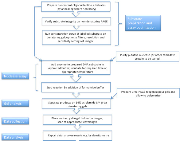
Figure 1. Flow chart of procedures undertaken when conducting a fluorescence-based exonuclease assay.
Click here to view larger image.
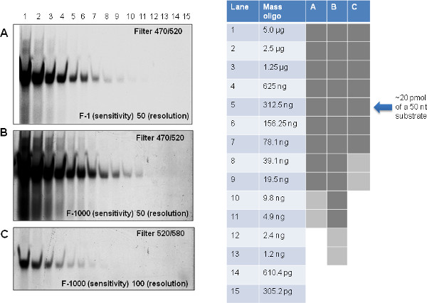
Figure 2. Optimizing detection of fluorescent substrate using the phosphorimager. A single gel of doubling dilutions of the 50-mer fluorescently labeled oligonucleotide substrate was exposed on the Phosphorimager using different filter conditions (A, B: 470 ex/520 em which is optimal for fluorescein, or C: 520 em/580 ex) and altering either sensitivity or resolution. Dark grey boxes indicate oligonucleotide concentrations that can be detected reliably with a high sensitivity and resolution; lighter grey boxes indicate those concentrations at which the signal can still be detected though the signal is much weaker. Unshaded areas of the table represent oligonucleotide concentrations that could not be detected using the filter, sensitivity and resolution settings indicated on the gel images A, B and C. Note that 20 pmol of a 50 nucleotide substrate is approximately equivalent to 312.5 ng. Click here to view larger figure.
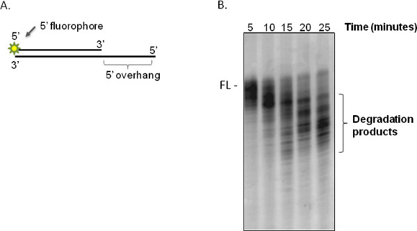
Figure 3. Typical fluorescently labeled substrate and exonuclease assay results. (A) Schematic representation of a duplex oligonucleotide substrate (5'OV) with 5' overhang on the unlabeled guide strand and 5' fluorescein label on the cleaved strand, as used in these assays. (B) Time course of degradation of the 5' overhang substrate by purified DmWRNexo at 37 °C. The enzyme binds to the 5' overhang of the guide strand and sequentially cleaves the labeled strand in a 3'-5' direction, resulting in a ladder of fragments of progressively smaller sizes that run more quickly on acrylamide gels. In this experiment, 10 pmol of DmWRNexo and 20 pmol oligonucleotide substrate were used in a 20 μl reaction. ('FL' represents the position of the nondegraded duplex 5'OV duplex fluorescent substrate).
Click here to view larger image.
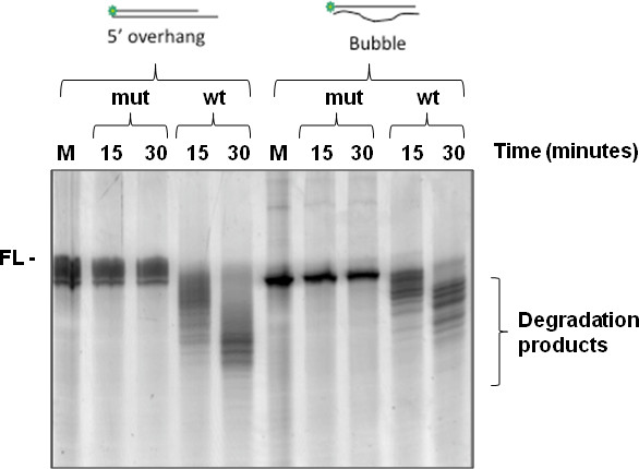
Figure 4. Testing the impact of point mutation of the exonuclease catalytic domain on ability of the enzyme DmWRNexo to cleave either duplex substrate with 5' overhang, or a bubble structure. In both cases, the substrate is labeled on one strand only at the 5' end with fluorescein. In a 20 μl reaction, 12.5 pmol purified DmWRNexo protein (wt or double point mutant D82A E84A) was incubated at 37 °C with 25 pmol oligonucleotide as shown in the schematic above the gels, and products analyzed on urea-acrylamide gels after 15 or 30 min. ('FL' represents the position of the nondegraded fluorescent substrate). 'wt' denotes wild type DmWRnexo protein, while 'mut' is a double point mutant version (D82A E84A). These results were useful in confirming the assignment of the enzyme's active site, which had been based on homology to human WRN exonuclease domain together with in silico structural modeling.
Click here to view larger image.
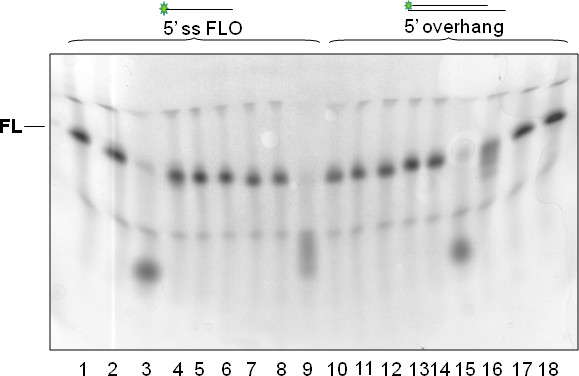
Figure 5. One example of a poor result. As a consequence of uneven polymerization of the gel, the fluorescently labeled substrate does not run at the same apparent mobility in all lanes. This can lead to incorrect interpretation of cleavage data, even though it is apparent that the substrates in lanes 3, 9 and 15 have been cleaved almost to completion, with partial cleavage in lane 16. Note that some lanes appear compressed in width in addition to the aberrant mobility leading to the 'smile' detected. Using identical conditions for polymerization (e.g. fresh APS, suitable concentrations of TEMED and a stable and consistent room temperature) can help to avoid such problems. 'FL' represents the position of the nondegraded fluorescent substrate.
Click here to view larger image.
Determination of exonuclease activity of purified proteins requires the analysis of DNA cleavage products. Sequential cleavage of DNA by exonucleases can be visualized by separation of labeled cleavage products on acrylamide gels. Historically this involved end-labeling of the DNA substrate with a radiolabel (e.g. 32P or 35S), but with the disadvantages inherent in use of radiolabel (cost, safety issues, and instability over time). To overcome these problems, we have developed an exonuclease assay that employs fluorescently labeled DNA substrates, and show the utility of the assay system in determining nuclease activity of the WRN exonuclease orthologue from Drosophila melanogaster, DmWRNexo10. The key benefit of our assay is that the fluorescently labeled substrates are stable over time thus providing greater reproducibility and standardization e.g. when comparing different batches of purified recombinant exonuclease. We have used the assay to undertake detailed analysis of DmWRNexo nuclease activity, permitting determination of nuclease polarity, processivity, and substrate and cation preferences (see also13,14).
Whilst this assay was developed specifically to determine the activity of DmWRNexo and related orthologous 3'-5' DNA exonucleases, it is equally applicable to nucleases of opposite polarity (e.g. we have tested it on Lambda exonuclease14). Moreover, the substrates are suitable for analysis of cleavage sites by endonucleases. Excitingly, since the system employs a fluorescently labeled substrate, it should also be possible to conduct protein-DNA interaction studies +/- nuclease activity (e.g. in the presence of nuclease inhibitors such as EDTA, or comparing DNA binding between wild type and mutant proteins to explore the basis of lack of activity of nuclease-dead mutant forms), either in a band shift format or quantitatively using fluorescence polarization assays in multiwell plates – i.e. there is adaptability for high throughput screening, especially using plate readers that include injectors for addition of inhibitors/activators. Extension of such studies to helicases would be possible by including a quenching agent on the strand complementary to the fluorescently labeled strand: helicase-dependent unwinding of the substrate would result in release of the fluorescent signal (an excess of unlabeled complementary strand may be necessary to prevent reannealing of the original duplex substrate). While DmWRNexo is a relatively small protein (~50 kDa) and is readily expressed as a soluble protein in E. coli, larger proteins (including many helicases) may need to be expressed in eukaryotic systems such as from baculovirus vectors in insect Sf9 cells, or even in human cells (e.g. human embryonic kidney 293 cells). Such systems are more efficient than E. coli for expression of larger proteins; they also permit post-translational modification, which may be critical for activity of some enzymes.
As for any biochemical assay, there are limitations as to the sensitivity of this nuclease assay, both in terms of the fluorophore used and the detection system. To obtain a fluorescent signal the fluorophore must be excited, and while the sensitivity can be modulated to some extent by changing the setting on the phosphorimager (e.g. Figure 2), it is not possible to enhance a signal by increasing time of exposure, as is the case with X-ray film exposed to radiolabel. Since exonucleases may be inhibited by large moieties bound to the end of the DNA substrate18, care should be taken to choose a small fluorophore to minimize inhibition or modulation the enzyme being assayed. It is also important to test cleavage of substrates labeled at either end (5' or 3') and to quantify all data using ImageJ or similar to assess how much substrate is cleaved. The concentrations of both substrate and enzyme are higher in the fluorescence assay described here than in published radioactivity-based assays; however, the relative proportions of DNA to nuclease are the same, validating our assay. Given increasing global problems in sourcing radiolabel, together with safety issues in its use and disposal, we believe that the relative safety, ease of use and stability of the fluorescent substrate outweigh the minor point of using more protein and DNA: concentrations are still low at ≤20 pmol substrate and 1-20 pmol enzyme. There is also the added benefit that there is no need to regularly label and gel-purify substrate as labeling takes place at the time of synthesis and is stable over time.
Alternative methods to the fluorescence based assay described here include use of radiolabeled substrates (as discussed above e.g.4,8,15,18) and a relatively recent addition to the range of assays available, based on nonradioactive isotope dilution mass spectrometry (LC-MS/MS) analysis of the final single nucleotide product of cleavage16. However, this latter procedure requires access to highly specialized equipment, and measures only the end-point of the nuclease reaction. We believe that the use of fluorescently labeled DNA substrates permits reproducible analysis of, and comparison between, exonucleases from different sources and between different batches of the same nuclease, permitting robust biological replication of experiments. The stability of the labeled substrate together with increasingly sophisticated methods of detecting fluorescence (e.g. in fluorescence polarization-based interaction studies) should allow this assay to be developed in the future for moderate to high throughput screening, particularly for inhibitors of nucleases.
The authors have nothing to disclose.
We thank the cross-council New Dynamics of Ageing Programme for funding this work [ES/G037086/1] and Prof Dave Sherratt (Department of Biochemistry, University of Oxford) for access to the Fuji FLA-3000.
Reagent/Material | |||
Custom oligonucleotides | Eurogentec | It is necessary to obtain these at high purity e.g. with PAGE purification. | |
5'FLO | fluoresescein-5' GAACTATGGCTCTC GAGTGCTAGGACATGTCTGA CTACGTACAAGTCACC – 3' |
||
bubble | 5'- GGTGACTTGTACGT AGTCAGACATGTCCTAGCAC TCGAGAGCCATAGTTC-3' |
||
40% 19:1 Acrylamide solution | Severn Biotech | 20-2400-05 | CAUTION: potent neurotoxin so gloves should be worn at all times |
His-Trap columns (1 ml) | GE Healthcare | 17-5247-01 | |
All other reagents | any reputable supplier | Molecular biology grade is necessary (DNase-free); microfuge tubes similarly should be DNase- and RNase-free | |
Equipment | |||
Hoefer SE400 gel apparatus | Hoefer | SE400-15-1.5 | |
FLA-3000 (phosphor and fluorescence imager) | Fuji | ||
Image Reader V2.02 | FujiFilm | ||
Image Gauge V3.3 | FujiFilm |