Here, we present a protocol to locally induce apical periodontitis in mice. We show how to drill a hole in the mouse's tooth and expose its pulp, in order to cause local inflammation. Analysis methods to investigate the nature of this inflammation, such as micro-CT and histology, are also demonstrated.
The mechanisms involved in local induced inflammation can be studied using several available animal models. One of these is the induction of apical periodontitis (AP). Apical periodontitis is a common pathology of an inflammatory nature in the periodontal tissues surrounding the tooth root. In order to better understand the nature and mechanism of this pathology it is advantageous to perform the procedure in mice. The induction of this odontogenic inflammation is achieved by drilling into the mouse tooth until the dental pulp is exposed. Next, the tooth pulp remains exposed to be contaminated by the natural oral flora over time, causing apical periodontitis. After this time period, the animal is sacrificed, and the tooth and the jaw bone can be analyzed in various ways. Typical analyses include micro-CT imaging (to evaluate bone resorption), histological staining, immunohistochemistry, and RNA expression. This protocol is useful for research in the field of oral biology to better understand this inflammatory process in an in vivo experimental setting with uniform conditions. The procedure requires a careful handling of the mice and the isolated jaw, and a visual demonstration of the technique is useful. All technical aspects of the procedures leading to induced apical periodontitis and its characterization in a mouse model are demonstrated.
The goal of this method is to induce apical periodontitis in a mouse by contaminating the apex with the natural microflora, and to then study various characteristics of this pathological process.
Apical periodontitis (AP) is a common pathology of an inflammatory nature in the periodontal tissues surrounding the tooth root. This dental disease can cause severe pain and must be treated by a dentist. The treatment options include root canal treatment (primary or secondary), endodontic surgery, tooth extraction, or follow-up depending on the clinical and radiographic findings, and the opinion of the clinician. The mechanism of this inflammatory process, although studied for several decades1,2,3, is still not comprehensively understood. Considering the severity of this pathology, there is thus a clear need for research addressing its fundamental nature. Thus, systems where the study of AP is possible are of great scientific interest.
Since AP is a complex pathological process involving the local tissues and the immune system, in vitro studies are insufficient for a complete understanding of the processes. Study of clinical samples of this disease are also problematic due to ethical limitations and significant variability between different people and different clinical stages4,5, and hence the necessity of in vivo models. These models are based on the concept of exposing the dental pulp to contamination and observing the inflammatory reaction of the body to this stimulus in the periapical tissues6,7. Common in vivo models include rodents or larger animals such as dogs. Despite the clinical challenge in treating mice, which are very small animals with miniature teeth, the advantages of the mouse model are significant: practically, working with mice is technically simple in terms of facilities and is most cost effective, and scientifically, the mouse is a well-studied animal model with readily available genetic and molecular tools and a well-studied genome. Indeed, previous studies used a mouse model for studying inflammatory and bone resorption signals and cells involved in apical periodontitis8,9,10,11. Therefore, a clear protocol on how to use a mouse model for the study of AP is needed. Here, we describe such a protocol.
The protocol described here is has the big advantage of being appropriate to study knock-out (KO) mice and learn how the lack of a specific gene affects dental inflammation7,12. Other useful applications of this protocol include the study of effects of medications and systemic conditions on the development of apical periodontitis13, the effect of apical periodontitis on the development of osteonecrosis of the jaws14,15 and stem cell therapy for bone regeneration16.
This protocol can also be generalized as a model to study local inflammation. To study the inflammatory process, several mouse models have been developed, which include for example induced colitis or arthritis17,18. These models have systemic effects and have no built-in control in the same animal. Models for induced apical periodontitis, which include a contralateral control without inflammation, have the advantage of overcoming these limitations14,19.
The protocol described below is therefore useful for researchers who are interested in local inflammatory processes. The controlled nature of this inflammation, its confinement to a specific location, and the contralateral control tooth, all make this protocol valuable for studying the mechanisms involved in this process. Moreover, the protocol is useful for researchers interested in the clinical aspects of periapical inflammation. The mouse model is ideal to study different variables of the disease, in addition to the advantage of being able to easily perform genetic manipulations in the mouse model, to investigate the activity of specific genes in periapical inflammation.
Technically, the clinical procedure is challenging to carry out due to the small dimensions of the mice teeth. It will be beneficial to visualize this procedure in order to learn about positioning, equipment needed, and performance.
All methods described here have been approved by the Institutional Animal Care and Use Committee (IACUC) of The Hebrew University (Ethics no. MD-17-15093-5).
1. Animal anesthesia and positioning
2. Pulp exposure
3. End of clinical procedure
4. Post procedure follow-up (42 days)
5. Experiment termination and analysis
A flow chart of the experimental steps is presented in Figure 1. As mentioned in the protocol, the mice are anesthetized, and their first mandibular molar on one side is drilled until pulp exposure, while the contralateral tooth is left as a control. Next, the teeth are left to be contaminated by the oral flora for 42 days, during which they are monitored and receive pain medication. After 42 days, mice are euthanized, and the teeth and adjacent jaw are taken for analysis.
Figure 2 demonstrates clinical images of the mouse mandible after first right molar pulp exposure. In Figure 2A the whole mandible is shown, while the inset in Figure 2B shows an enlargement of the treated first right molar. The left first molar is used as a control. Exposure of both mesial and distal pulp horns and the entrance to the canals can be seen in Figure 2B. Note that the tooth was mechanically lowered to sub-occlusion in order to reduce pain.
After dental pulp exposure to the oral flora, the dental pulp is eventually contaminated6,7 and becomes necrotic. According to the literature22, Gram negative bacteria then secrete lipopolysaccharide (LPS) to the apical region of the tooth. Through a complex reaction of the immune system, one of the outcomes is bone resorption in the periapical region, which is the hallmark of apical periodontitis23. This bone resorption can be identified and quantified by micro-CT. In micro-CT images, radiolucent periapical regions indicate areas where the hard bone tissue became a soft periapical lesion due to the inflammatory process. In Figure 3, representative micro-CT images demonstrate a treated tooth compared to a control. The arrow in Figure 3AIII points at the mesial pulp exposure (the distal pulp exposure does not appear on this slice of the sample), and the mesial and distal periapical radiolucent areas of bone resorption are surrounded by a dashed line. A boxplot graph shown here quantifies the significant periapical bone resorption in the treated teeth compared to the controls.
While micro-CT is an excellent technique for evaluation of the 3-dimensional size of the lesions, it is lacking information regarding their biological composition. Histological staining, as presented in Figure 4, provides this information. H&E staining is demonstrated in a control tooth compared to a treated tooth. In the treated tooth (Figure 4B,C,D), the dental pulp itself presents necrosis which can be seen clearly in the H&E staining (Figure 4C), compared to the control organized pulp tissue (Figure 4A). Additionally, and importantly, in the periapical region of the treated tooth a periapical lesion, composed of immune cells (dominantly macrophages and lymphocytes), is visualized (Figure 4D) (compared to the healthy periodontal ligament (PDL) and bone tissue in the periapical region of the control tooth). This periapical lesion is the desired outcome of the induced apical periodontitis technique presented here.
On the other hand, Figure 5 presents an unsuccessful contamination which can occur in rare cases (at least 85% of the animals show bone resorption in the micro-CT), i.e., a tooth which was exposed to the oral flora for 42 days, yet did not present pulp necrosis, and therefore did not demonstrate bone loss and periapical lesion in the micro-CT (Figure 5A) and histological (Figure 5B) analyses.
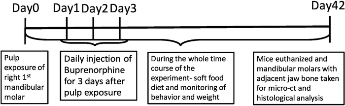
Figure 1: Time axis of experimental process. Schematic representation of the temporal progression of the experimental procedure. Please click here to view a larger version of this figure.
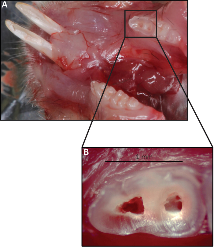
Figure 2: Clinical images of mouse mandible after right first molar pulp exposure. (A) Mouse mandible, first right molar with exposed pulp, first left molar serves as an untreated control. (B) Enlargement of the right molar with the exposed pulp. The entrance to the canals can be visualized. Please click here to view a larger version of this figure.
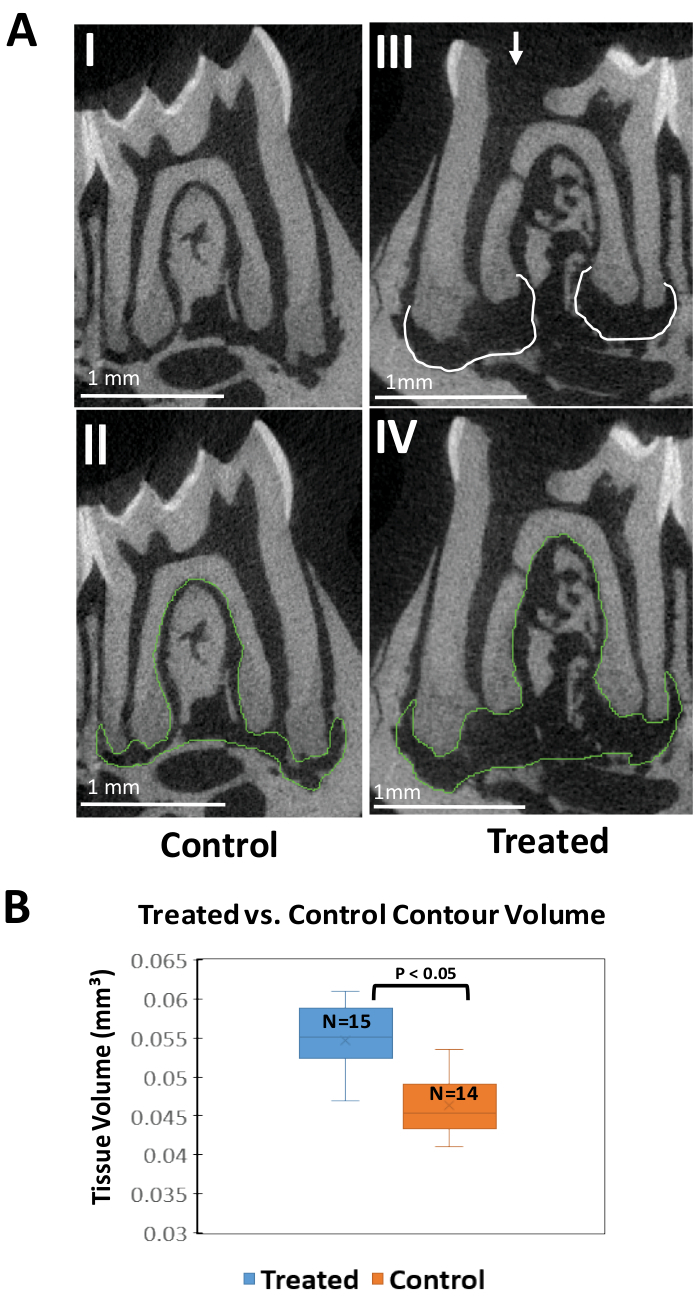
Figure 3: Micro-CT analysis. (A) Representative micro-CT scans (slices of 6µm) of treated (AIII, AIV) and control (AI, AII) teeth. Arrow points at mesial pulp exposure (distal pulp exposure is not present in this slice). Mesial and Distal periapical areas of bone loss adjacent to the treated tooth are surrounded by dashed lines. AII, AIV- Representative images of contour marking. (B) Box-plot quantifying the tissue volume (as measured by the contour marked for 11 slices of each sample) of the control (orange, n=14 animals) compared to the treated (blue, n=15 animals) samples. The analysis was performed using the Scanco evaluation software for micro-CT. Samples were aligned on the sagittal axis oriented in a way that the coronal pulp, root canals and apical foramen were visualized in the same slice (see protocol for detailed information about orientation). Contours were marked for 5 slices on either side of the mid-tooth area (all together 11 slices). Tissue volume for the contour of each sample was calculated using the software. Please click here to view a larger version of this figure.
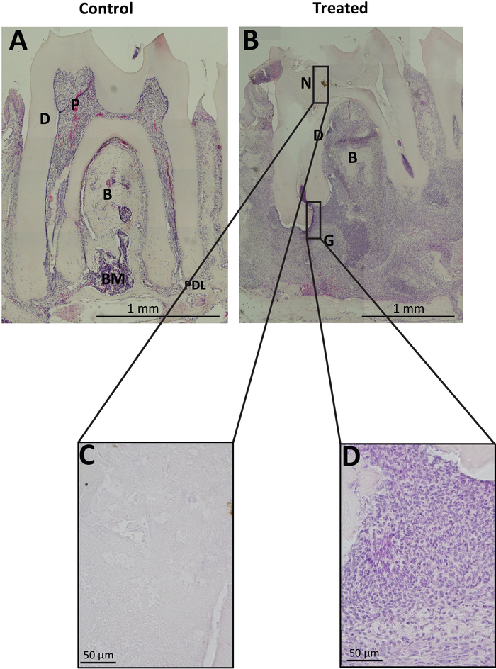
Figure 4: Histological H&E representative images: (A) Histological slice of a control tooth. (B) Histological slice of a treated tooth, with a severe periapical lesion presented. (C) enlargement of necrotic tissue. (D) enlargement of periapical granuloma tissue P=pulp, D=dentin, B=bone, BM=bone marrow, PDL=periodontal ligament, N=necrotic pulp, G= granuloma. Please click here to view a larger version of this figure.
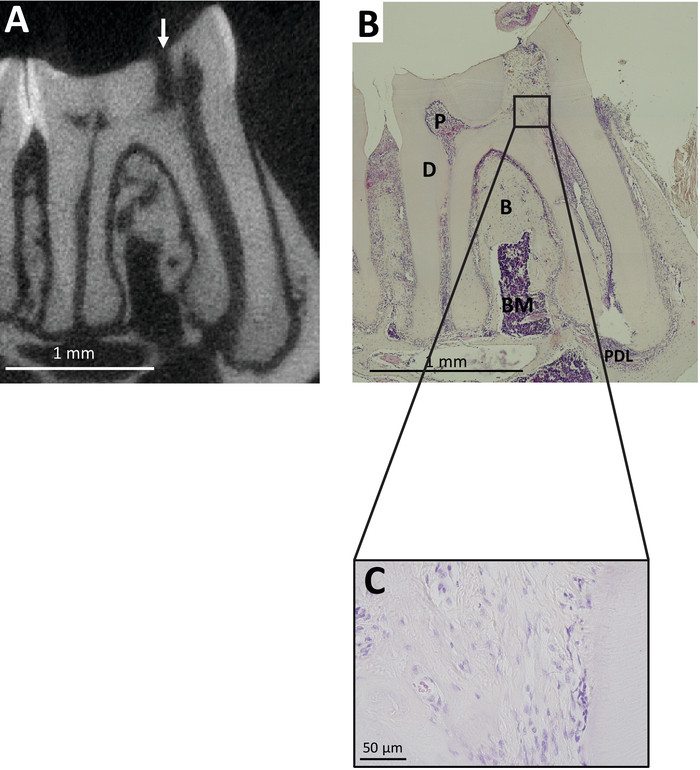
Figure 5: Example of unsuccessful contamination: (A) micro-CT representative image of a tooth with pulp exposure (white arrow), but no significant periapical bone loss, correlated to (B) Histological H&E representative image of the same tooth revealing vital (not necrotic) pulp, and normal apical tissues. P=pulp, D=dentin, B=bone, BM=bone marrow, PDL=periodontal ligament. (C) Enlargement of calcified tissue presented in B. Please click here to view a larger version of this figure.
A method is introduced here for the induction of apical periodontitis in mice. The goal of the method is to exploit the apical periodontitis condition for studying mechanisms and consequences of this inflammatory process. Apical periodontitis was induced in 6-8 week old mice, an age in which the roots are fully developed24. In order to cause apical periodontitis in this model, the tooth pulp of mouse mandibular molars is exposed using a dental burr. Bacteria from the oral flora of the mice (in SPF conditions) invade the pulp and root canals of the teeth, causing pulp necrosis, and secreting LPS to the apical tissues, which triggers apical inflammation.
When carrying out this procedure, pay attention to these following critical steps. Correct positioning of the animals during pulp exposure is critical for the success of the procedure. A critical step is the proper exposure of the pulp. Insufficient canal exposure can result in a Dentin bridge formation, which may lead to the failure of the procedure and the inability to create AP. During pulp exposure, attention is required to minimize soft tissue injury during pulp exposure to avoid unwanted pain and inflammation in other regions which may cause side effects. Proper alignment of the samples in the micro-CT analysis software is critical in order to obtain interpretable results. When preparing the jaw tissue for histology, it is recommended to peel off the soft tissue surrounding the bone for achieving correct orientation when preparing molds for histology. The samples must be kept in wet medium during the entire process (from the harvesting stage, through the micro-CT, until the histological block procedure). When cutting the tissue in a microtome, the angle of the sample should be fixed in order to achieve sagittal slices of the tooth. It is also important to take into account ethical issues regarding the welfare of the animals. For example, proper anesthesia is critical during the procedure (both systemic and local, see25,26). Pain medication is essential, especially for the first few days following the procedure, and monitoring weight and behavior as well as a supplying the animals with soft food should be implemented. From previous experience, the overall reaction of the mice is to tolerate well the procedure and they do not exhibit extreme distress.
A failure of the method is considered in cases where an apical inflammation failed to form: there is no evidence for bone resorption in the periapical region of the tooth on the micro-CT scan, and the PDL tissue of the tooth is intact in histological staining (as presented in Figure 5). The possible reasons for not achieving inflammation include stochastic differences in the behavior of the animals as well the specific exposure of each animal to bacteria. Although good results are achieved in causing apical periodontitis by using this method (at least 85% of animals show bone resorption), follow the protocol closely to reduce any variation between the samples. If any mice show signs of distress during the follow-up period, consider removing them from the experiment to prevent suffering. The decision to do so may be aided by looking at the loss or gain of weight of the animals during the follow-up period. Animals that lose a lot of weight suffer and should be removed. We have also observed that the animals suffering the most are those which started the experiment at a low weight or poor health condition. Overall, the procedure, if performed correctly, should yield consistent results.
Several limitations of this technique are to be noted. It is important to note that this model was developed in order to study apical periodontitis caused by bacterial contamination and does not mimic several other possible mechanisms by which apical periodontitis may arise, such as traumatic injury and autoinflammatory disease. Moreover, significant differences between this model and the clinical situation in human patients do exist. In mice, spontaneous periapical lesions have not been described and this is entirely an artificial situation. The anatomy of rodent teeth also differs from that of humans; the importance of these differences being unknown. Nonetheless, the mouse is an extremely useful model for understanding basic mechanisms and phenomena. Moreover, a root canal healing model in a mouse (by treating with a root canal) has not been reported so far, but potentially has high importance in this field of research should it prove to be feasible. A method of pulp capping has been reported for mice, which shows great potential27. A root canal healing model has indeed been recently developed in rats, a model animal often used in endodontic research28. The method reported here could serve as a preliminary procedure for developing this root canal healing model.
Using an in vivo method is crucial for the study of such a complex immune reaction, and a mouse model, although clinically challenging due to the small size of the animal, has many advantages: it is cost effective, involves easily accessible facilities, and a set of available genetic and molecular tools (such as available knock-outs, CRISPR libraries and a multitude of genomic data). This model is most suitable for endodontic research, as it induces the clinical presentation of AP in sterile lab conditions, with minimal variations (as opposed to clinical studies). Moreover, this model also has advantages in other research fields, such as chronic inflammation, due to the ability to cause a chronic but local inflammation with minimal systemic involvement, and the use of the contralateral side of the same animal as a control.
This work reports the use of micro-CT and histology for analysis of the lesion. Potentially, other types of analyses can be used in this model which were not performed here, such as isolating DNA and testing DNA methylation on different genes in the inflammatory cells; isolating RNA and performing RNA-seq to see the pattern of gene expression in different immune cells or different types of mice; or isolating cells and characterizing them by FACS20,21,29. Collectively, there is a great potential in the model reported here, which will hopefully be facilitated by this reported protocol and demonstration.
The authors have nothing to disclose.
We would like to acknowledge Dr. Oded Heyman for his help with animal positioning, Raphael Lieber for help with micro-CT analysis, and Prof. Andiara De Rossi Daldegan for advice on preforming the experiment. We would also like to acknowledge Dr. Sidney Cohen for critical reading and editing.
This work was supported by a grant from the Dr. Izador I. Cabakoff Research Endowment Fund to MK and IA, and a Yitzhak Navon fellowship from the Israel Ministry of Science and Technology to EG.
Atipamezole hydrochloride | Eurovet Animal Health | CAS 104075-48-1 | |
ATR | dentsply | tecnika | |
blocking machine | Leica | EG1150H | |
buprenorphine | vetmarket | 163451 | |
clinical microscope/binocular | Olympus | Sz61 | |
dental bur | Komet dental | ZR8801L 315 008 | |
dental spatula | Premier | 1003737 | |
EDTA | J.T Baker | 8993 | |
entelan | mercury | 1.07961 | |
Eosin Y solution, alcoholic | SIGMA | HT110116 | |
hematoxylin solution, Mayer's | SIGMA | MHS 16 | |
Ketamine hydrochloride | Vetoquinol | CAS 1867-669 | |
Medetomidine hydrochloride (cepetor) | CP-pharma GmbH | CAS 86347-15-1 | |
Mepivacaine HCl 3% | Teva | CAS 96-88-8 | |
microbrushes- adjustable precision applicators | PARKELL | S379 | |
micro-ct scanner | scanco | uCT 40 | |
parafin | Leica | 39602004 | |
PBS | SIGMA | D8537 | |
PFA | EMS | 15710 | |
Chloramphenicol eye ointment (5%) | Rekah pharmaceutical | CAS 56-75-7 | |
tweezers | WAM | Ref-CT | |
xylazine | Eurovet Animal Health | CAS 7361-61-7 | |
xylene | Gadot | CAS 1330-20-7 |