Two-Dimensional Semi-Denaturing Agarose Gel Electrophoresis: A Technique to Separate Polymorphic Amyloids Fibers Based on Size Heterogeneity

Published: April 30, 2023

Abstract

Source: Hanna-Addams, S., et al. Use of Two Dimensional Semi-denaturing Detergent Agarose Gel Electrophoresis to Confirm Size Heterogeneity of Amyloid or Amyloid-like Fibers. J. Vis. Exp. (2018).

This video demonstrates the two-dimensional semi-denaturing detergent agarose gel electrophoresis-based separation of amyloid fibers. The technique separates the polymorphic amyloid aggregates based on their heterogeneous size.

Protocol

1. Prepare Samples

  1. Culture 2 x 10amyloid producing HT-29 colon cancer cells in a 10-cm tissue culture dish in 10 mL of Dulbecco's Modified Eagle Medium (DMEM) containing 10% fetal bovine serum and penicillin-streptomycin. Culture cells overnight in a 37 °C incubator with 5% CO2.
    1. After the cells grow to 80% confluency, wash the cells with 10 mL of phosphate-buffered saline (PBS). Add 3 mL of Trypsin solution and incubate at 37 °C for 3 min.
    2. After the cells are totally dissociated from the dish, add 10 mL of culture medium and transfer the cells with a 10-mL pipette to a 15-mL conical tube. Centrifuge the cells at 1,000 x g for 3 min at room temperature. Aspirate the medium, resuspend the cells in 5 mL of culture medium, and count the cells using a cell counter. Plate 2 x 10cells in each of two 10-cm dishes.
    3. Allow the cells to adhere and recover overnight in a 37 °C incubator with 5% CO2. Apply treatment to one dish to induce the formation of amyloids with 20 ng/mL Tumor Necrosis Factor-Alpha (TNF-α), 100 nM Smac-mimetic, and 20 µM pan-caspase inhibitor Z-VAD-FMK. The combination is abbreviated as TSZ. Treat the other dish with vehicle as a control.
  2. After the appropriate length of time, usually 6 h, harvest the cell lysate.
    1. Scrape the cells off the plate with a plastic scraper and use a 10-mL pipette to transfer into a 15-mL conical tube. Centrifuge the cells at 1,000 x g for 3 min at 4 °C.
    2. Wash the cells 2 times by resuspending in 10 mL of ice cold PBS and centrifuging at 1,000 x g for 3 min at 4 °C. Aspirate the PBS solution.
      NOTE: The process can be paused here by freezing the cell pellet in liquid nitrogen and storing at ˗80 °C for up to 1 month.
    3. Transfer the cell pellet to a 1.5-mL microcentrifuge tube and incubate in 0.3 mL of lysis buffer for 30 min on ice. Centrifuge at 20,000 x g for 15 min at 4 °C. The supernatant is the whole cell lysate.
      NOTE: The process can be paused here by storing the sample at -20 °C for up to several months.
    4. Measure the protein concentration by a Bradford assay. Add 4x SDD-AGE loading buffer to prepare 20 µL of 3 µg/µL sample and incubate at room temperature for 10 min.

2. Prepare and Run Gels

  1. Add 2 g of agarose to 200 mL of 1x Tris-acetate buffer (TAE) in a glass beaker and heat in a microwave to melt the agarose. Add 1 mL of 20% SDS for a final concentration of 0.1% SDS. Carefully swirl to mix. Take care not to generate bubbles after the SDS addition.
  2. Pour the agarose solution into a 15 cm x 14 cm gel slab. Use a 1-mL pipette to eliminate any bubbles. Place one 20-well comb at the top.
  3. First dimension: Pipette 60 µg of whole cell lysate in the far-right lane. Run the gel at 60 V for about 4 h (until the dye front is about ¾ through the gel) using the TAE containing 0.1% SDS as the running buffer.
  4. Second dimension: Carefully rotate the gel 90° counter-clockwise (Figure 1A). Run the gel at 60 V for about 4 h.
    NOTE: The general running condition is 4 V/cm gel length. It is important that the running conditions are exactly the same for the first and second dimensions.

Representative Results

Figure 1
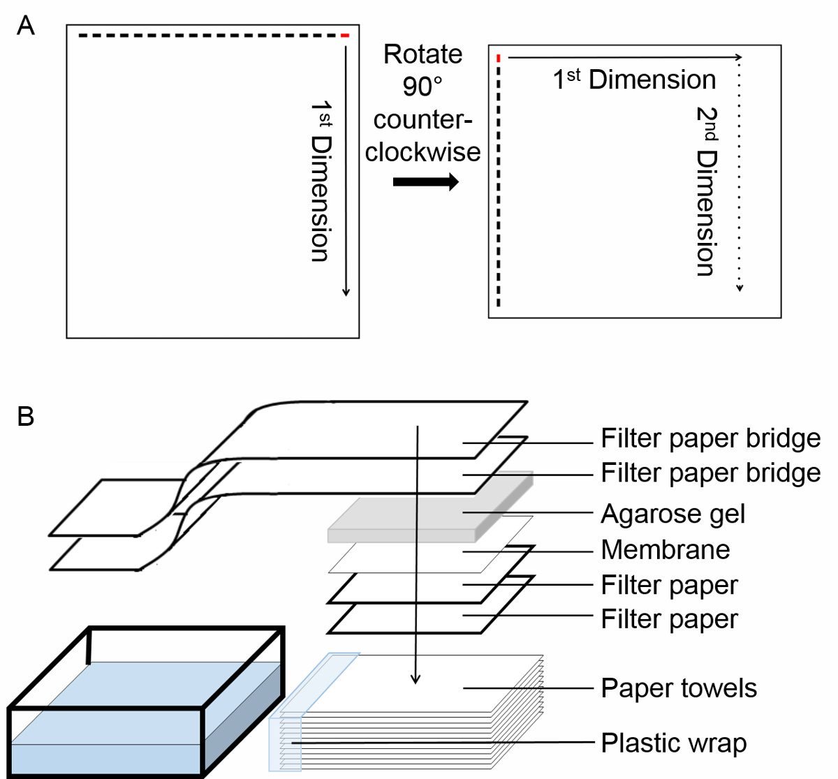
Figure 1. Experimental protocol. (A) Schematic of the two-dimensional semi-denaturing detergent agarose gel electrophoresis (2D SDD-AGE). Begin by loading the sample in the right most lane, labeled in red. After termination of the first dimension run, rotate the gel 90° counter-clockwise. Run the second dimension electrophoresis. Solid arrow indicates the direction of the first dimension run, dotted arrow indicates the direction of the second dimension run. (B) Schema of transfer by capillary action

Divulgazioni

The authors have nothing to disclose.

Materials

gel electrophoresis unit Fisher HE99XPRO appratus for gel running.
agrose VWR 97062-250 For agarose gel.
DMEM Sigma D6429 for cell culture
fetal bovine serum Sigma F4135 for cell culture
penicilin-streptomycin Sigma P4333 for cell culture
Trypsin solution Sigma T4049 for cell culture
PBS for tissue culture Sigma D8662 for cell culture
recombinant TNF made in our lab for inducing necroptosis. See reference 11.
smac-mimetic gift from Dr. Xiaodong Wang for inducing necroptosis. See reference 11.
ZVAD-FMK ApexBio A1902 for inducing necroptosis. See reference 11.
Cell Counter Bio-Rad 1450102 Model TC20; for counting cells
Pierce™ BCA Protein Assay Kit Thermo Scientific 23225 for measuring protein concentration in cell lysates
Cell lifter Fisher 07-200-364 to remove cells from dish
Lysis Buffer (1 L) 20 mL 1 M Tris pH 7.4
10 mL glycerol
30 mL 5 M NaCl
840 mL ddH2O
10 mL Triton-X100
(protease and phosphates inhibitors as desired)
10X TAE (1 L) 48.4 g Tris base
11.42 mL glacial acetic acid
20 mL 0.5M EDTA pH 8
ddH20 to 1 L
4X SDD-AGE loading buffer (50 mL) 5 mL 10X TAE
10 mL glycerol
4 mL 20% SDS
0.5 mL 10% bromophenol blue
31 mL ddH2O
PBST Wash Buffer (1 L) 100 mL 10xPBS
800 mL ddH2O
1 mL Tween20
10X PBS (10 L) 800 g NaCl
20 g KCl
144 g Na2HPO4·2H2O
24 g KH2PO4
add ddH2O to 10 L
check_url/it/21109?article_type=t

Play Video

Citazione di questo articolo
Two-Dimensional Semi-Denaturing Agarose Gel Electrophoresis: A Technique to Separate Polymorphic Amyloids Fibers Based on Size Heterogeneity. J. Vis. Exp. (Pending Publication), e21109, doi: (2023).

View Video