Harvesting an Inguinal Draining Lymph Node from a Murine Model

Published: August 31, 2023

Abstract

Source: Rezende, R. M. et al., Visualizing Lymph Node Structure and Cellular Localization using Ex-Vivo Confocal Microscopy. J. Vis. Exp.  (2019)

This video demonstrates the surgical isolation procedure of the inguinal draining lymph node from a mouse model. Incision of the abdominal region provides access to the inguinal lymph node, which is surgically harvested. The harvested translucent inguinal lymph node can be used for immunological studies.

Protocol

All procedures involving animal models have been reviewed by the local institutional animal care committee and the JoVE veterinary review board.

1. Mice used for the experiment

  1. Use 8-week-old male and female mice on the B6 background.

2. Surgery procedure to remove the inguinal draining lymph node

  1. Euthanize mice using CO2 asphyxiation followed by cervical dislocation.
  2. Immobilize mice on the acrylic stage with adhesive tape and apply mineral oil with a cotton swab to the abdominal skin to prevent fur deposition around the incision (Figure 1B). Fur removal is not necessary.
  3. Perform a midline incision using microsurgery curved scissors (11.5 cm) and microsurgery curved forceps (12.5 cm) in the abdomen from the pubis to the xiphoid process (Figure 1C).
  4. Dissociate the abdominal musculature from the skin (Figure 1D).
  5. Make horizontal skin incisions at the top and bottom of the vertical incision line to create skin flaps on the side of interest (according to the side of the antibody mix injection) and flap the skin to visualize the lymph node (Figure 1E).
  6. Tape the skin flap on the acrylic plate (Figure 1F).
  7. Remove the inguinal draining lymph node using microsurgery curved forceps (Figure 1F). Lymph nodes will appear as translucid, usually bilobular, spheres under the skin.

Representative Results

Figure 1
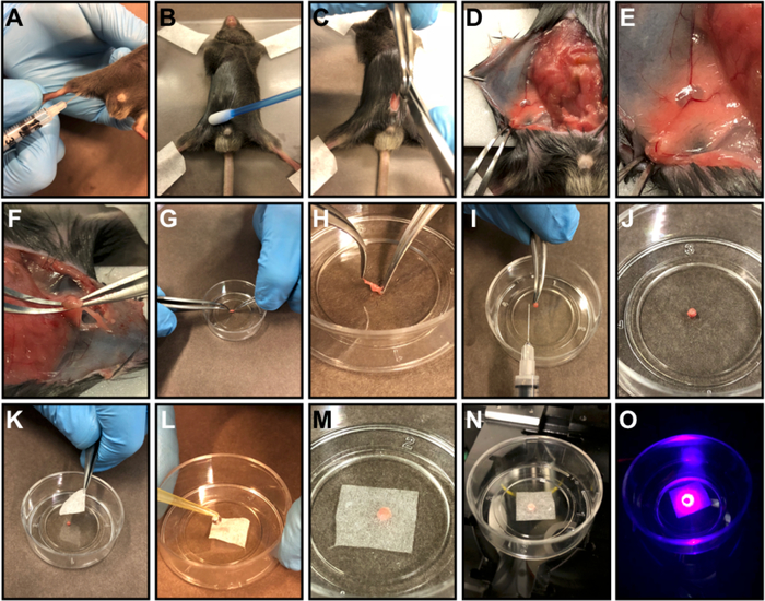
Figure 1: Inguinal lymph node preparation. 
(A) Subcutaneous injection of FACS antibody master mix into the inner thigh. (B) 3 h after the injection, euthanize the mouse, immobilize the mouse on an acrylic plate with adhesive tape, and apply mineral oil to the abdominal skin to prevent fur deposition around the incision. (C) Perform a midline incision in the abdomen from the pubis to the xiphoid process. (D) Dissociate the abdominal musculature from the skin and do a skin flap. (E) Tape the skin flap on the acrylic plate. (F) Remove the inguinal lymph node using microsurgery curved forceps. (G-H) Place the organ in a culture dish (G) and remove the fat that surrounds the organ (H). (I) Illustrative picture showing the organ size after cleaning. J-M) Centralize the organ in the middle of a petri dish (J), cover the organ with a piece of delicate task wipers (K) and keep soaked with warm 0.9% saline or 1x PBS (L, M). (N) Position the Petri dish in the microscope slot. (O) Scan the organ.

Divulgazioni

The authors have nothing to disclose.

Materials

Microsurgery curved forceps WEP Surgical Instruments Custom-made 12.5 cm
Microsurgery curved scissors WEP Surgical Instruments Custom-made 11.5 cm

Tags

check_url/it/21577?article_type=t

Play Video

Citazione di questo articolo
Harvesting an Inguinal Draining Lymph Node from a Murine Model. J. Vis. Exp. (Pending Publication), e21577, doi: (2023).

View Video