A Multiplex Bead-Based Assay for Analyzing Cytokines in Human Tear Samples
Published: January 31, 2024
Abstract
Source: Balne, P. K., et al. Bead Based Multiplex Assay for Analysis of Tear Cytokine Profiles. J. Vis. Exp. (2017)
This video illustrates a bead-based multiplex assay for analyzing cytokines in human tears. The method allows for the simultaneous analysis of multiple cytokines from a single sample, providing enhanced sensitivity for studying immune responses in various pathophysiological conditions.
Protocol
All procedures involving sample collection have been performed in accordance with the institute's IRB guidelines.
1. Tear Cytokine Analysis
- Collection of tears:
- Ask the subject to sit comfortably on an examination chair and place his/her head against the headrest.
- Ask the subject to look up, carefully open the Schirmer strips (made of Whatman filter paper no. 41) place the rounded end of the strip on the inferior fornix of the eye, and instruct the subject to close the eyes for 5 min. While collecting the tears take great care to minimize ocular surface contact.
- Ask the subject to open his/her eyes to remove the strip and place it in a sterile 1.5 mL microcentrifuge tube. Immediately transfer the tube to the laboratory or store it at -80 oC until testing for cytokine profiling.
- Elution of tear sample from tear flow strip:
- Cut the tear flow strip to a length of 0.5 cm (to standardize the quantity of tears to be tested) and place it in a sterile 1.5 mL microcentrifuge tube.
- Add 30 µL of assay buffer and incubate at room temperature for 5 min followed by centrifuging the tube at 14,000 x g for 1 min.
- Transfer the supernatant into another 1.5 mL microcentrifuge tube and discard the strip. Place the sample containing the tube on ice and use the eluted tear sample immediately for cytokine profiling.
- Preparation of reagents:
NOTE: The forty one analytes to be tested in each sample by bead based multiplex assay are interleukin (IL)-1α, IL-1β, IL-Rα, IL-2, IL-3, IL-4, IL-5, IL-6, IL-7, IL-8, IL-9, IL-10, IL-12 p40, IL-12 p70, IL-13, IL-15, IL-17A, interferon-alpha (IFN- α) 2, IFN-γ, IFN-gamma-inducible protein 10 (IP-10, CXCL10), macrophage-derived chemokine (MDC), macrophage inflammatory protein (MIP)-1α and MIP-1β, monocyte chemotactic protein (MCP)-1, MCP-3, tumor necrosis factor-alpha (TNF-α), TNF-β, growth-regulated oncogene (GRO), tumor growth factor alpha (TGF-α), vascular endothelial growth factor (VEGF), epidermal growth factor (EGF), fibroblast growth factor (FGF)-2, platelet derived growth factor (PDGF)-AA, PDGF-AB/BB, granulocyte colony-stimulating factor (G-CSF), granulocyte-macrophage colony-stimulating factor (GM-CSF), eotaxin, fractalkine, soluble CD40 ligand (sCD40L), Fms-like tyrosine kinase 3 ligand (Flt-3L) and regulated upon activation, normal T-cell expressed and secreted protein (RANTES).- Bring the antibody bead solution (50X) vials to room temperature (20 – 25 oC) and vortex the vials for 1 min.
- Count the number of wells required for the assay, and calculate the quantity of total antibody bead cocktail solution (25 µL per well) and each antibody bead solution (50X) required for the assay.
- Prepare the cocktail solution by adding the calculated amount of each antibody bead solution and fill to the desired final volume by adding the remaining amount of bead diluent. Ensure that the final concentration of each antibody in the cocktail is 1X. Always prepare at least 20% extra volume of cocktail solution in case of pipetting errors.
NOTE: As an example, for a 96-well assay prepare 3000 µL of antibody bead cocktail solution. The required amount of each antibody bead solution = 3000/stock concentration of each antibody bead solution; 3000/50 = 60 µL of each antibody bead solution- Add 60 µL of each of the 41 antibody bead solutions (60 X 41 = 2460 µL) into a cocktail bottle and add 540 µL of bead diluent solution to the antibody bead mixture to make 3000 µL of final working solution (1X).
- Prior to use, mix the bead cocktail solution properly. After the assay store the remaining volume at 2-8 oC for up to 30 days.
- Prepare the quality controls (QC) by adding 250 µL deionized water to QC 1 and QC 2 stocks.
- Mix properly by inverting the bottles several times and vortexing for 10 seconds. Transfer the solution to properly labeled polypropylene microcentrifuge tubes and store the remaining solution at -20 oC which can be used for up to 30 days.
- Prepare the wash buffer by adding 270 mL deionized water to 30 mL of 10X wash buffer solution and mix well (before dilution, bring the 10X buffer to room temperature and dissolve all salt precipitates by mixing). Store the unused wash buffer (1X) at 2-8 oC which can be used for up to 30 days.
- Prepare the human cytokine standard stock (10,000 pg/mL of all analytes) by adding 250 µL deionized water to the stock vial. Mix well by inverting several times and vortexing the vial for 10 seconds. Allow it to stand for 10 min and transfer the stock solution to properly labeled polypropylene microcentrifuge tubes. After the assay, store the remaining solution at -20 oC, which can be used for up to 30 days.
- Prepare the working human cytokine standards by 5-fold serial dilutions (50 µL -> 200 µL) with assay buffer to get 2000, 400, 80, 16, and 3.2 pg/mL.
- After standard preparation, use the working standards within 60 min and use the assay buffer as blank/background (-pg/mL).
- Cytokine profiling by bead-based multiplex assay:
- Bring all reagents to room temperature and vortex for 5-10 seconds before adding them to the 96-well microtiter plate. If eluted tear samples are stored at -80 oC prior to the assay, thaw the frozen tear extracts on ice and centrifuge at 1000 X g for 5 min.
- Prepare an assay worksheet in a vertical configuration for working human cytokine standards [0 (Blank), 3.2, 16, 80, 400, 2,000, and 10,000 pg/mL], QC1, QC2 and samples.
- Add 200 µL of 1X wash buffer to each well of the plate, seal it with a plate sealer, and keep it on a plate shaker at room temperature (20-25 oC) for 10 min.
- Decant the 1X wash buffer by inverting the plate and taping it onto absorbent towels several times to remove any residual amount of wash buffer in the wells.
- Add 25 µL of each working human cytokine standard, QC1, QC2, blank (assay buffer), and samples into the appropriate wells.
- Add 25 µL of assay buffer into each well.
- Add 25 µL of 1X antibody bead cocktail solution into each well. As the antibody bead solution is light sensitive, seal the plate with a plate sealer and cover it with aluminum foil to protect it from light during the assay.
- Incubate the plate at 4 oC overnight on a shaker.
- Place the plate on the plate rack of an automatic magnetic plate washer. Let it sit for 1 minute to settle the magnetic beads at the bottom of the well and aspirate the well contents. Add 200 µL of wash buffer per well, let it sit for 1 min and then aspirate the well contents. Repeat the wash once again (for plate washing, follow the kit manufacturer's instructions).
- Add 25 µL of detection antibodies solution into each well, seal the plate, cover it with aluminum foil, and incubate at room temperature for 60 min on a shaker.
- Add 25 µL of Streptavidin-Phycoerythrin solution into each well, seal the plate, cover it with aluminum foil, and incubate at room temperature for 30 min on a shaker.
- Place the plate on a magnetic plate washer. Let it sit for 1 minute and then aspirate the well contents. Add 200 µL of wash buffer per well. Let it sit for 1 minute and then aspirate the well contents. Repeat the wash once again.
- Add 150 µL of sheath fluid to each well and place the plate on a shaker for 5 min at room temperature to resuspend the antibody beads.
- Read the plate immediately using the bead-based multiplex assay plate reader and analyze the cytokine concentrations using a 5-parameter curve-fitting algorithm. A schematic flow diagram of tear cytokine profiling is shown in Figure 1.
- Reading the assay plate (instrument set up) and data analysis:
- Switch on the bead-based multiplex assay reader and pre-warm the laser for 30 min.
- Launch the bead-based multiplex assay software. Under the 'Automated Maintenance' tab, select the 'Calibration-verification' option. Vortex each reagent vial of the calibration and verification beads for 30 seconds. Place 5 drops of each reagent into the designated wells. Fill the designated reservoirs with deionized water and 70% ethanol.
Representative Results
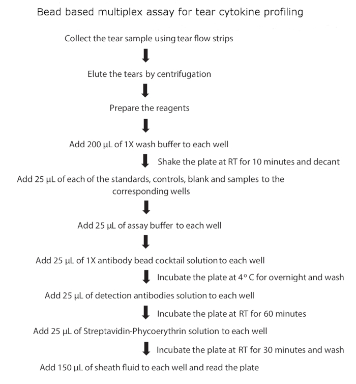
Figure 1: Schematic flow diagram of tear cytokine profiling.
Divulgazioni
The authors have nothing to disclose.
Materials
| Milliplex MAP human cytokine / chemokine magnetic bead panel -1 kit | Merck, USA | HCYTOMAG-60K-41 | |
| Flexmap 3D luminex instrument | Luminex Corp, Austin, TX, USA | ||
| xPonent software | Luminex Corp, Austin, TX, USA | ||
| RBXGenerator software | BIO-RAD, France | ||
| Bio-Plex Manager 6.1 | BIO-RAD, France | ||
| Plate shaker | Corning, USA | ||
| TECAN Microplate Washer | Tecan, Switzerland | HydroSpeed | |
| Pipettes | Mettler Toledo, CA, USA | ||
| 1.5 ml microcentrifuge tube | Axygen, USA | MCT-150-C | |
| Schirmer tear flow test strip | Eye Care and Cure, USA | 101657 | |
| Flexmap 3D Calibration Kit | Luminex Corp, Austin, TX, USA | 40-028 | |
| Flexmap 3D Verification Kit | Luminex Corp, Austin, TX, USA | 40-029 | |
| Ocular examination chair |
Tags

Citazione di questo articolo
A Multiplex Bead-Based Assay for Analyzing Cytokines in Human Tear Samples. J. Vis. Exp. (Pending Publication), e21906, doi: (2024).