1. Collagen Bead Preparation
- Prior to loading EVTs for 3-D cell culture, one needs to prepare the Cytodex-3 microcarrier beads:
- Weigh out the appropriate amount of Cytodex-3 beads required for the experiment. This protocol is adapted for the 10ml RCCS vessel, in which 0.05g of beads are needed. For a 50ml RCCS vessel, scale accordingly. In a 50mL autoclavable conical tube, mix 250 mg Cytodex-3 beads with 12mL Dulbecco phosphate buffered solution (DPBS). This amount is sufficient for 5 RCCS vessels.
- Ensure adequate volume is present in the conical tube, as the autoclave process will result in evaporation. Loosely cap the tube and autoclave for 10 min at 110°C.
- Remove the conical tube at the completion of the autoclave cycle and allow the Cytodex-3 bead solution to cool.
- After the conical tube has cooled to room temperature, use sterile technique to bring the total volume to 12.5mL using 1X DPBS.
- Cap and store the prepared Cytodex-3 beads at room temperature. Allow the prepared beads to swell and cool to room temperature prior to use. The above preparation of Cytodex-3 beads will provide for five 10mL RCCS vessels. We have observed that extended storage of Cytodex-3 beads at room temperature will result in diminished performance, as the collagen will destabilize after approximately one month. Cytodex-3 beads range in size from 133-215 μm.
2. Media Preparation
Prepare 1L of RCCS optimized GTSF-2 media (adapted from 22), which consists of 40% MEM alpha, plus supplements and 60% L-15 Leibovitz’s media (adapted from 22). First, prepare 400mL in total volume of MEM alpha supplemented with:
21.2mM sodium bicarbonate
0.06% Peptone
0.7mM Fructose
1.4mM Galactose
5.6mM Glucose
1% HEPES
1% L-glutamine
0.5% ITS
10% FBS- To prepare a stock solution of ITS, dissolve in 5mL sterile acidified H2O prepared by addition of glacial acetic acid (approx. 0.05mL). Swirl to dissolve, follow with 45mL sterile water.
- Weigh out enough L-15 Leibovitz’s powder for 600ml of medium by dissolving in tissue-culture grade H2O.
- Bring the total volume of cell culture medium to 1L with L-15 Leibovitz’s medium. Filter-sterilize and store at 4°C in the dark. Add 1% Penicillin-Streptomycin individually to each aliquot of medium used.
- Many media formulations other than GTSF-2 media have been successfully tested in the RCCS. Individual laboratories must decide for themselves which medium is optimal for the experiment being performed.
3. Cells and Bead Incubation
- Propagate the EVTs to ~80% confluency, trypsinize and count using accepted cell culture practices.
- Suspend 1×10⁶ EVTs in 4mL of warmed media.
- Gently mix prepared Cytodex-3 beads. Using sterile techniques, remove 2.5mL of prepared beads using a wide tip, 10mL serological pipette and transfer to an unused 15mL conical tube. Note that there may be a small loss of beads as they attach to the pipette.
- Allow the Cytodex-3 beads to settle on the bottom of the tube. After sedimentation, use a pipettor to remove the top layer of DPBS without disturbing the Cytodex-3 beads.
- Mix the 1×10⁶ prepared EVTs in media with the prepared Cytodex-3 beads.
- Incubate the cell-bead mixture at room temperature for 30 min. Periodically mix gently. Incubate for a further 30 min at 37°C and 5% CO2. Periodically mix gently.
4. Loading the RWV
- In a laminar flow cabinet, remove a 10mL RCCS from the packaging and place it in a sterile, 6-well culture plate for stability. Refer to Figure 1 for a labeled diagram of the RCCS.
- Remove the large stopper from the port.
- Bring the total volume of the incubating cell-bead mixture to 10mL with warmed media.
- Load the cell/bead-mixture into the RCCS through the large port. The RCCS should be tilted at a 45° angle (large port up) when loading to aid in removal of potential air bubbles. Prevent build-up of positive pressure by filling the vessel slowly and steadily.
- Replace the stopper in the large port.
- Remove the pistons from the 3-mL syringes and place the empty syringes onto the small ports. Add 1-3mL of media to each syringe. Slowly open the two valves. Replace the syringe pistons on the syringes gently. Add media from one syringe until all bubbles are removed from the inside chamber.
- Pick up the RCCS and gently tap the side while rotating it in front of you to check for bubbles. If there are any bubbles, you must get them out of the chamber, as air bubbles will interfere with formation of cell aggregates and introduce shear forces. Remove bubbles by rotating the RCCS until the bubbles are under the small port. Then gently push down on the syringe on the opposite side to force the bubbles into the port and out of the chamber.
- Close one side port. Gently push down on the other syringe piston to introduce a small amount of positive pressure into the vessel, which prevents bubbles from forming. Close second valve.
- Load the RCCS onto the rotor. Start the rotation at 19rpm in a 37°C CO2 incubator.
5. Changing the Media
- Change the media every other day for the first three days, then every day thereafter.
- Turn off the rotor and remove the RCCS. Pull the pistons up to create some suction, remove the syringes from each small port and place the RCCS on an angle so that the beads settle opposite of the large port.
- After the beads have all settled, open one of the small valves and allow the media to flow out of the RCCS and into a waste container. Remove 2/3 of the media in this manner. Be sure to not disturb the beads and to not discard any aggregates.
- Close the small valve and open the large port. Add media back into the RCCS through the large port. Replace the stopper in the large port.
- Repeat steps 4.6 – 4.9.
- As the aggregates increase in size, you need to increase the rotation speed to keep them in suspension at all times, ideally in a small circulatory pattern at optimal speed. Generally, increase the speed between 0.3-0.7 rpm after each feeding once the aggregates start growing visibly.
6. Collecting Propagated Cells
- Remove the RCCS from the rotor. Remove the syringes from each small port and place the RCCS on an angle so that the beads may settle opposite of the large port.
- After the beads have all settled, open one of the small valves and allow the media to flow out of the RCCS and into a waste container. Remove 1/3 of the media in this manner.
- Close the small valve and open the large port. Gently swirl the vessel to disperse the aggregates back into solution. Empty liquid mixture into a sterile 50ml conical tube. Allow the collected aggregates to settle in the conical tube before using supernatant to thoroughly wash the culture vessel to maximize recovery of aggregates. The aggregates may be used for downstream assays immediately, such as flow cytometry, invasion assays, immunofluorescence and others.
7. Representative Results
An example of EVT-like cells (SGHPL-4 trophoblast cell line) grown in the RCCS on Cytodex-3 beads is shown in Figure 2. The EVT-like cell line displays projections extending away from the main cluster and attaching to neighboring clusters. Many of the beads are completely covered with propagating cells. Once removed from the RCCS and plated on an extracellular matrix, the EVT-like 3-D grown cells aggressively invade and/or migrate (Figure 3). RT-PCR data confirms the increased expression of MMPs seen in 3-D aggregates as opposed to traditional cell culture monolayers (Figure 4). Interestingly, genes not associated with invasion are also upregulated in the RCCS (Table 1) and may aid in delineating such areas as immune interactions with invading trophoblasts cells.
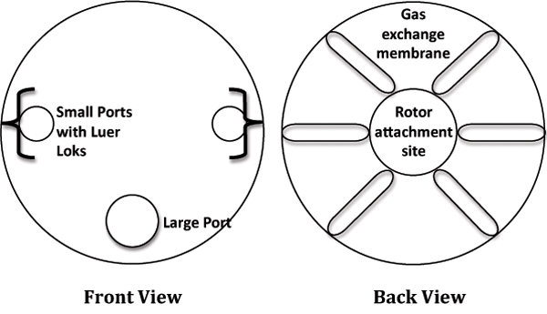
Figure 1. Cartoon depiction of the Rotating Cell Culture System (RCCS).
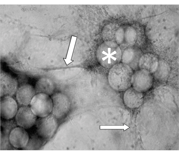
Figure 2. Phase contrast micrograph of a representative aggregate after 5 days growth in a RCCS. Arrows indicate invading cells and * denotes the Cytodex-3 microcarrier beads.
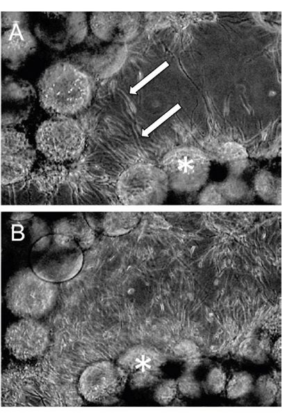
Figure 3. RCCS Grown aggregates were embedded in fibrin gels. Invasion through the fibrin gel was observed as early as (A) 24 hours post-fibrin embedding and (B) continued through 48 hours. Arrows indicate invading cells and * denotes the Cytodex-3 microcarrier beads (Adapted, with permission, from Ref. 7).
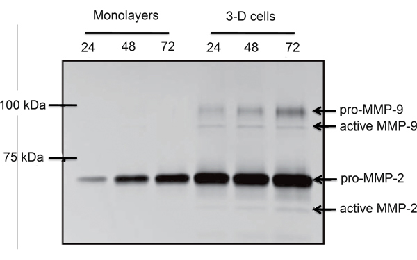
Figure 4. Comparative gelatin zymogram of monolayer and RCCS propagated SGHPL-4 EVT-like cells. Pro- and active forms of MMP-2 and MMP-9 were secreted by the RCCS grown trophoblast cells (Adapted, with permission, from Ref. 7).
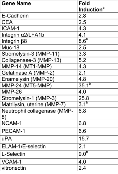
Table 1. Summary of microarray results from SGHPL-4 cells grown in a RCCS (Adapted, with permission, from Ref. 7).
- Fold induction in the 3-D aggregates was calculated as follows: normalized 3-D gene value/normalized monlayer gene value.
- Indicates genes that were below the limit of detection in monolayers.
