1. Cell Culture
- Culture MDA-MB-231 human breast cancer cells (American Type Culture Collection) in RPMI-1640 (Invitrogen, Germany) supplemented with 10% FCS (Sigma, Germany). Keep all cultures under standard conditions (37 °C, humidified atmosphere, 5% CO2) and passage the cells 2-3 times a week to keep them in logarithmic growth. For the animal model described below, there is no need for usage of bone-specific sublines of MDA-MB-231 cells as the tumor take rate is over 90%1.
- Harvest sub-confluent tumor cells after using 2 mM EDTA in PBS- (phosphate-buffered saline without Ca2+ and Mg2+) and 0.25% trypsin (Sigma, Taufkirchen, Germany). Count MDA-MB-231 cells in a Neubauer’s chamber and suspend them in RPMI-1640 (5×10⁵ cells in 1 ml).
2. Nude Rat Model of Bone Metastasis
- All experiments were approved by the responsible governmental animal ethics committee.
- Use nude rats at an age of 6-8 weeks and keep them at pathogen-free conditions in an appropriate small animal system (e.g. mini-barrier system). Keep animals under controlled conditions (21 +/- 2 °C room temperature, 60% humidity, and 12 hr light-dark rhythm) and offer autoclaved feed and water ad libitum to the rats.
- Before animal surgery, inject an analgesic drug (e.g. Carprofen 4 mg/kg s.c.; due to its single administration and short half-life (approx. 8 hr), carprofen should not affect tumor growth). Anesthetize rats with a mixture of oxygen (0.5 l/min) and isoflurane (1-1.5 vol. %) and make sure the rat is anesthetized properly and breathes regularly before beginning the following procedure.
- Place the anesthetized animal under an appropriate binocular operating microscope (e.g. Leica) and work with a magnification of 16-fold.
- Begin the surgical procedure by cutting the skin and subcutaneous tissue in the inguinal region at a length of 2-3 cm using scissors (BC060r Iris scissors 108 mm). All arteries branching off the femoral artery (FA) have to be dissected including the superficial epigastric artery (SEA), descending genicular artery (DGA), popliteal artery (PA) and saphenous artery (SA).
- Put clips on the femoral artery proximal of the SEA’s origin as well as on the DGA, PA and SA to temporally occlude local blood flow. Ligate the SEA at its distal part to allow opening of this vessel without bleeding (Figure 1A).
- Cut the SEA using scissors (Vannas Micro-SCRS-Grooved STR 85 mm) proximal of the ligation (Figure 1B) and administer a 1% papaverin solution onto the SEA, to facilitate the subsequent insertion of a needle due to relaxation of the vessel (Figure 1C).
- Cut approximately half of the SEA’s diameter with scissors and insert a needle (0.3 mm diameter and 42 mm length) into the lumen of the SEA while holding the cut-end of the vessel with a forceps (Figure 1D, E). When available, fix the needle in an external device to reduce irregular movements that could result in perforation of the vessel wall. Connect a syringe to the needle. Remove the clip from the distal FA and place it on the saphenous artery (Figure 1F).
- Inject the MDA-MB-231 cells suspended in 0.2 ml media slowly into the SEA. By virtue of the clips MDA-MB-231 cells are directed to the DGA and PA. Remove the needle and ligate the SEA to prevent bleeding before taking off the artery clips. Close the wound using surgical clips and terminate inhalation anesthesia.
- For post-procedure monitoring, rats are usually euthanized 7-8 weeks after tumor cell inoculation to avoid severe skeletal complications. During this time, animals should be monitored daily to assess tumor size and any evidence of pain (e.g., behavioral deviations, weight loss, motor defects). If animals show a tumor size exceeding the ethically allowed limit or evidence of pain during tumor growth, they have to be euthanized.
- Immuno-deficient (nude) rats were used for xenogenous transplantation of human MDA-MB-231 breast cancer cells. Nude rats were not chosen to better visualize the growing tumor.
3. Magnetic Resonance Imaging (MRI)
- After tumor cell inoculation, allow approximately 25-30 days of tumor growth before beginning with the imaging. For MR imaging use a dedicated experimental scanner or a human MR system with an appropriate animal coil. We used a human MR system (Symphony, Siemens, Germany) and a home-built coil for radiofrequency excitation and detection, designed as a cylindrical volume resonator with an inner diameter of 83 mm and a usable length of 120 mm (Figure 2A).
- Anesthetize the rat with oxygen and isoflurane as given above. Place a catheter in the tail vein and fix it on the tail using a tape. Connect a syringe containing the contrast agent (e.g. 0.1 mmol/kg Gd-DTPA in approximately 0.5 ml; Magnevist, Bayer-Schering, Germany).
- Place the rat in the MR system maintaining the inhalation anesthesia. Begin with a morphologic MR sequence to locate the bone metastasis (e.g. T2 weighted: turbo spin echo sequence, TR 3240 ms, TE 81 ms, matrix 152 x 256, FOV 90 x 53.4 mm2, slice thickness 1.5 mm, 3 averages, scan time 3:40 min).
- Determine a slice of the bone metastasis with the largest diameter and start the sequence for DCE-MRI (e.g. saturation recovery turbo flash sequence, TR 373 ms, TE 1.86 ms, matrix 192 x 144, FOV 130 x 97.5 mm2, slice thickness 5 mm, measurements 512, averages 1, scan time 6:55 min). After approximately 30 sec, begin to inject the contrast agent over a time period of 10 sec. The total time for the above-mentioned procedures to perform a MRI examination is approximately 15-20 min per animal.
4. Volumetric Computed Tomography (VCT)
- Choose an appropriate CT system, either a human or an experimental scanner. Here, we used a prototype of a flat panel equipped volumetric computed tomograph (Figure 2B; Volume CT, Siemens, Germany).
- Anesthetize the rat with oxygen and isoflurane as given above. Place a catheter in the tail vein and fix it on the tail using a tape. Connect a syringe containing the contrast agent (e.g. 1 g iodine per kg in approximately 0.5 ml; Imeron 400, Bracco, Germany).
- Place the rat on the scanner under inhalation anesthesia. Use the following scan parameters for VCT: tube voltage 80 kV, tube current 50 mA, scan time 51 sec, rotation speed 10 sec, frames per second 120, matrix 512 x 512 and slice thickness 0.2 mm. Inject the contrast agent during the second rotation of the flat panel system. The total time for the above-mentioned procedures to perform a VCT examination is approximately 5-10 min per animal.
- Reconstruct images with a modified FDK (Feldkamp-Davis-Kress) cone-beam reconstruction algorithm (kernel H80a, Afra, Germany).
5. Ultrasound (US)
- Experimental and clinical US systems are available for this purpose. We used the clinical system Acuson Sequoia 512 ultrasound system with a 15L8 linear transducer (Figure 2C; Siemens-Acuson, Mountain View, CA).
- Anesthetize the rat with oxygen and isoflurane as given above. Place a catheter in the tail vein and fix it on the tail using a tape. Connect a syringe containing a microbubble contrast agent (e.g. 1.6 ml/kg in approximately 0.5 ml; SonoVue, Bracco, Italy). Fix the US transducer on the respective hind leg using a tripod and apply US gel between the transducer and the hind leg.
- Perform B-mode imaging (transmission frequency: 17 MHz; mechanical index: 0.51) to determine the largest diameter of the bone metastasis and fix the transducer in this position. Add Doppler signal on B-mode images for information on tissue perfusion. Please bear in mind that only lesions that disrupt cortical bone are accessible to US waves.
- For dynamic contrast-enhanced US (DCE-US) set the US device in cadence contrast pulse sequencing (CPS) mode (transmission frequency: 7 MHz; mechanical index: 0.18), inject the microbubbles and record a cine loop of 90 sec length. The total time for the above-mentioned procedures to perform a US examination is approximately 10-15 min per animal.
6. Postprocessing of Imaging Data
- Use the morphological information from MRI, VCT and US to characterize the soft tissue tumor (MRI, US) and the skeletal destruction (VCT) of bone metastases and determine location, lesion size and volume of the lesions with a dicom viewer (e.g. Osirix Dicom Viewer).
- To obtain the branching pattern of vessels in bone metastases (angiography), the VCT data can be used. Reconstruct 2D or 3D images using the information of the arterial phase with or without subtraction techniques (e.g. Osirix Dicom Viewer).
- In order to quantify parameters of vascularization from DCE-MRI, DCE-VCT and DCE-US, use software tools specific for the modalities. For DCE-MRI, determine the vascular parameters amplitude A (associated with blood volume) and exchange rate constant kep (associated with perfusion and vessel permeability) in bone metastases with Dyna Lab (Mevis Research, Bremen, Germany) based on the two-compartment model of Brix8,9. Alternative pharmacokinetic models for evaluation are available, e.g. the Tofts model10.
- To quantify DCE-VCT data, perform a descriptive analysis of data to calculate parameters such as area under the curve (AUC) or peak enhancement (PE) with Dyna Lab (Mevis Research, Bremen, Germany).
- Quantify information from real-time DCE-US using quantitative analysis software (e.g. Qontrast, Bracco, Italy) by analyzing cine-loops according to the implemented bolus injection model. Place the region of interest (ROI) over the bone metastasis, determine either descriptive factors such as area under the curve or quantitative parameters from color-coded maps, e.g. regional blood volume, regional blood flow and filling time.
7. Representative Results
Following intraarterial injection of MDA-MB-231 cells into the SEA (Figure 1), site-specific bone metastases develop in the respective hind leg of the nude rat. Osteolytic lesions confined to the femur, tibia and fibula can be imaged non-invasively by MRI, VCT and US (Figure 2) beginning approximately 25-30 days post injection and followed-up for several weeks. When combining MRI, VCT and US including native and contrast-enhanced techniques, complementary information can be assessed in bone metastases that are composed of a soft tissue tumor (tumor cells and stroma) and the respective osteolytic lesion (bone destruction). For comparison of the respective data between the techniques, all three imaging modalities can be used sequentially in the same rat. MRI displays morphology of the bone metastatic soft tissue that is initially confined to the bone marrow cavity and subsequently exceeds cortical bone in the course of development. Functional parameters such as regional blood volume, perfusion and vessel permeability can be obtained from DCE-MRI and quantified (Figure 3). Bone structure, and in particular osteolytic changes in the metastases are assessed in high resolution by VCT. Complementary to MRI findings, osteolytic lesions are located adjacent to intramedullary tumor growth. VCT angiography reveals the altered macrovessel architecture of bone metastases, and DCE-VCT displays respective aspects of microcirculation (Figure 4). Due to local destruction of cortical bone in metastatic lesions, US is applicable to assess morphological and functional features of the soft tissue tumor by the use of B-mode and Doppler techniques. Upon application of microbubbles, DCE-US allows for real-time imaging of vascularization in bone metastases (Figure 5).
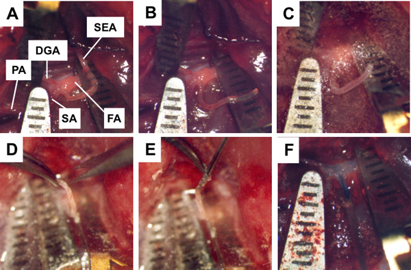
Figure 1. Hind leg of a nude rat prepared for tumor cell inoculation as imaged through an operation microscope. A, branching pattern of the femoral artery (FA) including the superficial epigastric artery (SEA), descending genicular artery (DGA), popliteal artery (PA) and saphenous artery (SA). Arterial clips placed on the SA, PA and proximal FA as well as ligation of SEA; B, SEA was cut proximal of the ligation; C, muscle relaxation of the SEA after addition of papaverine; D, incision of the SEA (taken up by a forceps); E, insertion of needle into SEA; F, fixated needle in the SEA (external fixating device) and injection of MDA-MB-231 tumor cells via the SEA into the DGA and PA by virtue of the clips.
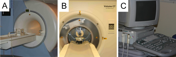
Figure 2. A, human MR system (Symphony, Siemens, Germany) and a home-built coil for radiofrequency excitation and detection placed in the scanner; B, flat panel equipped volumetric computed tomograph (Volume CT, Siemens, Germany); C, clinical ultrasound system Acuson Sequioa 512 (Siemens-Acuson, Mountain View, CA).
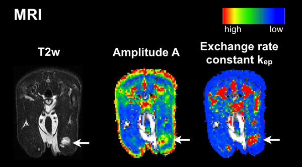
Figure 3. Axial MR sections. Left panel, T2w MRI; middle panel, amplitude A (DCE-MRI); right panel, exchange rate constant kep (DCE-MRI). Arrows point at bone metastases. The color map for DCE-MRI data ranges from red (high values) to blue (low values).
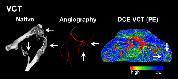
Figure 4. 3D VCT reconstructions of the osteolytic bone metastasis (left panel) and an angiography (middle panel) as well as an DCE-VCT section in axial orientation from the parameter peak enhancement (right panel). The color map for DCE-VCT data ranges from red (high values) to blue (low values).
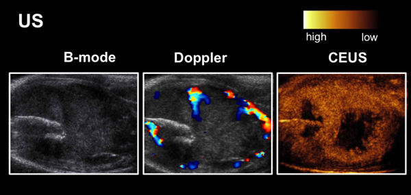
Figure 5. US images from B-mode (morphology, left panel), Doppler (perfusion, middle panel) and CEUS (right panel, peak enhancement after injection of microbubbles from real-time imaging of vascularization) of a bone metastasis.
Supplemental movie 1. Click here to view supplemental movie.
