Biocompatible pH responsive sol-gel nanosensors can be incorporated into poly(lactic-co-glycolic acid) (PLGA) electrospun scaffolds. The produced self-reporting scaffolds can be used for in situ monitoring of microenvironmental conditions whilst culturing cells upon the scaffold. This is beneficial as the 3D cellular construct can be monitored in real-time without disturbing the experiment.
Culturing cells in 3D on appropriate scaffolds is thought to better mimic the in vivo microenvironment and increase cell-cell interactions. The resulting 3D cellular construct can often be more relevant to studying the molecular events and cell-cell interactions than similar experiments studied in 2D. To create effective 3D cultures with high cell viability throughout the scaffold the culture conditions such as oxygen and pH need to be carefully controlled as gradients in analyte concentration can exist throughout the 3D construct. Here we describe the methods of preparing biocompatible pH responsive sol-gel nanosensors and their incorporation into poly(lactic-co-glycolic acid) (PLGA) electrospun scaffolds along with their subsequent preparation for the culture of mammalian cells. The pH responsive scaffolds can be used as tools to determine microenvironmental pH within a 3D cellular construct. Furthermore, we detail the delivery of pH responsive nanosensors to the intracellular environment of mammalian cells whose growth was supported by electrospun PLGA scaffolds. The cytoplasmic location of the pH responsive nanosensors can be utilized to monitor intracellular pH (pHi) during ongoing experimentation.
A key strategy in tissue engineering is the use of biocompatible materials to fabricate scaffolds whose morphology resembles the tissue that it is going to replace and is also capable of supporting cell growth and function1,2. The scaffold provides mechanical support by allowing cell attachment and proliferation yet allows cell migration throughout the interstices of a 3D cellular construct. The scaffold must also allow for the mass transport of cell nutrients and not inhibit removal of metabolic waste3.
Electrospinning has emerged as a promising method for the fabrication of polymeric scaffolds capable of supporting cellular growth4-6. The nonwoven electrospun fibers produced are suitable for cell growth as they are often porous and allow cell-cell interaction as well as cell migration throughout the interstices of a 3D cellular construct7. It is important to monitor cell viability during the period of culture and to ensure that cell viability is maintained throughout the whole of the 3D construct. For example, culture conditions such as oxygen and pH require careful control, as gradients in analyte concentration can exist within the 3D construct. Bioreactors or perfusion systems can be employed to mimic in vivo conditions of interstitial flow and as a result increase nutrient transfer and metabolic waste removal8. The question of whether such systems are ensuring constant microenvironmental conditions can be addressed by assessing the cellular microenvironment in real-time.
Key microenvironment metrics that could be monitored in real-time include: temperature, chemical composition of cell media, concentration of dissolved oxygen and carbon dioxide, pH, and humidity. Of these metrics, temperature can be most readily monitored using in situ probes. Methods for monitoring the remaining listed metrics commonly involve removal of an aliquot for sampling and therefore disturb the cell culture and increase the contamination risk. Continuous, real-time methods are being sought. Current monitoring methods usually rely upon instruments that physically probe the cellular construct such as a pH monitor or oxygen probe. However, these intrusive methods can damage the cellular construct and disturb the ongoing experiment. Noninvasive monitoring of analyte concentrations within the 3D construct could enable real-time monitoring of various environmental aspects such as nutrient depletion9. This would allow assessment of nutrient supply to deeper regions within the structure and determine whether metabolic waste was being removed effectively10,11. Systems that attempt to address the issue of invasiveness generally involve the use of a perfusion chamber that passes culture medium through both the culture vessel and to external sensors to monitor pH, oxygen and glucose12. There is increasing interest in developing sensors that can be directly integrated into the culture vessel that do not require removal of an aliquot for sampling and as such would provide in situ monitoring.
To address such shortcomings for in situ and noninvasive monitoring of microenvironmental conditions we have incorporated analyte responsive nanosensors into electrospun scaffolds to produce self-reporting scaffolds13. Scaffolds that act as sensing devices by monitoring fluorescence activity have been prepared previously, where the sensing device was either the actual polymeric scaffold created by electrospinning or through the use of an analyte responsive dye which is incorporated into the polymer prior to scaffold formation14,15. However, these sensing devices have the potential to give erroneous optical outputs caused by possible interference from other analytes. The use of a ratiometric sensing device such as those prepared in the described protocol holds the potential to eliminate these possible adverse effects and provide a response specific to the analyte in question.
The electrospun scaffolds presented here have been prepared from the synthetic co-polymer poly(lactic-co-glycolic acid) (PLGA), selected due to having Food and Drug Administration (FDA) approval, owing to its biodegradable and biocompatible properties and a track record of supporting the growth and function of various cell types16-18. The prepared ratiometric analyte responsive nanosensors are responsive to pH. The nanosensors incorporate two fluorescent dyes into a biocompatible sol-gel matrix where one dye, FAM is responsive to pH and the other, TAMRA acts as an internal standard as it is not responsive to pH. Furthermore the fluorescence of both FAM and TAMRA can be analyzed separately as they do not significantly overlap. Determining the ratio of the fluorescence emission of both dyes at specific wavelengths gives a pH response independent of other environmental conditions. The self-reporting scaffolds could allow repeat assessment of pH in situ and in real-time without disrupting the developed 3D model. We have demonstrated that these scaffolds are capable of supporting cell attachment and proliferation and remain responsive to the analyte in question. The kinetics of acidic by-products in engineered constructs remains understudied and as such using the pH responsive scaffolds could greatly facilitate such studies19. Furthermore, the use of the self-reporting scaffolds for tissue engineering applications presents the opportunity to fully understand, monitor and optimize the growth of 3D model tissue constructs in vitro, noninvasively and in real-time.
The pH responsive nanosensors have also been delivered to the intracellular environment of fibroblasts cultured upon electrospun PLGA scaffolds. The ratio of the fluorescence emission from the dyes were used to monitor pHi and compared to a self-reporting scaffold incorporating pH nanosensors. The delivery of nanosensors to cells cultured in a 3D environment could enable monitoring of analyte concentration deep within the construct in a nondestructive manner. Therefore nanosensors may be a viable imaging tool to nondestructively assess cell behavior throughout 3D constructs allowing long-term analysis. Screening the analyte concentration of individual cells within a 3D construct could ensure that they are receiving sufficient nutrient and oxygen concentrations. Monitoring process parameters could assist in the development of standardized techniques for the effective mass transport of oxygen and nutrients. The delivery of nanosensors to the intracellular environment and incorporation of nanosensors into polymeric scaffolds could be combined to allow assessment of cell viability as well as scaffold performance within 3D constructs during the tissue growth process. This may lead to increased knowledge of these constructs and progress the fabrication of biologically relevant tissue substitutes.
Panoramica
Section 1 describes the preparation of pH responsive nanosensors and characterization of nanosensor response to pH using fluorescence spectrometry and their size using SEM.
Section 2 describes the preparation of electrospun polymer scaffolds and characterization of their morphology and size using SEM. Section 2 also describes the preparation of self-reporting scaffolds which are electrospun scaffolds with the inclusion of the pH responsive nanosensors. The resulting self-reporting scaffolds are characterized by SEM to assess whether any morphological changes in the electrospun fibers has occurred. The fluorescence from the self-reporting scaffolds resulting from the incorporated nanosensors is assessed by confocal microscopy.
Section 3 describes the culture of cells upon the electrospun scaffold and the self-reporting scaffold. The scaffolds are first sterilized in preparation for cell culture following which the scaffolds are assessed to determine whether sterilization and the culture of cells affect the fluorescence capability of the self-reporting scaffolds. The protocol also describes the delivery of nanosensors to cells that are cultured upon electrospun scaffolds. Lysotracker dye is used to ensure that nanosensors have not been taken into cellular acidic compartments.
1. Preparation and Analysis of pH Responsive Sol-gel Nanosensors
1.1 Preparation of pH Responsive Sol-gel Nanosensors
Note: This preparation should be performed in a fume hood.
1.2 Nanosensor Response to pH
1.3 SEM of Nanosensors
2. Preparation and Analysis of PLGA Scaffolds
2.1 Electrospinning PLGA Scaffolds
2.2 Electrospinning pH Responsive PLGA Scaffolds
2.3 SEM of PLGA Scaffold and pH Responsive Scaffold
2.4 Calibration of pH Responsive Scaffold
3. Cell Culture upon PLGA Scaffolds and pH Responsive PLGA Scaffolds
Note: The following should be performed in a cell culture cabinet.
3.1 Preparation of PLGA Scaffolds for Cell Culture
3.2 Nanosensor Delivery to Cells Cultured upon PLGA Scaffold
3.3 Nanosensor Location within Mammalian Cells
The size distribution of the prepared pH responsive nanosensors was characterized using SEM, where the population of nanosensors imaged were measured and found to have nanometer dimensions in the range of 240-470 nm (Figure 1A). The achievement of a narrow and reasonably small diameter is consistent with using the Stöber method to prepare nanoparticles. It has been found that using a basic pH environment during the synthesis of nanoparticles i.e. nanoparticles prepared using the Stöber method, allows good control of the size, whereas preparation using acidic conditions produces a wide dispersion in particle size. Representative SEM micrographs of the PLGA electrospun fibers fabricated using the described methods are shown in Figure 1B demonstrating that the produced PLGA scaffold consists of nonwoven fibers displaying minimal beading or breakage where the fibers have nanometer dimensions. Figure 1C demonstrates that the addition of nanosensors to the PLGA solution at a concentration of 5 mg/mL produces fibers that are comparable to the control sample in Figure 1B, The SEM micrographs provide evidence of nanosensor association on the surface of the fibers however they may also be incorporated within the scaffold fibers, degradation studies of these scaffold/nanosensor scaffolds could be carried out in conjunction with SEM and/or confocal microscopy to determine if this affects nanosensor performance.
The capability of the nanosensors to retain their optical activity following incorporation into the PLGA fibers was verified by examining the self-reporting scaffolds using confocal microscopy. Retention of the ability to remain optically and chemically active when incorporated into the scaffold fibers could enable analyte concentrations to be monitored. The fluorescence emitted from the nanosensors demonstrated that the nanosensors retained their optical activity and are associated with the scaffold fibers. Fluorescence emitted from the dyes FAM (green) and TAMRA (red) incorporated in the pH responsive nanosensors is shown in Figures 2A and 2B respectively. This is a positive result that makes it feasible for analyte concentrations to be monitored in situ. It is also the first time that optically active ratiometric nanosensors have been incorporated into PLGA scaffold fibers using the electrospinning process. Having confirmed that the nanosensors can be visualized using fluorescence microscopy following incorporation into PLGA fibers, the nanosensor response to changes in analyte concentration was verified. Immersing the scaffold into buffers of differing pH resulted in a change of fluorescence intensity from the FAM dye whilst the fluorescence emitted by the TAMRA reference dye did not noticeably change. The ratio of fluorescent intensity of both dyes can be used to produce a calibration curve as shown in Figure 2C. The calibration curve for nanosensors assessed alone and when incorporated into electrospun PLGA scaffold is comparable and demonstrates that the nanosensors retain their optical activity following incorporation into the self-reporting scaffolds. The ability of the self-reporting scaffold to reversibly respond to different pH values was assessed by alternately immersing the scaffold in buffers with pH values of 5.5 and 7.5. The reversibility was found to be very good as shown in Figure 2D where the self-reporting scaffold can respond to numerous cyclic changes in pH. Long-term studies have not been performed to assess the effect of PLGA degradation upon the nanosensor incorporation; it is thought that because the nanosensors provide a ratiometric response the quantity of nanosensors present will not affect the result.
Analyte responsive nanosensors were delivered using a liposomal transfection agent to the intracellular environment of 3T3 fibroblasts whose growth was supported by blank PLGA scaffolds. Confocal microscopy images of 3T3 fibroblasts cultured upon blank PLGA scaffolds demonstrate that nanosensors are associated with the fibroblast cells with fluorescence observed from FAM and TAMRA shown in Figures 3A and B respectively. Overlaying the fluorescence from these channels shows that the fluorescence from the two dyes is co-localized, suggesting that the dyes have remained entrapped within the nanosensors’ biocompatible matrix (as depicted by yellow fluorescence in Figure 3C). The nanosensors are thought to be within the cytoplasm of the 3T3 fibroblasts and not contained within acidic compartments as evidenced by the lack of co-localized fluorescence from FAM only nanosensors and Lysotracker Red dye depicted in Figure 3D. The fluorescence intensity of pH responsive nanosensors delivered to 3T3 fibroblasts was monitored whilst the cells were subjected to changes in pH the results of which are shown in Figure 3E. The graph produced demonstrates that the cells cultured upon the electrospun PLGA scaffold have maintained a pHi in the range 6.8-7.0.
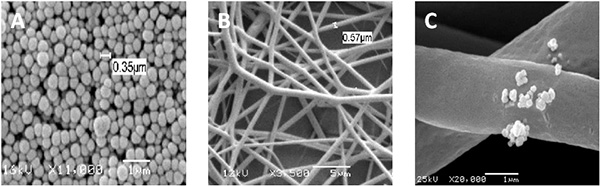
Figure 1. SEM micrographs of nanosensors, PLGA fibers and self-reporting scaffolds. (A) pH responsive sol-gel nanosensors showing spherical nanometer sized particles (B) PLGA electrospun nonwoven fibers where fiber morphology is regular with minimal beading and breakages (C) pH responsive self-reporting scaffolds where the nanosensors can be observed to be protruding from the PLGA yet fiber morphology remains comparable with control PLGA fibers that do not contain nanosensors. Click here to view larger image.
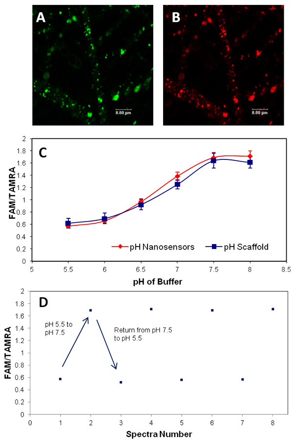
Figure 2. Confocal microscopy images of pH responsive self-reporting scaffolds where fluorescence can be observed from the nanosensors associated with the PLGA scaffold fibers. (A) FAM (green) the pH responsive dye and (B) TAMRA (red) the reference dye. (C) Calibration graphs of pH responsive sol-gel nanosensors and pH responsive scaffolds demonstrate that the optical and chemical response of the nanosensors has not been affected when incorporated within the PLGA scaffold fibers. (D) Reversibility of the self-reporting scaffold performed by immersing the scaffold in alternate buffer solutions of pH 5.5 and pH 7.5. Click here to view larger image.
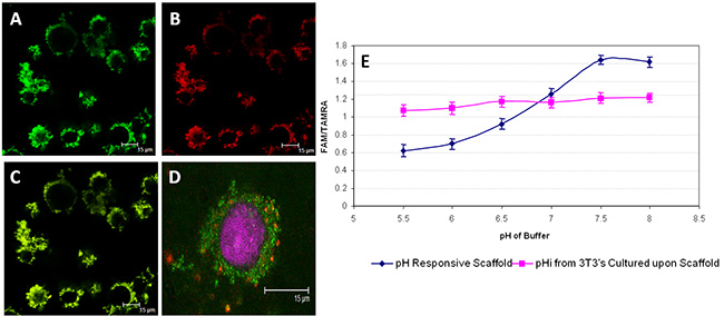
Figure 3. Confocal microscopy images of cell internalized nanosensors and graphs of pH and pHi response. Fluorescence from the internalized nanosensors (A) FAM (green) (B) TAMRA (red) (C) co-localization of fluorescence from FAM and TAMRA (yellow) (D) FAM only nanosensors (green) delivered to 3T3 fibroblasts cultured upon PLGA scaffold with Draq5 (magenta) labeling of the nucleus and LysoTracker Red (red) labeling of lysosomes. The fluoresence of FAM and Lysotracker red are not co-localized therefore demonstrating that it is unlikely that nanosensors have been internalized into cellular acidic compartments (E) pHi measurements taken from nanosensors delivered to 3T3 fibroblasts cultured upon a blank PLGA scaffold compared to the pH calibration of a pH sensing scaffold without the culture of cells. This also demonstrates that the pH measurements performed using the internalized nanosensors are not acidic as the cells cultured upon the electrospun PLGA scaffold have maintained a pHi in the range 6.8-7.0. Click here to view larger image.
Tissue engineering aspires to create biological substitutes that can be used both as in vivo like in vitro tissue models and in tissue replacement therapy to repair, replace, maintain or enhance the function of a particular tissue or organ. Synthetic substitutes have been used for many years to replace or aid repair of tissues but these often fail due to poor integration with the host tissue and/or infection, which ultimately leads to rejection or further revision surgery. Generating living tissue in the laboratory prior to implantation may address the issue of achieving full integration and reduce the need for revision surgery. However, for this goal to be realized appropriate in vitro propagation and manipulation of cells within the 3D constructs should be coupled with appropriate physical conditioning of the tissue in vitro. This can be achieved in a number of ways one being to continue research into the fundamental biology involved but also to research the engineering and manufacturing issues related to the production of synthetic substitutes. It is this latter issue that the described protocol aims to contribute.
The intended use of the described PLGA scaffolds is to provide a temporary support to mammalian cells to assist their growth into a biologically relevant structure. Incorporation of analyte responsive nanosensors into the PLGA scaffolds could allow local analyte concentrations to be monitored throughout a 3D construct during the tissue growth process. The use of pH responsive scaffolds could allow the determination of whether acidic by-products produced as the scaffold degrades and cells grow are being effectively removed from within a 3D construct. This is a step towards addressing the current lack of process monitoring and control of in vitro tissue regeneration processes. The utilization of scaffolds that are themselves sensing devices may enable in situ, real-time microenvironmental assessment. Monitoring the construct microenvironment without damaging the tissue is essential and will allow process monitoring at all stages of tissue production.
The capability of the produced pH responsive scaffold to report the local analyte concentration was assessed by monitoring the fluorescence output from the nanosensors using confocal microscopy. A calibration experiment was performed for pH sensing scaffold and proved comparable to that of the pH responsive nanosensors not incorporated into PLGA fibers. The ability to produce a calibration curve demonstrates that the nanosensors remain responsive to changes in pH when incorporated into the polymer scaffold and also that the process of sterilization of the scaffolds did not cause any detrimental effects to the sensing ability of pH nanosensors. Furthermore, nanosensors were delivered to the intracellular environment of cells cultured upon PLGA scaffolds where cellular response to changes in pHi can be monitored. The ratio of the fluorescence intensities from FAM and TAMRA were used to monitor the pHi and compared to a pH responsive scaffold, suggesting that the 3T3 fibroblast cells have maintained a pHi in the range pH 6.8-7. Incubating 3T3 fibroblasts transfected with nanosensors with the nuclear stain Draq5 indicated that the majority of cell associated nanosensors were not located in the nuclear region. Nanosensor location does not appear to be within acidic compartments such as endosomes or lysosomes as determined by incubating the nanosensor transfected 3T3 fibroblasts with LysoTracker Red, where co-localization of FAM and LysoTracker Red would be depicted by yellow pixels in confocal images. Further studies such as analysis by transmission electron microscopy (TEM) and identification of biological markers for internalization pathways could verify this further. In addition, long-term studies such as time-resolved confocal microscopy could determine the long-term fate of the nanosensors within cells cultured upon self-reporting scaffolds. If nanosensors were to be delivered to other cell types then their location should be identified as the result may not be the same as that shown here.
The use of self-reporting scaffolds have the potential to noninvasively monitor microenvironmental conditions within tissue engineered constructs. Having the capability to perform in situ measurements could enable optimizing the growth of 3D tissue constructs in vitro noninvasively and in real-time. The cytoplasmic location of the pH responsive nanosensors can be utilized to monitor pHi during ongoing experimentation. The nanosensors delivered to the internal environment of cells or self-reporting scaffolds can both be used within static or dynamic tissue culture systems and can lead to determining the culture conditions required for optimum growth of tissue engineered constructs in vitro. Further development can lead to an on-line system that act upon nutrient depletion ensuring a consistent supply of nutrients and oxygen throughout the construct, ultimately giving a highly reproducible method for the culture of tissue constructs.
The described protocols have prepared scaffolds with dimensions suitable to allow imaging using confocal microscopy however, to enable larger 3D constructs to be imaged multi-photon excitation may be required to allow imaging within deeper constructs. The optical equipment used such as the microscope lens should be selected to determine the optimal working distance for the lens and the construct dimensions.
The authors have nothing to disclose.
Funding from the BBSRC is kindly acknowledged (grant number BB H011293/1).
Ethanol | Fisher | 32221 | |
Anhydrous dimethylformamide (DMF) | Sigma | 270547 | |
Ammonium hydroxide 50% (v/v) aqueous solution | Alfa Aesar | 35574 | diluted to 30% (v/v) with pure water |
TEOS | Sigma | 13190-3 | |
3-Aminopropyltriethoxysilane (APTES) | Sigma | A3648 | |
5-(and-6)-carboxyfluorescein, succinimidyl ester (FAM-SE) | Invitrogen | C1311 | |
6-carboxytetramethylrhodamine, succinimidyl ester (TAMRA-SE) | Invitrogen | C1171 | |
Sodium phosphate monobasic (0.2 M) | Sigma Aldrich | S-9638 | |
Sodium phosphate dibasic (0.2 M) | Sigma Aldrich | S-0876 | |
NaOH | Sigma Aldrich | S8045 | |
Trypsin/EDTA | Sigma Aldrich | T4174 | |
Penicillin/Streptomycin | Sigma Aldrich | P0781 | |
PBS | Sigma Aldrich | D8537 | |
DMEM | Sigma Aldrich | D6046 | |
FBS | Source Bioscience | Batch-213-101992 | |
L-Glutamine | Sigma Aldrich | G7513 | |
Lipofectamine 2000 | Invitrogen | 11668-019 | |
Optimem | Invitrogen | 11058-021 | |
LysoTracker Red | Invitrogen | L-7528 | |
Draq5 | Biostatus Ltd | DR50050 | |
Nitrogen gas | BOC | ||
DCM | Sigma Aldrich | 320269 | |
TFA | Sigma Aldrich | T6508 | |
Confocal microscope | Leica TCS-SP | equipped with argon and krypton lasers and a 63X 0.9NA water immersion lens | |
UV light | UVLS28 UVP, USA | ||
Stirrer plate | SB161-3 Jencons-PLS | ||
pH meter | Jenway model 3510 | ||
Rotary Evaporator | Buchi Rotary Evaporator R200 | ||
Centrifuge (nanosensors) | Hermle Z300 | ||
Centrifuge (cell culture) | Thermo Scientific Heraeus Biofuge Primo | ||
Vortex | Whirlimixer Fisherbrand | ||
Ultrasonicator | FB11021 Fisherbrand | ||
Aluminum sheet | Nottingham University | ||
35mm cell culture plate | Iwaki | 3000035 | |
10 ml syringe | Becton Dickenson | ||
3T3 Fibroblast cells | European Collection of Cell Cultures | ||
PLGA | Lakeshore Biomaterials | 7525 DLG 7E | |
Pyridinium formate | Sigma Aldrich | P8535 | |
Trypan blue | Sigma Aldrich | T8154 | |
Sodium phosphate monobasic | Sigma Aldrich | S9638 | |
Sodium phosphate dibasic | Sigma Aldrich | S5136 | |
HCl | Sigma Aldrich | 320331 |