A microfluidic vortex assisted electroporation platform was developed for sequential delivery of multiple molecules into identical cell populations with precise and independent dosage control. The system’s size based target cell purification step preceding electroporation aided to enhance molecular delivery efficiency and processed cell viability.
Electroporation has received increasing attention in the past years, because it is a very powerful technique for physically introducing non-permeant exogenous molecular probes into cells. This work reports a microfluidic electroporation platform capable of performing multiple molecule delivery to mammalian cells with precise and molecular-dependent parameter control. The system’s ability to isolate cells with uniform size distribution allows for less variation in electroporation efficiency per given electric field strength; hence enhanced sample viability. Moreover, its process visualization feature allows for observation of the fluorescent molecular uptake process in real-time, which permits prompt molecular delivery parameter adjustments in situ for efficiency enhancement. To show the vast capabilities of the reported platform, macromolecules with different sizes and electrical charges (e.g., Dextran with MW of 3,000 and 70,000 Da) were delivered to metastatic breast cancer cells with high delivery efficiencies (>70%) for all tested molecules. The developed platform has proven its potential for use in the expansion of research fields where on-chip electroporation techniques can be beneficial.
In recent years, the use of electric pulses to facilitate cytosolic delivery of extracellular molecules has become an attractive means of manipulating mammalian cells.1 This process, also known as electroporation, reversibly permeabilizes the cellular membrane, allowing for inherently membrane impermeable molecules to gain access to the cells’ intracellular milieu. Because virtually any molecule can be introduced into the cytosol via temporary created pores in the membrane of any type of cells using electroporation, the technique has been reported as being more reproducible, universally applicable, and more efficient than other methods including virus-mediated, chemical and optical approaches.2-3 This technique has been utilized to introduce fluorescent molecules,4 drugs5 and nucleic acids6-7 while keeping cells viable and intact. Given these benefits, electroporation has been adopted as a common laboratory technique for DNA transfection, in vivo gene therapy8 and cell vaccination studies. It is, however, still difficult for conventional electroporation systems to simultaneously achieve practical efficiency and viability for samples with large heterogeneity in size because the electric field strength required for successful electroporation closely correlates with the cell’s diameter. Moreover, those systems do not allow precise control of the multiple molecular amounts being delivered due to reliance on bulk stochastic molecular delivery process.9 In order to address these issues, many groups have developed microfluidic electroporation platforms, offering the advantage of lower poration voltages, better transfection efficiency, a large reduction in cell mortality, and ability to deliver multiple molecules.10-13 These advantages were made possible owing to the small footprints of microscale electroporation systems whose electrode pitch lengths are sub-millimeters, dramatically decreasing voltages required for successful delivery. Moreover, these microscale electroporation systems can achieve uniform electric field distribution and rapidly dissipate generated heat, yielding reduced cell mortality while enhancing delivery efficiency. The utilization of transparent materials for these microchips further allows in situ observation of the electroporation process for prompt parameter modifications.2,12 However, precise dosage control and molecular- and cellular-dependent parameter control, required for emerging research and therapeutic applications,6,14-16 still remain unresolved.
This work presents a microfluidic vortex-assisted electroporation system, capable of delivering multiple molecules sequentially into a pre-selected identical population of target cells. Cells with uniform size distribution are isolated prior to electroporation using previously reported size-selective trapping mechanism.17-18 By having a uniform size distribution, less variation in electroporation efficiency and enhanced viability per given electric field strength were achieved.19 Furthermore, continuously agitating trapped cells using microscale vortices allowed for uniform delivery of molecules across the entire cytosol, in agreement with the results previously reported using another vortex-assisted electroporation platform.20 To demonstrate that this system would be applicable to a broad range of molecules commonly utilized in biological applications, macromolecules with a wide range of molecular weights were delivered to metastatic breast cancer cells. In addition, with the aid of real-time process monitoring, this work provides more evidence to put an end to the long standing debate regarding the mechanism of molecular delivery during electrporation, being predominantly electrophoresis-mediated versus diffusion-mediated.14 Unlike other electroporation systems, this platform uniquely provides the combined advantages of precise multi-molecule delivery, high molecular delivery efficiency, minimal cell mortality, a wide span of size and charges of delivered molecules, as well as real-time visualization of the electroporation process. Given these capabilities, the developed electroporation system has practical potential as a versatile tool for cellular reprogramming studies,6,14,21-22 drug delivery applications10,19 and applications requiring for in-depth understanding of electroporation molecular delivery mechanisms.
1. Cell Preparation
2. Device Design and Fabrication
NOTE: The mask, master mold fabrications and the microchannel enclosing process are to be conducted inside a clean room while the polydimethylsiloxane (PDMS) microchannel casting process can be performed on a regular laboratory benchtop.
3. Flow Experiments
The developed parallel microfluidic electroporator delivered macromolecules with varied sizes and electrical charges into living metastatic breast cancer cells. Successful molecular delivery was qualitatively determined by monitoring changes in fluorescent intensity of electroporated orbiting cells in situ and confirmed by quantitative measurements via flow cytometry analysis. Figure 4A shows that 90% of treated cells uptake the 70,000 Da neutral dextran. For the statistical analysis, an intensity threshold for each fluorophore was established such that the majority (>99%) of unprocessed living cells is counted below the threshold (see Figure 4 (c)). The efficiency is defined as the ratio of the number of cells successfully taking up the molecules of interest to the total number of processed cells. Figure 4B illustrates that the efficiency does not substantially vary depending on molecular weight or electrical charges (P >0.1). All tested dextran molecules were delivered into the cytosol with efficiency greater than 70%. In addition, Figure 4D exhibits that sequential molecular delivery was successfully performed with a dual molecule delivery efficiency of 56% using the anionic and neutral dextrans with identical molecular weight (MW = 3,000 Da). The current system can process cells with 10-fold higher throughput and multi-molecule delivery efficiency than the previously reported system18 and this improvement does not affect single-molecule delivery (82% and 70% for anionic and neutral dextran, respectively).
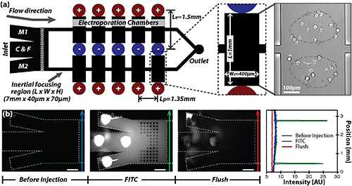
Figure 1. (A) A schematic of the microfluidic electroporation system, consisting of inlets for cells (denoted as C), molecules (denoted as M1 and M2) and a flush solution (denoted as F), two straight channels where inertial focusing occurs, 10 electroporation chambers with electrodes and an outlet. (B) Solution exchange demonstration at the inlet using a 1μM FITC solution and a flush solution (DPBS) indicates that the active solution can be uniformly injected to all arrays of chambers downstream. Image contrast is enhanced by adjusting look-up table (LUT). Scale bars are 250 μm. Please click here to view a larger version of this figure.
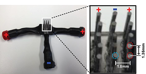
Figure 2. A photograph of the 15-pin electrode utilized for short-pulse high voltage application, consisting of 10 positive (+) and five negative electrodes (-). Each positive electrode is spaced 2 mm apart from a negative electrode, and each electrode of the same polarity is spaced 1.35 mm apart. Please click here to view a larger version of this figure.
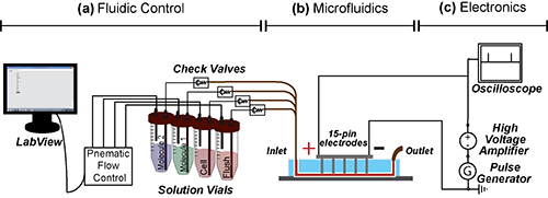
Figure 3. Schematic of the experimental apparatus, consisting of (A) the fluid control unit, (B) the microfluidic electroporator, and (C) the electrical equipment. (A) Vials containing solutions with molecules, cells and clean buffer are individually pressurized on demand using the LabView controlled pneumatic flow system. The solution from the pressurized vial is injected into the microfluidic electroporator through PEEK tubing with a check valve installed. (B) and (C) The electric signals are sent to 15-pin electrodes in contact with the flowing solution in the microfluidic system during the electroporation step. The electric pulses with the programmed duration are generated using the pulse generator and the magnitude of the electric pulse is amplified to 100 V by the high voltage amplifier. All applied electrical parameters are monitored in real time using an oscilloscope. Please click here to view a larger version of this figure.
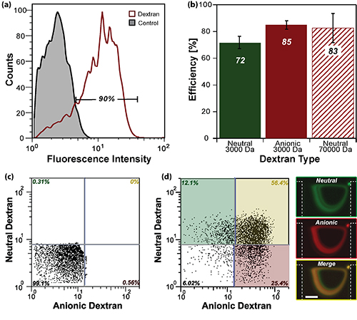
Figure 4. Representative flow cytometry data. (A) Fluorescent signals of MDA-MB-231 cells, which successfully took up the 70,000 Da anionic dextran molecule, compared to that of the control counterpart. (B) The efficiencies for each transferred dextran molecule do not exhibit significant molecular dependent variation (p >0.1). (C) Representative flow cytometry profiles for cells, which were not treated with electroporation (control). The fluorescent threshold indicating successful molecular delivery is set from the data such that the signals from control samples are found below the threshold. (D) Representative flow cytometry data for sequentially electroporated cells. Green, red and yellow boxes in the flow cytometry plot and fluorescent streak images on the right-side represent fluorescent signals from cells uptaking 3,000 Da neutral dextran-only, anionic dextran-only and both dextran molecules, respectively. Scale bar is 100 m. Please click here to view a larger version of this figure.
With the new parallelized electroporation platform, 10-fold enhancement in throughput and efficiency of multi-molecule delivery was achieved in addition to all the merits that the previously developed single-chamber system provides.18 Previously available merits include (i) pre-purification of target cells with uniform size distribution for viability enhancement, (ii) precise and individual molecular dosage control, and (iii) low operational electrical current. Fluorescently labeled dextrans were chosen as molecules of interest because they are readily available in a wide range of molecular weights, conjugated fluorophore types, and electrical charges. These macro-molecules are good candidates for such a study since they are inherently membrane impermeable for living cells and do not exhibit cytotoxicity.24 Optimum concentration of fluorescent dextran, incubation time, and electrical parameters were identified for the current system prior to the electroporation experiments. The optimization process was rather simple and efficient due to the platform’s ability to monitor the molecular uptake process in real-time. Visualization of this process is very beneficial if a molecule or cell type with little-to-no knowledge of electroporation parameters is being tested. The incubation time for all molecules tested was set to be 100 s by which cells trapped in all 10 chambers exhibit detectable fluorescent intensity signals in real time, indicating the completion of successful molecular delivery. Note that this is not the minimum incubation duration required for molecular delivery.
There has been a long-standing debate regarding the molecular delivery process using electroporation, whether it is solely mediated by diffusion4,19,25 or by electrophoresis.26 Identification of the dominant mechanism of molecular delivery involves a series of laborious individual tests, comparing the outcomes depending on molecular sizes, electrical charges and electroporation parameters. To the best of the authors’ knowledge, these laborious optimization processes were not reported with conventional electroporation systems.4,26-28 The developed system’s simple parameter optimization capability allowed for identification of the dominant molecular delivery mechanism, by examining variations in the electroporation efficiency depending on molecular and electric parameters. Results showed that the efficiency of anionic 3,000 Da dextran was not significantly different than that of its neutral counterpart (83 and 75%, respectively), suggesting that the molecular uptake observed was possibly mediated by diffusion. Unlike other electroporation systems, processed cells were continuously agitated throughout the electroporation process in this system. Gradual increase in fluorescent signals of orbiting cells was observed, suggesting that diffusion would be the dominant delivery mechanism. High efficiency molecular uptake (90%) of neutral 70,000 Da dextran molecules further implies that even at high molecular weights the delivery process occurs as a result of diffusion. Lastly, successful sequential delivery of 3,000 Da anionic and neutral dextran molecules solidifies the claim that molecular delivery occurs despite the molecules charge. This result also suggests that membrane resealing is not completed within 3 min of electroporation (100 sec per molecular delivery).
The presented molecular delivery platform can sequentially deliver wide ranges of molecular sizes regardless of their electrical charges. Timely fine-tuning and modification of individual parameters can be achieved with little effort aided by the real-time process monitoring capability. The system’s size-based cell purification process eliminates the need for laborious sample preparation and time-consuming centrifugation pre- and post-electroporation steps. Furthermore, the system’s low operational current substantially reduces cell mortality.
The authors have nothing to disclose.
This work is supported by the Rowland Junior Fellow program. The authors would like to express gratitude to the scientists and staff at the Rowland Institute at Harvard: Chris Stokes for his help in the development of the custom-built, computer-assisted pressure control setup, Diane Schaak, Ph.D. for her input for biological sample handling, Winfield Hill for developing the electrical setup, Alavaro Sanchez, Ph.D. for granting access to the flow cytometer, Scott Bevis, Kenny Spencer and Don Rogers for machining mechanical plumbing components required for the pressure setup. Microfluidic masters were fabricated at the Center for Nanoscale Systems (CNS) at Harvard University.
MDA-MB-231 cancer cell line | American Type Culture Collection (ATCC) | HTB-26 | |
Leibovitz’s L-15 Medium | Cellgro, Mediatech, Inc. | 10-045-CV | |
fetal bovine serum (FBS) | Gibco, Life Technologies | 16000-044 | |
penicillin-streptomycin | Sigma-Aldrich | P4333 | |
Dulbecco's phosphate buffered saline (DPBS) | Cellgro, Mediatech, Inc. | 21-030 | |
Trypsin | Gibco, Life Technologies | 25200-056 | |
Flow Cytometer easyCyte HT | Millipore | 0500-4008 | |
Oxygen Plasma Cleaner | Technics Micro-RIE | ||
Dektak 6M surface profiler | Veeco | ||
KMPR 1050 | Microchem | ||
SYLGARD 184 SILICONE ELASTOMER KIT | Dow Corning | ||
Compressed Nitrogen gas | Airgas | NI 300 | |
High Pressure Regulator | McMaster-Carr | 6162K22 | |
Downstream regulator | McMaster-Carr | 4000K563 | |
high-speed 3/2way-8 valve manifold | Festo | ||
Inline Check Valve | Idex Health and Science | CV3320 | |
5/32" OD x 3/32"ID Polyurethan tubes | Pneumadyne | PU-156F-0 | |
1/4" OD X 0.17" ID Polyurethan tubes | Pneumadyne | PU-250PB-4 | |
1/16" PEEK tubings | Festo | P1533 | |
1/32" PEEK tubings | Idex Health and Science | P1569 | |
PEEK tubing unions | Idex Health and Science | P881 | |
Pulse Generator | HP | 8110A | |
Aluiminum Wire | Bob Martin Company | 6061 ALUM | |
oscilloscope | Agilent | DSO3062A | |
50 mL centrifuge tubes | VWR | 21008-178 | |
15 mL centrifuge tube | VWR | 21008-216 | |
T75 culture flask | VWR | 82050-862 | |
Dextran, Tetramethylrhodamine, 3000 MW, Anionic | Gibco, Life Technologies | D3307 | |
Dextran, Tetramethylrhodamine, 70,000 MW, Neutral | Gibco, Life Technologies | D1819 | |
Dextran, Texas Red, 3000 MW, Neutral | Gibco, Life Technologies | D3329 |