This article describes a detailed protocol to produce a forebrain stab injury in adult mice. The stab injury induces severe reactive gliosis and glial scar formation which can be subsequently examined by standard immunohistochemistry methods.
Following injury to the CNS, astrocytes undergo a broad range of biochemical, morphological, and molecular changes collectively referred to as reactive astrogliosis. Reactive astrocytes exert both inflammatory and protective effects that inhibit and promote, respectively, neural repair. The mechanisms underlying the diverse functional properties of reactive astrogliosis are not well understood. Achieving a greater understanding of these mechanisms is critical to developing therapeutic strategies to treat the injured CNS. Here we demonstrate a method to trigger reactive astrogliosis in the adult mouse forebrain using a forebrain stab lesion. This lesion model is simple, reliable, and requires only a stereotaxic device and a scalpel blade to produce the injury. The use of stab lesions as an injury model in the forebrain is well established and amenable to studies addressing a broad range of neuropathological outcomes, such as neuronal degeneration, neuroinflammation, and disruptions in the blood brain barrier (BBB). Thus, the forebrain stab injury model serves as a powerful tool that can be applied for a broad range of studies on the CNS response to trauma.
A major challenge for developing successful therapies to treat the injured CNS is an incomplete understanding of the complex multicellular events that are triggered by the trauma. Reactive astrocytes are gaining increasing recognition as a promising target for novel therapies1. Though historically regarded as hostile to neural repair, reactive astrocytes are now recognized as critical components of a complex, multicellular neuroprotective response that includes attenuation of inflammatory processes and limiting secondary damage and neurodegeneration2-6. Although the neuropathological characteristics of reactive gliosis have long been well defined, the cellular and molecular mechanisms regulating reactive gliosis, and the diverse array of downstream consequences remain poorly understood. Understanding the mechanisms that drive reactive gliosis, as well as the subsequent cellular and molecular events, is an important step towards developing strategies aimed at promoting the neuroprotective properties of reactive gliosis, while attenuating the detrimental effects.
Here we demonstrate a method to induce severe reactive astrogliosis in the forebrain of adult mice using a stab injury. In contrast to other traumatic brain injury (TBI) models, such as controlled cortical impact (CCI) or fluid percussion injury (FPI), which require specialized equipment to produce an injury, the forebrain stab requires only a stereotaxic device to stabilize the head and a No. 11 scalpel blade. Thus the forebrain stab lesion model is more broadly accessible to a wide range of laboratories that do not have access to the specialized devices necessary for creating an FPI or CCI injury. The method described here enables investigators to reliably and reproducibly trigger a robust gliosis response to investigate subsequent cellular and molecular events. Once recovered from surgery, animals that have received a forebrain stab injury can survive for prolonged periods without the need for specialized care and can be returned to the colony for acute, intermediate, or chronic studies. Though less clinically translatable than FPI or CCI models of TBI, a forebrain lesion produced by a stab injury serves as a simple yet useful experimental model to investigate basic biological mechanisms underlying reactive gliosis and other neuropathological events following trauma to the CNS.
Adult (3-4 months old) male mice on a mixed C57BL/6 background were used in this protocol. Animals were kept on a 12 hr light/dark cycle, and allowed free access to food and water. All procedures performed in this protocol were conducted according to protocols approved by the Drexel University Institutional Animal Care and Use Committee.
1. Preparing Surgical Area
2. Prepping Mouse for Surgery
3. Surgical Procedure
4. Post-surgical Care
Because animals undergoing this procedure do not require specialized post-operative care, short or long-term time survival periods are easily incorporated into the study, depending on the need to investigate acute or chronic pathology following injury. Principal features of reactive gliosis, such as upregulation of GFAP and hypertrophy of soma, can be observed as early as 2-3 days following injury. The peak phase of proliferation for reactive astrocytes is during days 3-5 following injury10. The representative results shown below are from animals that received a stab wound lesion 7 days earlier.
The general morphology and cytoarchitecture of the forebrain following a forebrain stab injury can be visualized by Nissl staining (Figure 2). Although the blade track is most prominent throughout the center of the lesion, the disrupted cortical cytoarchitecture reveals the rostral and caudal extent of the damaged tissue. Reactive astrocytes can be observed by immunohistochemistry for GFAP (Figure 3). Note that many cortical astrocytes do not exhibit immunohistochemically detectable levels of GFAP in the absence of injury. However, GFAP expression is dramatically upregulated in the hemisphere ipsilateral to the injury while remaining at relatively low levels in the contralateral hemisphere (Figure 3), suggesting that reactive astrocytes are restricted to the ipsilateral hemisphere. Note that while the cortical tissue ipsilateral to the lesion demonstrates marked upregulation of GFAP, other astrocytic markers, such as S100β are constitutively expressed in the absence of an injury (Figure 4), and maintain similar expression levels following injury (Figure 4). In addition to increased GFAP expression, reactive astrocytes undergo cellular hypertrophy. Cell bodies and processes become enlarged and show intense staining for GFAP (Figure 3).
The proliferation of reactive astrocytes can be observed by administering the thymidine analog, 5-bromo-2'-deoxyuridine (BrdU), or by immunostaining for the proliferative markers Ki67 or PCNA. We routinely administer 200 mg/kg BrdU, i.p., to animals during days 3-5 following injury, the peak of reactive gliosis10 (Figure 3). However the precise dosage and timing of BrdU should be independently considered for each study, bearing in mind that BrdU will permanently label cells undergoing proliferation, as well as their progeny, at the time of administration, but that cells that enter the cell cycle before BrdU starts, or after BrdU administration is complete, will not be marked. In Figure 4, we show extensive co-localization between GFAP and BrdU at 1 week post injury, indicating that many reactive astrocytes proliferated during the time course of BrdU administration. Note that proliferating reactive astrocytes are predominantly localized adjacent to the lesion core, whereas reactive astrocytes localized distal from the lesion core are largely non-proliferative (Figure 4).
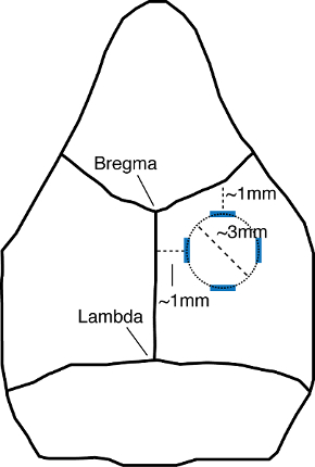
Figure 1: Schematic of the mouse skull, depicting the area of the craniotomy. Blue lines depict the initial markings identifying the boundaries of the area to be drilled. The top and left marks are measured at 1 mm below or lateral to the coronal or sagittal sutures, respectively. The bottom and right marks are measured at 4 mm from the coronal and sagittal sutures, respectively. The craniotomy is performed by drilling a circle within the marked boundaries (dotted line), creating a craniotomy that is roughly 3 mm in diameter (dashed line). Schematic is not to scale.
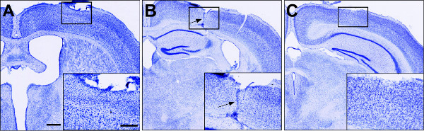
Figure 2: Nissl staining throughout the rostral-caudal extent of the lesion volume. (A–C) Coronal slices of injured brains 1 week post injury, showing the hemisphere ipsilateral to the lesion. Insets depict zoomed in images of the boxed regions. The blade track is most prominent in the center of the lesion, ~2.5 mm from Bregma (arrow in B). Note the disrupted cortical cytoarchitecture (insets) in sections anterior (A) and posterior (C) to the lesion center. Scale bar 500 μm, inset, 250 μm. Please click here to view a larger version of this figure.
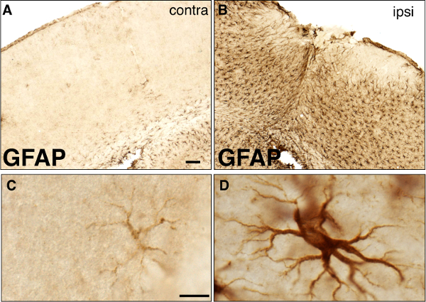
Figure 3: Brightfield immunohistochemistry for GFAP 1 week following a forebrain stab injury. (A–B) Low magnification images of GFAP staining in the contralateral (A) and ipsilateral (B) hemispheres of the same tissue section from an injured animal. The lesion site is shown in (B), and the corresponding region in the uninjured contralateral hemisphere is shown in (A). Scale bar, 100 μm. (C–D) High magnification images of normal (C) and reactive (D) astrocytes from the contralateral and ipsilateral hemispheres, respectively. Note the dramatic hypertrophy of the cell body and processes of reactive astrocyte in (D), compared to (C). Scale bar, 10 μm.
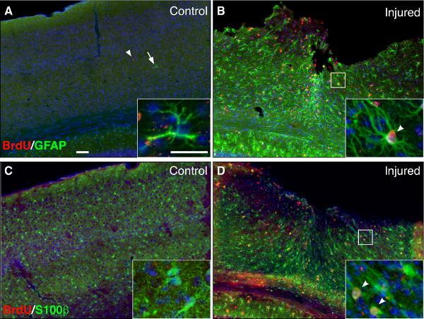
Figure 4: Reactive astrocytes proliferate following a forebrain stab injury. (A–B) Immunofluorescent staining for BrdU (red) and GFAP (green) in uninjured, control (A) and injured (B) brains, 1 week following stab injury. (C–D) Immunofluroescent staining for BrdU (red) and the astrocytic marker S100β (green) in the uninjured (C) and injured (D) hemispheres, 1 week following stab injury. Animals received BrdU over days 3-5 post injury. Note that many astrocytes are proliferating at the lesion site in the injured cortex (B and D, insets, arrowheads), whereas astrocytes in the uninjured cortex are not proliferating (A and C, insets). Counterstaining with DAPI (blue). Scale bars, 100 μm, inset, 25 μm.
It is critical that the skull or underlying dura are not damaged during the drilling. Use light pressure while drilling to ensure the skull is not punctured. In addition, care should be taken while lifting the skull piece to ensure the dura is not lifted off with the bone.
The forebrain stab injury described here models a penetrating injury to the CNS. Though less clinically translatable than TBI models such as FPI or CCI, the forebrain stab lesion model serves as a useful tool for a broad range of studies aimed at investigating various biochemical, cellular, or molecular events triggered by a discrete CNS insult. Although cognitive deficits have been reported in rats, 3 weeks after a bilateral stab injury11, it should be noted that the unilateral stab lesion model, as described here, is best suited for studies addressing the cellular response to injury. Fundamental aspects of various neuropathological processes, such as reactive gliosis and scar formation can be readily observed and studied. In contrast to FPI or CCI which produce diffuse and widespread neuropathology, the localized glial activation and pathology, facilitate intra-animal comparisons, between the injured and uninjured hemispheres. In addition, the infiltration of meningeal cells into the CNS at the lesion site presents an opportunity to investigate the interactions between these cells and local reactive astrocytes. Indeed, such interactions are critical in the formation of scar tissue, and have been shown to negatively regulate the permissive properties of reactive astrocytes to axonal regeneration12.
Here we use standard immunohistochemical procedures to demonstrate some of the principal features of reactive astrogliosis such as hypertrophy of cell bodies and processes, increased GFAP expression, and injury-induced proliferation. Note that although cavities are not observed in mice at 1-2 weeks following this procedure, studies using rats report cavity formation, 3 weeks following the injury11. Differences in neuropathology between mice and rats are also observed in spinal cord injuries. Whereas rats that undergo a spinal cord injury exhibit cysts or cavity formation at the lesion site, such cavities do not form in mice13,14.
The procedure is simple, reliable, easily reproducible, and requires minimal equipment. It can be easily modified to perform rat or mouse studies. In particular, use of this model with various transgenic mouse lines or in pharmacological studies can provide novel insight into mechanisms regulating the CNS response to injury. Lesion size and severity can be modified by adjusting the depth of the blade and travel distance of the stereotaxic arm holding the blade to produce discrete punctures rather than longitudinal mechanical lesions. Thus, the forebrain stab injury model can serve as an excellent experimental platform with which to study specific aspects of various neuropathological responses to injury.
Injury to the CNS triggers a complex response that is dynamic and multicellular6. In addition to reactive astrocytes, microglia mobilize quickly to phagocytose debris and initiate both pro and anti-inflammatory signaling cascades15-18. Activated microglia and invading macrophages produce cytokines and chemokines, creating a hostile environment for normal cell function and survival19-21. Extracellular matrix (ECM) molecules, such as chondroitin sulfate proteoglycan (CSPG), are produced from a variety of cell types, including reactive astrocytes and fibroblasts, and create a hostile environment for the regeneration and structural reorganization of surviving neurons22,23. Here we demonstrate some of the principal features of reactive astrogliosis that occur following a forebrain stab injury. Upregulation of intermediate filaments such as GFAP, astrocyte proliferation, and glial scar formation, followed by subsequent tissue remodeling are readily assessed using standard immunohistochemistry procedures.
It should be noted that while some features of reactive gliosis are emphasized here, reactive gliosis is highly context dependent, with varying features and gene expression profiles that emerge depending on the specific trigger24. Nevertheless, a number of fundamental properties regarding the CNS response to injury and attempts at repair can be modeled and studied in forebrain stab lesioned tissue. Indeed, it has been shown that targeted ablation of proliferating reactive astrocytes following a forebrain stab injury leads to increased leukocyte infiltration into the CNS and increased neuronal degeneration, demonstrating the neuroprotective properties of reactive astrocytes2. More recently, reactive astrocytes isolated following a penetrating stab lesion, but not a non-invasive injury, demonstrate neural stem cell potential in vitro25,26. Thus the forebrain stab is a powerful injury model for studying a broad range of biochemical, cellular, and molecular events triggered by injury to the CNS. The ease and simplicity of this injury model will facilitate further studies that can lead to novel insights into the CNS response to injury and repair mechanisms.
The authors have nothing to disclose.
We thank Katherine Clark for technical assistance. A.D.R.G. is funded in part by 5K01MH097957-03
Stereotax | Harvard Apparatus | 726049 | |
High speed micro drill | Harvard Apparatus | 724950 | |
stainless steel scalpel blade, #11 | MedVet | JOR581S | |
5/45 angled forceps | Fine Science Tools | 11251-35 | |
Gelfoam sponge 12cmx7mm | Fisher | NC9841478 | |
Antibodies and other reagents | Manufacturer | Catalog Number | Dilution (brightfield) Dilution (fluorescence) |
Rb anti-GFAP | DAKO | Z033429-2 | 1:20k 1:1k |
Shp anti-BrdU | Abcam | ab1893 | 1:2k 1:500 |
Biotinylated goat anti-rabbit | Vector Laboratories | BA-1000 | 1:400 |
Biotinylated rabbit anti-sheep | Vector Laboratories | BA-6000 | 1:400 |
Alexafluor 488 goat anti-rabbit | Life Technologies | A-11008 | 1:400 |
Alexafluor 568 donkey anti-sheep | Life Technologies | A-21099 | 1:1000 |
DAPI Nucleic Acid Stain | Life Technologies | D3571 | 1:1000 |
Cresyl Violet Acetate | Sigma Aldrich | C5042-10G | 1% |