Note: The goal of this protocol is to construct and utilize a vascular muscular thin film (vMTF) with the structure shown in Figure 1 to assess contractility during extended culture of vascular smooth muscle cells (VSMCs) on PDMS substrates. To prolong VSMC viability, we utilize the crosslinker compound genipin. The substrates for these vMTFs are designed to analyze tissue contractility as developed by Grosberg et al.8 Other vMTF methods5 may also be used, with subtle changes to the presented substrate fabrication protocol.
1. Substrate Fabrication
- Coverslip Cleaning
- Place 25 mm diameter glass coverslips into a coverslip staining rack. Place the rack into a large beaker or container (e.g., an empty 100 – 1,000 µl pipet tip container).
- Add 70% ethanol to the container to fully immerse the coverslips. Sonicate them for at least 30 min.
- Remove the coverslip rack from the ethanol solution. Allow the coverslips to air dry by hanging the rack in a sterile culture hood (to prevent particle accumulation on coverslips) for 1- 2 hr.
Note: The coverslips must be completely dry prior to the following steps.
- Poly(N-iso-propylacrylamide) (PIPAAm) Strip Isolation on Coverslip
- Using adhesive tape, tape off the sides of a cleaned coverslip, leaving an exposed strip centered on the coverslip (Figure 1A). Modify the width of this exposed strip based on application and/or microfluidic design.
- Mark the edges of tape strips on the coverslip using a lab marker for later reference (Figure 1A).
- Cut around the perimeter of the coverslip to release from the dish (Figure 1A, dotted red line).
- Poly(N-iso-propylacrylamide) (PIPAAm) Coating
- Using an analytical balance, weigh 1 g of PIPAAm powder. Do not use unpolymerized N-iso-propylacrylamide, which is a carcinogen.
- Transfer the PIPAAm to a 50 ml centrifuge tube. Add 10 ml of 1-butanol inside a chemical hood to yield a 10% w/v solution. CAUTION: The flash point of 1-butanol is 37 oC. Store the resulting solution in a flammable cabinet and avoid heating.
- Allow the PIPAAm to dissolve for 10 min. If powder is still visible, mix the solution using a vortex-mixer until all powder dissolves.
Note: The following steps require the use of a spin coater. For each coverslip: - Place the taped coverslip on the spin coater chuck with forceps.
- Transfer 150 µl of PIPAAm solution onto the coverslip by placing droplets along the exposed glass in the center of the coverslip. Ensure complete coverage of the exposed area.
- Spin coat PIPAAm using the following recipe:
- Ramp 10 sec to 3,000 rpm. Dwell for 5 sec.
- Ramp 10 sec to 6,000 rpm. Dwell for 60 sec.
- Ramp 10 sec to 3,000 rpm. Dwell for 5 sec.
- Place the coverslip into a covered Petri dish with the PIPAAm facing up. Allow to air dry for at least 15 min.
- Carefully remove the adhesive tape from all coverslips, leaving the full coverslip exposed with a thin layer of PIPAAm coating in a center strip.
- PDMS Coating
- Mix and degas 15 g of PDMS in a 10:1 base:crosslinker ratio. Add 7 – 8 drops of sonicated 0.2 µm fluorescent microbeads prior to mixing. Cover the cup of PDMS with aluminum foil when not in use to prevent dust and other particles from contaminating the PDMS.
Note: The following steps require the use of a spin coater. For each coverslip: - Place a PIPAAm-coated coverslip on the spin coater chuck with forceps.
- Transfer PDMS onto the coverslip, covering at least one-third of the coverslip area.
- Spin coat using the following recipe:
- Ramp 5 sec to 500 rpm. Dwell 5 sec.
- Ramp 5 sec to 1,000 rpm. Dwell 5 sec.
- Ramp 10 sec to 3,000 rpm. Dwell 10 sec.
- Ramp 10 sec to 4,000 rpm. Dwell 60 sec.
- Ramp 10 sec to 2,000 rpm. Dwell 15 sec.
- Ramp 10 sec to 1,000 rpm. Dwell 10 sec.
- Ramp 5 sec to 500 rpm. Dwell 5 sec.
- Place the coverslip into a covered Petri dish with the PDMS facing up. Record the time when the coverslip was spin-coated. Keep track of the time associated with each coverslip throughout the experiment for later use in determining the PDMS substrate thickness.
- Place the Petri dish containing the coverslips in a 90 °C oven for at least 1.5 hr to ensure proper PDMS curing. If an oven is not available, let the coverslips cure for at least 48 hr at RT.
- Remove the coverslips from the oven and store them in a dark drawer until ready to use.
- Set aside every fourth coverslip for later measurement of substrate thickness, as a function of spin coating time, with a profilometer.
- Mix and degas 15 g of PDMS in a 10:1 base:crosslinker ratio. Add 7 – 8 drops of sonicated 0.2 µm fluorescent microbeads prior to mixing. Cover the cup of PDMS with aluminum foil when not in use to prevent dust and other particles from contaminating the PDMS.
2. Microfluidic Patterning for Engineering Tissues
- Fabrication of Microfluidic Devices
- Design of Tissue Microfluidic Photomask
- Use any appropriate computer-aided design program to design microfluidic patterns. For arterial lamellae composed of human umbilical cord artery vascular smooth muscle cells, use an alternating pattern of 10 µm channels with 10 µm walls.
- Use binary channel branching if possible17, but other branching designs may be used. Decrease the width and length of channels for each branching iteration until attaining the desired tissue pattern spacing (walls and channels, Figure 1B).
- Design the device to have a single inlet for surface treatment solution placement and a single outlet for application of vacuum.
- Fabricate a photomask containing the microfluidic design(s), as previously described18.
- Photolithographic Wafer Fabrication
Note: Perform photolithography in a suitable cleanroom or similar facility. To make silicon wafers with patterns for soft lithographic fabrication of tissue microfluidic devices (~20 – 25 µm channel height) using photolithography:- Clean a silicon wafer in acetone, methanol, and isopropyl alcohol for 1 min each. Dry the wafer with a nitrogen gun.
- Prebake the wafer on a hot plate for 5 min at 115 °C to remove excess moisture.
- Spin coat the wafer with SU-8 3025 photoresist using the following recipe to yield features 20 – 25 µm in height:
- Ramp 5 sec to 500 rpm. Dwell 5 sec.
- Ramp 15 sec to 4,000 rpm. Dwell 15 sec.
- Soft bake the wafer on a hot plate at 95 °C for 15 min.
- Load a photomask, and expose the wafer for 16 sec using a vacuum contact program on a contact mask aligner.
- Hard bake the wafer on a hot plate at 95 °C for 4 min.
- Develop the wafer for 6 min in developer. Then, wash the wafer twice for 2 sec in fresh developer and rinse the wafer with isopropyl alcohol.
- Silanate the patterned wafer O/N by placing 2 – 3 drops of tridecafluoro-trichlorosilane in an empty dish within a vacuum desiccator. Prop the wafer up using Petri dishes so that both the bottom and top of the wafer are exposed.
CAUTION: Tridecafluro-trichlorosilane is a flammable and corrosive liquid. Proper personal protective equipment and local exhaust is necessary for use.
- Tissue Microfluidic Device Fabrication
- Place a silanated, patterned wafer feature-side up in a Petri dish.
- Mix and degas 100 g of PDMS with a 10:1 base:crosslinker ratio. Pour the PDMS into the dish, completely and evenly covering the wafer.
- Place the dish in a vacuum desiccator until all air bubbles are removed from the uncured PDMS, approximately 30 min. Cure the PDMS in the dish at 90°C for at least 1.5 hr. Time and temperature can be adjusted as dictated by manufacture guidelines to obtain a complete cure.
- Once the PDMS has cured, cut out the PDMS around the wafer with a razor blade and carefully release the PDMS-covered wafer from the dish. Remove excess PDMS underneath wafer and slowly peel PDMS away from the top of wafer.
- Place the PDMS disk feature-side up in a clean dish and store the wafer away from light after use.
- Cut away the excess PDMS from around the patterns using a razor blade. Cut devices into rectangular shapes (Figure 1C) to ease peeling of device from substrates in later steps. Precise cuts are not necessary, as long as ample space exists for the inlet, outlet, and tissue pattern area (Figure 1C).
- Punch inlet and outlet holes (Figure 1C) using a 1 mm surgical biopsy punch.
- Design of Tissue Microfluidic Photomask
- Microfluidic Device Deposition
Note: In this protocol, microfluidic delivery is used to deposit patterned genipin, the key crosslinking agent for long-term tissue culturing, as well as fibronectin. Steps prior to penicillin/streptomycin sterilization (2.2.3) do not need to take place in sterile conditions, but limiting contamination and dust collection is encouraged throughout the protocol. All steps occurring after coverslip sterilization with penicillin/streptomycin (2.2.3) should utilize sterile technique. Note: This portion of the protocol must be started one day prior to cell seeding.- Substrate and Microfluidic Device Preparation
- Sonicate the microfluidic devices in 70% ethanol for at least 30 min.
- Dry the sonicated microfluidic devices using pressurized air or nitrogen, and place them in a Petri dish with channel features face-up to prevent unnecessary wear on the features.
- Place up to 10 vMTF substrate coverslips in a UVO cleaner (cover removed on dish so the surface is functionalized) for 8 min.
- Remove the UVO-treated coverslips, and place the microfluidic devices feature-side down onto each slip one at a time (orientation should be similar to Figure 1C). Press down firmly on the devices to ensure a tight seal to the PDMS-coated coverslips.
- Deposition of Genipin and Fibronectin
- Prepare a 5 mg/ml genipin solution by adding 1 ml of sterile ddH2O to a 5 mg container of lyophilized genipin. Mix the solution using a vortex-mixer. Set aside at RT for at least 30 min.
Note: The powder is difficult to solubilize at RT, so repetitive mixing for at least a minute is often necessary. - Quickly, place a drop of 70% ethanol at the inlet of each device for device priming. Note: The ethanol should wick through the devices.
- After 5 – 10 min, carefully aspirate the excess ethanol at the inlet, immediately replacing it with 1X phosphate buffered saline (PBS) at the inlet. From this point forward, be sure to not allow inlet to become completely dry to avoid introduction of air to the device.
- Place a vacuum aspirator tip at the outlet of each device. Draw 1X PBS through devices to rinse ethanol away. Leave a small amount of 1X PBS at the inlet. Note: If inlet appears nearly dry, add more 1X PBS.
- Aspirate excess 1X PBS so that only a small amount remains at the inlet prior to application of genipin solution.
- Place 60 μl of the 5 mg/ml genipin solution at each inlet (Figure 1D). Draw the genipin solution through the devices by placing a vacuum aspirator tip at the outlet (Figure 1D). Be sure not to draw all of the solution through, leaving a small amount of solution at the inlet.
- Place drops (approximately dime-sized) of 1X PBS at both the inlet and outlet to maintain wetting during incubation. Move the dish containing devices to a humidified oven or incubator (sterile environment is not necessary) set to 37 °C, and incubate for 4 hr. The dish does not need to be covered.
- During incubation, resuspend fibronectin to a concentration of 50 μg/ml in sterile ddH2O on ice for at least 30 min prior to application to microfluidic device.
- After the incubation of genipin, aspirate all remaining 1X PBS at the device outlets. Continue to apply a vacuum aspirator at each device outlet, pulling through the remaining 1X PBS at the inlet.
- Place 100 μl of 50 μg/ml fibronectin solution at each inlet, adding to a minimal amount of remaining 1X PBS at inlet (Figure 1D).
- Draw the fibronectin solution through the devices using a vacuum aspirator tip at the outlet (Figure 1D). Be sure to not draw all of the solution through. Note: Fibronectin drop will look different than the 1X PBS and genipin due to differences in surface tension.
- Move the uncovered dish containing the devices to an oven or incubator set to 37 °C, and incubate for 24 hr. Note: The fibronectin step does not require wetting of the inlet and outlet with 1X PBS. The remaining pool of fibronectin at the inlet will dry out. This is expected.
- Prepare a 5 mg/ml genipin solution by adding 1 ml of sterile ddH2O to a 5 mg container of lyophilized genipin. Mix the solution using a vortex-mixer. Set aside at RT for at least 30 min.
- Sterilization and Preparation for Cell Seeding
- Prepare a solution of penicillin/streptomycin for sterilization of patterned vMTF coverslips. Add 5 ml of penicillin/streptomycin (10,000 units/ml; 10,000 μg/ml) to 500 ml of sterile 1X PBS.
- Place the dish containing the devices in a sterile biosafety hood.
- Carefully remove the devices from the coverslips by slowly peeling the device at a corner, while lightly grasping the coverslip in opposite hand. Note: This step requires practice to reduce coverslip damage in the removal process. An alternative is using a syringe to inject 1X PBS at the inlet and/or outlet to aid in device release.
- Place the coverslips in sterile six-well dishes. Add at least 5 ml of penicillin/streptomycin solution to each well. Place the dishes in a sterile incubator at 37 °C for at least 30 min.
- After sterilization, aspirate the penicillin/streptomycin solution, and seed the coverslips with cultured human umbilical artery vascular smooth muscle cells19 (Figure 1D). The seeding concentration for VSMCs is ~80,000 cells per cm2. To reduce the number of cells needed for each sample, use a reducer to decrease the seeding area. One example of a reducer is the cut top of a 15 ml conical tube attached to the coverslip with sterile vacuum grease prior to seeding.
- Incubate the seeded coverslips in a sterile incubator at 37 °C and 5% CO2 and allow the seeded cells to attach and form aligned arterial lamellae mimics O/N (Figure 2A-B).
- Long-term vMTF Tissue Culture
- One day after seeding, remove the cell medium and reducers. Rinse the tissues with 1X PBS. Add 4 ml of serum-free cell medium to induce a contractile phenotype in the VSMCs20.
- Repeat the 1X PBS rinse and addition of fresh serum-free medium every other day as desired for long-term culture.
- Substrate and Microfluidic Device Preparation
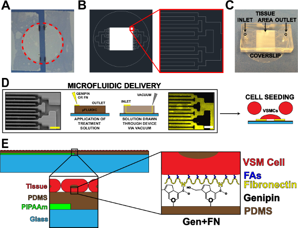
Figure 1. Microfluidic Protein Delivery Device. (A) Taped off coverslip for PIPAAm coating. Red dotted circle: cutting path to release coverslip. (B) Representative AutoCAD drawing of tissue microfluidic mask pattern. Inset: Detail of binary branching to alternating 10 µm x 10 µm tissue pattern. (C) Placement of microfluidic device on a coverslip substrate with inlet and outlet indicated. (D) Schematic of microfluidic protein patterning and delivery. Left-to-right: scanning electron microscope image of microfluidic channels (scale bar: 50 µm); Detailed schematic of method for protein deposition; Immunohistochemistry stained fibronectin (scale bar: 50 µm); Cell seeding with vascular smooth muscle cells. (E) Schematic of fabricated tissue. 1st inset: Detail of layered construct. 2nd inset: Detail of genipin modification of PDMS substrate after microfluidic deposition. © IOP Publishing. Reproduced and/or modified with permission. All rights reserved.19 Please click here to view a larger version of this figure.
3. Tissue Function Analysis with vMTF Contractility Assay
Note: The MTF contractility assay presented here is modeled after the technique developed in Grosberg et al.8
- vMTF Contractility Experiment
- Place a tissue sample in a 100 mm dish. Add sterile 1X Tyrode’s solution8 at pH 7.4 warmed to 37 °C to cover the sample.
- Use a razor blade to make several parallel cuts perpendicular to the PIPAAm edge. Make cuts in a manner that yields wider tissue sections that will be the vMTFs (with width ~2 mm) alternating with thin strips (Figure 3A, side cuts). To make clean cuts, place a razor blade in contact with the sample and firmly drag to the side.
- Rotate the dish 90° and make two straight, parallel cuts in the middle of the tissue, parallel to the strip of PIPAAm (Figure 3A, end cuts). Remove and dispose of the loose strip of tissue between these cuts and the thin strips in between vMTFs (cut in the previous step) to prevent adjacent films from making contact.
- Allow the sample to rest at RT for 10 min, or until all the PIPAAm has dissolved. Note: If PIPAAm remains in future steps, sample may be returned to the cutting dish to dissolve residual PIPAAm. A gentle scraping of the underside of vMTF can aid in PIPAAm removal, as needed.
- Place a small dot of vacuum grease in a clean 35 mm Petri dish. Add 5 ml of fresh, sterile 1X Tyrode’s solution at 37 °C. Transfer the coverslip with cut films from the 100 mm dish to the 35 mm dish, and press onto vacuum grease to prevent movement of the coverslip.
- Place the dish in a temperature-controlled platform on the stereomicroscope stage.
- Capture time-lapse transmitted and fluorescent light images at desired intervals (e.g., 30 sec) throughout treatment assay.
- Serially treat vMTFs with 50 nM endothelin-1 for 20 min (induced contraction) and 100 µM HA-1077 for 30 min (tissue relaxation). Add concentrated solutions of each treatment to the experimental dish containing 5 ml of sterile 1X Tyrode’s solution at specified time points, yielding the desired treatment concentration in the 5 ml volume. Make treatment additions during the interval between time-lapse image acquisitions to avoid capturing pipette in images.
- vMTF Contractility Analysis
- Using coverslips set aside in 1.4.8, measure PDMS substrate thickness with a profilometer21. Create a thickness vs. spin time curve for each set of coverslips. Use this curve to estimate the vMTF thickness for each coverslip used in a contractility experiment.
- Measure the vMTF projection lengths for each time point during the experiment, and calculate the associated radii of curvature (Figure 3B) using previously reported methods8.
- Calculate vMTF stress at each time point using previous vMTF methods5.
Note: Use the estimated vMTF thickness calculated from 3.2.1. Measure VSMC thickness using confocal images, as previously reported9. Obtain PDMS Young’s modulus from company data sheets.
The primary goal of this work was to extend the viability of micropatterned VSMCs on hydrophobic PDMS substrates. This was accomplished by incorporating a microfluidic delivery system to deposit patterned genipin and fibronectin on PDMS (Figure 1). Deposition of ECM proteins using microfluidic delivery yielded high fidelity transfer of the channel pattern with bare PDMS between lines of genipin and fibronectin (Figure 1D). The attached cells (Figure 1E) form confluent monolayers mimicking the in vivo structure of arterial lamellae (Figure 2), similar to previous microcontact printing methods5,10,16. These tissues yielded responsive, contractile constructs, whose stress was measured using vMTF technology (Figure 3).
Qualitative assessment of tissue viability over the course of two weeks showed minimal deterioration on genipin-modified substrates (Figure 2A). Tissue confluence and alignment were maintained over two weeks (Figure 2B, 2C and 2D). Two key stress values were calculated for every vMTF: 1) basal tone and 2) induced contractility (Figure 3C and 3D). Basal tone is the stress maintained by unstimulated VSMCs at equilibrium. Induced contractility is the additional stress induced by stimulation with endothelin-1. Both basal tone and induced contractility showed consistent behavior over the two-week time course, demonstrating vasoactive tissues throughout (Figure 3E and 3F). The slight drop in tissue contractility at the end of the assay is the direct result of the reduced number of cells composing the tissue, since serum-starved VSMCs do not proliferate19. The addition of a minimal basal level of serum to culture medium may alleviate this result in future work.
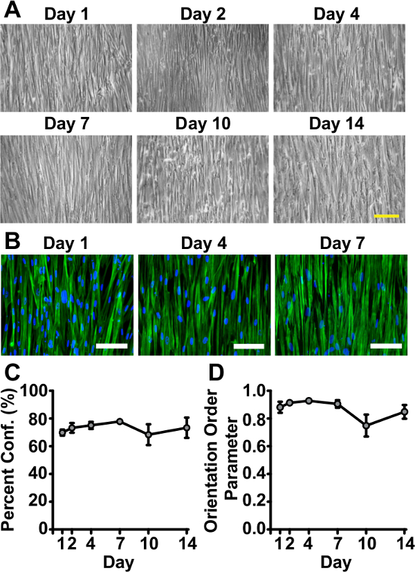
Figure 2. Tissues Remain Viable and Successfully Mimic in vivo Arterial Lamellar Structure for Two Weeks on Genipin-modified Substrates. (A) Representative phase contrast images of tissues at sacrifice time points throughout the course of two weeks (scale bar: 200 µm). (B) Representative immunohistochemistry images of tissues fabricated on genipin-modified substrates fixed at Day 1, Day 4, and Day 10 after serum starvation (green: f-actin filaments, blue: nuclei (shown to establish presence of cells), scale bar: 100 µm). (C) Percent confluence measured by f-actin coverage (error bars: standard error, n = 3 – 7). (D) Tissue alignment measured by f-actin orientation order parameter (OOP)22 (error bars: standard error, n = 3 – 7). © IOP Publishing. Reproduced and/or modified with permission. All rights reserved. All rights reserved.19 Please click here to view a larger version of this figure.

Figure 3. Tissue Contractility is Maintained Over the Course of Two Weeks. (A) Representative vMTF cutting scheme. (B) Schematic of cut vMTF. Upon cooling below 32 °C, PIPAAm dissolves, releasing vMTF. Stress in the active cell layer causes the passive PDMS layer to bend. Measurement of projection length can be converted to a radius of curvature according to methods in Grosberg et al.8 Radius of curvature is used to calculate the average cross-sectional stress in the tissue. (C) Sequential transmitted light images of representative contractility assay (scale bar: 1 mm). Tissues reach equilibrium, are stimulated with endothelin-1, then treated with HA-1077 to allow complete relaxation. Bottom: Schematic of side view of idealized tissue during the course of the assay. (D) Representative stress curve for the contractility assay. Two key stress values are calculated. Basal tone is the difference between the equilibrium stress state and the relaxed stress state. Induced contractility is the change in stress from the equilibrium state to the endothelin-1 stimulated state. (E) Basal tone (error bars: standard error, n = 5 – 12). (F) Induced contractility (error bars: standard error, n = 5 – 12). © IOP Publishing. Reproduced and/or modified with permission. All rights reserved.19 Please click here to view a larger version of this figure.
