Isolated Musculus gastrocnemius tightness is a common cause for foot and ankle pathologies. Currently no standardized examination procedure exists. This manuscript demonstrates that 20 degree of knee flexion eliminates the restraining effect of the M. gastrocnemius on ankle dorsiflexion and presents a video description of a standardized examination protocol.
Common foot and ankle pathologies have been linked to isolated Musculus gastrocnemius tightness (MGT). Various examination techniques have been described to assess MGT. Still, a standardized examination procedure is missing. Literature argues for weightbearing examination but the degree of knee flexion needed to eliminate the restraining effect of the M. gastrocnemius on ankle dorsiflexion (ADF) is unknown. This manuscript investigates the effect of knee flexion on ankle dorsiflexion and provides a detailed description of a standardized examination protocol. Examination on 20 healthy individuals revealed, that 20° of knee flexion is sufficient to fully eliminate the influence of the M. gastrocnemius on ADF. This builds the prerequisite for a standardized examination for MGT. Non-weightbearing and weightbearing examination of ADF has to be conducted with the knee fully extended and at least 20° flexed. Two investigators should conduct non-weightbearing testing with the subject in supine position. In order to obtain reliable results, the axis of the fibula should be marked. One examiner can conduct weightbearing examination with the subject in lunge stance. Isolated MGT is present if ADF is impaired with the knee fully extended and knee flexion results in a significant ADF increase. The herein presented standardized examination is the prerequisite for future studies aiming at establishing norm values.
Limited ankle dorsiflexion (ADF) alters the gait kinematics and is held responsible for common foot pathologies including Achilles tendinopathy, stress fractures, metatarsalgia and plantar heel pain 1-5. The most common cause for limited ADF is isolated Musculus gastrocnemius tightness (MGT) 3,6.
The joint kinematics of the ankle are influenced by knee flexion as the M. gastrocnemius bridges both joints. The muscle is under tension when the knee is fully extended, as the origin of the muscle is then furthest proximal. The M. gastrocnemius then restrains ADF. Knee flexion approximates the muscle's origin, thereby reducing the tension of the M. gastrocnemius, and consequently increases ADF. Ankle dorsiflexion is then limited by other anatomical structures of the ankle joint. Figure 1 illustrates this principle. In the case of MGT, ADF is limited with the knee fully extended but substantially increases by flexion of the knee 4.
Clinical examination for MGT takes advantage of the above outlined principled and was first published in 1923 by Silfverskiöld, a Swedish orthopedic surgeon 7. Since then, numerous examination techniques have been described, all of which compare the ADF with the knee extended and flexed. Published clinical tests can be categorized into non-weightbearing 5,8, weightbearing 9,10, and instrumented 11,12. Today, non-weightbearing examination is most commonly conducted 13. The patient is placed supine on the examination couch and ADF is assessed with the knee fully extended and typically 90° flexed (Figure 2A). On the contrary, weightbearing ADF measurements are conducted with the subject upright in lunge stance. The rear knee is extended or bent and the subject is asked to lean forward just before heel liftoff (Figure 2B). For both tests MGT is diagnosed, if the ADF with the knee extended is impaired and knee flexion results in a significant increase of ADF.
Although non-weightbearing tests are frequently performed, weightbearing testing has several advantages. First, only one investigator is needed for weightbearing examination, whereas two examiners are required to achieve reliable non-weightbearing measurements. Second, the weightbearing examination more closely reflects the load during gait. Third, the force applied to the ankle is independent of the examiner. Fourth, weightbearing examination features a higher intra- and inter- rater reliability 9,10,13-15.
The major limitation of all tests for MGT is that the minimal degree of knee flexion needed to eliminate the restraining effect of the M. gastrocnemius on ADF is unknown 8,15. Whereas 90° of knee flexion are typically applied in non-weightbearing testing 5,8,16, this is not feasible for the weightbearing examination. The broad population is unable to perform a Lunge with 90° of knee flexion without lifting the heel off the ground. Consequently, most studies conducting weightbearing examinations did not control for knee flexion 8,15. In order to perform reliable weightbearing examinations it is essential to identify the minimal degree of knee flexion needed to eliminate the restraining effect of the M. gastrocnemius on ADF.
Overall, literature argues for weightbearing testing for the diagnosis of MGT. In order to provide a valid weightbearing examination procedure, the minimal degree of knee flexion required to eliminate the ADF restraining effect of the M. gastrocnemius must be known. The aim of this study was to investigate the influence of knee flexion on ADF in non-weightbearing and weightbearing testing and to provide a step-by-step guide to conduct non-weightbearing and weightbearing examination for MGT.
Ethics statement: The study was approval by the local ethics committee of the University of Munich (# 007-14).
Note: An examination couch freely accessible on both sides and the foot end is required for non-weightbearing testing. A line (tape, approximately 2 meters) on the ground perpendicular the wall is needed for weightbearing testing. A standard goniometer with 2° increments and 20 cm length is used. We recommend documenting the results using a standardized fourfold table containing ADF measurement for the knee extended and flexed for each leg separately.
1. Participant Preparation
2. Non-weightbearing Measurement (Figure 2A)
3. Weightbearing Measurement (Figure 2B)
4. Data Analysis and Interpretation
Both ankles of 20 healthy individuals (mean age of 27.1 ± 3.9 years), 50% female, were examined. Non-weightbearing and weightbearing tests at six different degrees of knee flexion (full extension, 20°, 30°, 45°, 60°, 75°) and the Lunge Test (uncontrolled knee flexion) were conducted. A functional brace was used to control knee flexion. Measurements were performed by two investigators blinded to each other's results. Between each individual, the order of the investigators and the order of knee flexion were altered. The D'Agostino and Pearson Test revealed no normal distribution. Multiple testing was accounted for by a Bonferroni correction (p <0.004). Standard descriptive statistics, a Mann-Whitney-U-Test for differences between the sides, gender and measurement technique (non-weightbearing vs. weightbearing), and a repeated measurement ANOVA for differences between degrees of ADF were calculated. An interclass correlation coefficient (ICC; 1,1) was used to assess inter-rater reliability.
The data presented are average values of both examiners. The inter-rater reliability ranged between 0.961 to 0.992. No gender or side differences were found. Figure 3 presents the pooled data of both sides for ADF for each step of knee flexion. All weightbearing measurements resulted in significant higher ADF values compared to non-weightbearing measurements (Mann- Whitney-U-Test, p <0.001).
The boxplots presented in Figure 4 show pooled ADF differences (delta) between each step of knee flexion. Significant differences were only observed between full knee extension and 20° of knee flexion (p <0.001). Further knee flexion did not result in an increase of ADF. No significant differences for ADF were found between the Lunge test and any weightbearing examination with the knee flexed (20° to 75°).
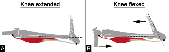
Figure 1. Schematic Illustration of the Functional Anatomy of Testing Isolated M. gastrocnemius Tightness. (A) With full extension of the knee the tensed M. gastrocnemius restrains ADF; (B) Knee flexion reduces the tension of the M. gastrocnemius and therefore increases ADF; Figure was adapted from 17. Please click here to view a larger version of this figure.
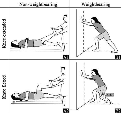
Figure 2. Schematic Illustration of the Test Procedures. (A) Non-weightbearing examination: The investigator applies force to the plantar aspect of the foot with the knee fully extended (A1) and 90° flexed (A2). The second investigator measures ADF. (B) Weightbearing examination: The subject gets into the lunge position with the rear leg centered on a line. ADF is measured with the knee fully extended (B1) and at least 20° flexed (B2). For both measurements take care that the foot measured is in a neutral pronation-supination position. Please click here to view a larger version of this figure.
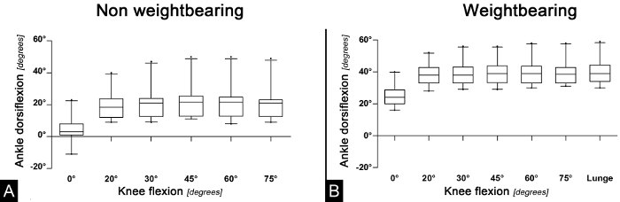
Figure 3. Boxplots (95% CI) of Pooled Ankle Dorsiflexion for Each Degree of Knee Flexion. (A) Non-weightbearing measurements: Mean ADF for each step of knee flexion; (B) Weightbearing measurements: Mean ADF for each step of knee flexion. Figure was adapted from 17. Please click here to view a larger version of this figure.
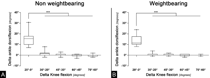
Figure 4. Boxplots of the Differences of Ankle Dorsiflexion Between Each Step of Knee Flexion (pooled values). (A) Non-weightbearing measurements: Difference of ADF between each step of knee flexion; (B) Weightbearing measurements: Difference of ADF between each step of knee flexion; ***: p <0.001; Figure was adapted from 17. Please click here to view a larger version of this figure.
Examinations on 20 healthy individuals revealed, that 20 degrees of knee flexion already eliminates the influence of the M. gastrocnemius on ADF. Further knee flexion resulted in no significant ADF increase. The herein presented video description of a standardized non-weightbearing and weightbearing examination for MGT builds the prerequisite for future studies establishing physiological norm values.
The study has limitations. First, no custom-made measurement device was used to assess ADF, which might contribute to measurement inaccuracy. This is for the investigator depended force applied during non-weightbearing testing, the use of a goniometer and adjacent joint movements. Moreover, the herein used goniometer had 2° increments, which could also contribute to measurements inaccuracy. Still, the herein used measurement device is available to every physician 18. Next, adjacent joint movements, i.e., in the subtalar and midtarsal joints, alter especially weightbearing ADF measurements. In order to reduce this effect we closely monitored for subtalar neutral position of the foot during testing, as recommended in the literature 19-22. Overall, the authors aimed at presenting a standardized examination routine, which is applicable in the daily routine and does not rely on special devices. The herein observed high ICC values argue for the validity of the presented examination procedure. Finally, the minimal degree of knee flexion (20°) was predefined by the orthesis used. Therefore, we could not investigate whether even less knee flexion already eliminates the ADF restraining effect of the M. gastrocnemius. Still, the 20° of minimal knee flexion identified are practical for weightbearing testing.
Various modifications of the herein presented techniques have been published. First, different anatomical landmarks have been employed. For non-weightbearing tests, the axis of the fibular (y-axis) and either the plantar surface of the foot 13,23,24 or the axis of the fifth metatarsal bone 8,25,26 have been used. For weightbearing measurements, the floor (x-axis) and the axis of the fibular 10,14, the Achilles tendon 27,28, or the tibia 29-31 have been assessed. Second, the devices used to measure ADF vary. Measurement devices applied in previous studies include the digital inclinometer 27,31, mobile app 27, acrylic plate 10, custom made devices 11,12,32, measuring tape 33,34, and a standard goniometer 5,15,29. Third, the weightbearing examination has initially been described as the "knee-to-wall" lunge test measuring the distance of the greater toe to the wall 33-35. Overall, the published variations are considerable and have been combined in various ways. This not only hinders the inter-study comparison but the techniques applied are often not practical for every day use. We aimed at defining a standardized and reproducible procedure that can be conducted non-weightbearing and weightbearing with little effort and resources. Therefore the axis of the fibula and the plantar aspect of the foot / floor where chosen as measurement landmarks. A standard goniometer was used as the authors consider this a tool available to every physician. Finally the weightbearing test was conducted with the rear leg being the one measured as the "knee-to-wall" principle can not be conducted with the knee extended.
Conducting the above outlined examinations, the authors experienced several pitfalls. First, the identification of the fibular during weightbearing testing is difficult due to the prominence of the peroneal tendons. In order to generate reproducible results, we recommend marking the axes of the fibular prior to testing. Second, for the weightbearing test with the knee extended, care must be taken that the knee is fully extended. We found minimal knee flexion to have a profound impact on the ADF. Third, the investigator has to ensure subtalar neutral position as adjacent joint movements alter the measurements. Finally, as outlined above, the authors recommend non-weightbearing examination to be conducted by two investigators. One investigator applies force on the plantar aspect of the forefoot and assures a neutral pronation-supination position of the rear foot. The other investigator conducts the actual measurement.
The major drawbacks we are currently facing are missing physiological and pathophysiological values 4,36. First, the values for ADF with the knee fully extended obtained by non-weightbearing and weightbearing testing vary significantly. In the literature weightbearing test has been shown to be more reproducible 9,10,13-15. Moreover, it more closely resembles the physiological loading conditions and partially eliminates inter-rater differences in the force applied to gain maximum ADF. Further, non-weightbearing testing requires two examiners whereas only one examiner can conduct weightbearing testing. In summary the weightbearing test seems to be superior. Until now it was unclear what degree of knee flexion is needed to eliminate the ADF restraining effect of the M. gastrocnemius in weightbearing testing. This study for the first time could demonstrate that 20° of knee flexion is sufficient. Therefore, the herein presented video description of a standardized weightbearing examination for MGT ensures valid testing and should be conducted in future studies.
Future studies should apply the herein presented weightbearing examination in order to define norm values of ADF with the knee extended and flexed. Previous studies in line with our results suggest a high variety in ADF. If this variation does not allow the definition of norm values a promising approach is to compare the ADF of both ankles.
The authors have nothing to disclose.
We would like to thank Jakob Binder for his help in subject acquisition and organization of the examinations, Mrs. Hella Thun for designing Figure 1 and 2 and finally Mr. Dipl.-Ing. (FH), M.Sc. M. Saller for his statistical support.
Examination couch | |||
Standard goniometer | MDF Instruments USA, Inc. Malibu, CA, USA | 2° increments |