This protocol describes a step-by-step method for the reproducible isolation and long-term culture of adult mouse cardiomyocytes with high yield, purity, and viability.
Cultured cardiomyocytes can be used to study cardiomyocyte biology using techniques that are complementary to in vivo systems. For example, the purity and accessibility of in vitro culture enables fine control over biochemical analyses, live imaging, and electrophysiology. Long-term culture of cardiomyocytes offers access to additional experimental approaches that cannot be completed in short term cultures. For example, the in vitro investigation of dedifferentiation, cell cycle re-entry, and cell division has thus far largely been restricted to rat cardiomyocytes, which appear to be more robust in long-term culture. However, the availability of a rich toolset of transgenic mouse lines and well-developed disease models make mouse systems attractive for cardiac research. Although several reports exist of adult mouse cardiomyocyte isolation, few studies demonstrate their long-term culture. Presented here, is a step-by-step method for the isolation and long-term culture of adult mouse cardiomyocytes. First, retrograde Langendorff perfusion is used to efficiently digest the heart with proteases, followed by gravity sedimentation purification. After a period of dedifferentiation following isolation, the cells gradually attach to the culture and can be cultured for weeks. Adenovirus cell lysate is used to efficiently transduce the isolated cardiomyocytes. These methods provide a simple, yet powerful model system to study cardiac biology.
Cultured cardiomyocytes are frequently used to monitor cell behavior in a well-controlled environment in vitro. For example, morphological, electrical, biochemical, or mechanical cell properties can be studied on engineered substrates,1,2 in defined media, and in response to small molecule drugs, peptides, gene regulation,3 or electrical stimulation.4 The cellular content can also be controlled using defined co-cultures.5 These in vitro experiments are useful in large drug or genetic screens and complement in vivo methods for various types of investigations involving cardiomyocyte biology.
Long-term culture enables experimental avenues that require extended periods of time to achieve phenotypic change. A timely example is that of adult mammalian cardiomyocyte proliferation, where dedifferentiation, cell cycle re-entry, and cell division is typically studied over several days to weeks.6,7 Here, the extended culture time facilitates genetic manipulation,7,8 functional dedifferentiation (e.g., sarcomeric disassembly)9 and potentially transcriptional dedifferentiation.6 Subsequent cell cycle re-entry and cell division requires even longer culture periods to observe, especially if multiple rounds of division are the experimental goal. The importance of the cardiomyocyte cell-cycle is central to several recent key scientific works in heart regeneration, where the dedifferentiation and proliferation of pre-existing cardiomyocytes has been shown responsible for heart regeneration in zebrafish and neonatal mice.10-12 Thus, the possibility to stimulate dedifferentiation and cell cycle re-entry in mammalian adult cardiomyocytes remains a key question in human heart regeneration13-15
In vitro experiments studying the cell cycle of mammalian cardiomyocytes have predominantly used rat sources, due to their relative ease of long-term culture compared to mouse models.16 However, murine systems offer a rich resource of well-characterized transgenic tools and disease models that are useful in both in vitro and in vivo protocols. For example, Cre-based lineage tracing has enabled the identification of pre-existing cardiomyocytes as a source of regenerating myocardium in the neonatal mouse heart in vivo.12 In vitro studies of lineage-traced neonatal mouse cardiomyocytes have enabled the examination of interactions with stromal cells through co-culture with fibroblasts.5 However, due to its challenges,17 few reports exist of the isolation and long-term culture of adult mouse cardiomyocytes.18,19
The isolation of viable adult mouse cardiomyocytes for short-term culture alone is known to be a challenging task. This protocol provides step-by-step instructions on how to achieve viable cardiomyocytes from adult mice that can be used for both short-term as well as long-term investigations. Cardiomyocytes isolated using this protocol can be efficiently transduced with adenoviral vectors20,21 and cultured for weeks. These methods provide a powerful system to study cardiomyocyte biology in vitro.
The methods described herein are based on several elements from previous works using variations of the Langendorff retrograde perfusion.18,22 Although several protocols have been published on the isolation of adult mouse cardiomyocytes for short-term culture and study,23-25 the advantage of this protocol is the ability to culture the isolated cardiomyocytes long-term. This will be useful in the study of cellular processes involving ectopic gene expression and requiring extended periods of time, such as in cellular reprogramming strategies.
All procedures outlined here have been approved by the Institutional Animal Use and Care Committee at the University of California, San Francisco.
NOTE: Briefly, after extracting the heart from the mouse thorax, coronary retrograde perfusion is used to efficiently digest the extracellular matrix with collagenase and protease XIV. The ventricles are then isolated, mechanically dissociated and filtered into a single cell suspension. Gravity sedimentation is performed to isolate cardiomyocytes, which are separated from stromal cells like fibroblasts and endothelial cells by their high density. The purified cardiomyocytes can be cultured for weeks on laminin-coated polystyrene and transduced with adenovirus gene vectors.
1. Cardiomyocyte Isolation
2. Cardiomyocyte Culture
3. Gene Transduction with Adenovirus
A wild type adult ICR (CD1) mouse heart typically yields 500,000 to 1 million cardiomyocytes from a successful isolation. Immediately after isolation, the cells maintain a mostly rod-shaped appearance (Figure 3A) with intact sarcomeres and can be used for functional studies involving cardiomyocyte contractility. A high percentage of rod-shaped cardiomyocytes (above 90%) is an indication of effective perfusion and digestion. Viable cardiomyocytes will be large (~ 100 – 200 μm in length) and appear to have a sharp outer membrane under brightfield illumination (Figure 3A). Immunostaining for cardiomyocyte-specific sarcomeric markers, such as alpha-actinin, will result in a distinct sarcomeric banding pattern (Figure 3C). In conjunction with morphological analysis and nuclear staining with DAPI, immunostaining can be used to assess the purity of the isolated cardiomyocytes. Fibroblasts will be small and round just after isolation, but will quickly attach and spread thin on cell culture plastic, thus enabling facile distinction from cardiomyocytes.
A low percentage of rod-shaped cardiomyocytes is an indication of an unsuccessful isolation. This can result from a failure to quickly cannulate the aorta after the heart is extracted from the animal. Inefficient perfusion, due to air embolism or incorrect cannulation for example, can also impact the morphology and viability of the cardiomyocytes.
After several days in culture, the cardiomyocytes assume a rounded morphology (Figure 3B). Within 1 – 2 weeks, live cardiomyocytes attach to the substrate and begin spreading (Figure 3D, E). Dead cells can be identified by trypan blue inclusion. A successful isolation and culture will yield approximately 50% live cardiomyocytes after 1 week. Contamination by non-cardiomyocytes, such as fibroblasts will result in a high percentage of these cells later in culture due to their proliferative potential. This can be avoided by increasing the number of gravity sedimentations and/or through pre-plating to increase the purity.28
Transduction with adenovirus can be used to achieve strong gene expression within 48 – 72 hr (Figure 3E). Adenovirus serotype 5 is efficient at transducing adult mouse cardiomyocytes in vitro due to the high expression of the coxackie-adenovirus receptor, but may depend on the type of media used.20
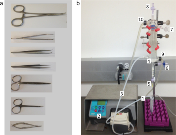
Figure 1. Cardiomyocyte Isolation Equipment and Instrumentation. (A) Surgical instruments used to extract the mouse heart and cannulate the aorta, from top to bottom: hemostats, tissue forceps, curved forceps, Dumont #5 fine forceps, small dissecting scissors, fine iris scissors, fine squeeze scissors. (B) The Langendorff perfusion system used to digest the mouse heart. Perfusion buffers are aspirated through the inlet tube (1) by the perfusion pump (2) and through the pump outlet tube (3) into the inlet port on the heated water jacket (4). The perfusion buffers are warmed by the heated water jacket as they travel through the spiral glass tube, the debubbling chamber, and then out of the cannulation needle attached to the perfusion outlet port (5), which is connected by a stopcock. The conical tube below catches the perfusion buffers, which are re-circulated through the inlet tube. The pressure port (6), compliance port (7) and vent (8) remain closed during perfusion in order to maintain a constant flow rate. The water jacket is heated by a circulating water bath entering through the jacket inlet (9) and outlet (10) ports. A ring stand is used to hold the heated water jacket in a vertical position (red clamps, black base below the purple conical tube rack). Please click here to view a larger version of this figure.
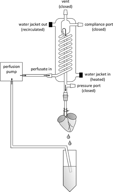
Figure 2. Schematic Overview of Perfusion System. Important Parts of the Perfusion Apparatus are Labeled as Referred to in the Manuscript. The flow direction of perfusion buffer is indicated by arrows. Note the positioning of the aortic cannula, the tip of which should be visible through the translucent aorta, ensuring it is placed above the aortic valve. Please click here to view a larger version of this figure.
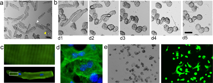
Figure 3. Isolated Adult Mouse Cardiomyocytes. Cardiomyocytes Were Isolated and Seeded onto Laminin-Coated 96 well Plates as Described in the Protocol Herein. (A) Freshly isolated cardiomyocytes, seeded at approximately 8,000 cells/cm2. Note that freshly isolated, viable cardiomyocytes appear approximately rod shaped with sharp edges (white arrowhead). However, cardiomyocytes that appear to have a diffuse outer membrane under brightfield are dead. During the first several days of culture, the cardiomyocytes assume a rounded shape (B). (C) Immunostaining for alpha-actinin (green) reveals characteristic sarcomeric banding in a cardiomyocyte at d1 post-isolation. Nuclear stain (DAPI) shown in blue. Top shows zoomed image of outlined region in lower image. (D) Organized sarcomeres can also be seen after redifferentiation in well spread cardiomyocytes at d11 post-isolation. (E) Brightfield and epifluorescent images show cardiomyocytes at day 11 post-isolation transduced with adenovirus carrying a GFP reporter under the control of a CMV promoter (a gift from Robert Gerard at UT Southwestern). Cells were seeded at approximately 18,000 cells/cm2 and were transduced on day 0 using a multiplicity of infection of approximately 800 plaque-forming units (PFU) per cell. Scale bars: 50 μm. Please click here to view a larger version of this figure.
Cardiomyocyte isolation buffer (CIB) | |||
Add 900 ml of ultrapure water to a clean beaker under magnetic stirring. | |||
Add the buffer components in the table as indicated. | |||
Component | MW | Final Conc (mM) | 1X (g/L) |
NaCl | 58.44 | 120 | 7.013 |
KCl | 74.55 | 5.4 | 0.403 |
Na2HPO4.7H2O | 268.07 | 0.33 | 0.088 |
MgSO4.7H2O | 246.48 | 0.5 | 0.123 |
Taurine | 125.1 | 30 | 3.753 |
BDM | 101.1 | 10 | 1.011 |
HEPES | 238.3 | 25 | 5.958 |
Glucose | 180.16 | 22 | 3.964 |
Adjust the pH with 5 M NaOH. | |||
Adjust the volume to 1 L with ultrapure water. | |||
Sterile filter w/ 0.22 μM vacuum filter. | |||
Add 0.5 μl of 0.1 U/ml insulin per liter of cardiomyocyte isolation buffer. | |||
Perfusion buffer | |||
Component | Final | Volume (ml) | |
CIB | – | 200 | |
EGTA (0.4M) | 0.4 mM | 0.2 | |
Total | – | 200 | |
Digestion buffer | |||
Component | Final | Amount | |
CIB | – | 15 ml | |
CaCl2 (1M) | 300 μM | 4.5 μl | |
Protease XIV | 0.2 mg/ml | 3 mg | |
Collagenase II | 2.4 mg/ml | 36 mg | |
Total | – | 15 ml | |
Stop buffer | |||
Component | Final | Volume (ml) | |
Perfusion buffer | 95% | 14.25 | |
CaCl2 (1M) | 1.5 mM | 22.5 | |
FBS | 5% | 0.75 | |
Total | – | 15 ml | |
Culture media | |||
Component | Final % | Volume (ml) | |
MEM media | 90% | 90 | |
FBS | 10% | 10 | |
Primocin | 0.2% | 0.2 | |
Total | 100% | 100 ml | |
Allow culture media to equilibrate at 37 °C, 5% carbon dioxide. |
Table 1: Solutions and Buffers.
The overall health of the isolated cardiomyocytes depends on several important aspects of this protocol. First, the time from heart extraction to perfusion is critical and should be performed in 5 min or less. Removal of calcium helps to dissociate cell-cell interactions, but can negatively impact cell health long-term.29-32 Thus, we find it sufficient to remove calcium during the initial few minutes of perfusion by EGTA (ethylene glycol tetraacetic acid) chelation,22 but quickly restore calcium during digestion and subsequent steps.
The use of protease XIV greatly improves digestion efficiency compared to collagenase alone. This reduces inconsistency due to collagenase batch variability,18,24 and increases the purity of cardiomyocytes by facilitating separation during gravity sedimentation. Nonetheless, over-digestion will prevent efficient attachment to the culture substrate, so a fine balance is required to optimize cell yield and purity while maintaining cell attachment and viability. Thus, the concentration of proteases and length of digestion can be modified to suit the experimental goals. In general, a lesser degree of digestion will enhance the attachment and survival of the cardiomyocytes. Use of trypsin has been reported in some cardiomyocyte isolation protocols.18 However, addition of trypsin to the protease cocktail used here (collagenase and protease XIV) reduces the long-term viability of the cardiomyocytes in culture, perhaps due to over-digestion or cleavage of essential surface and/or extracellular matrix proteins.
The inclusion of butanedione monoxime (BDM) during isolation inhibits cardiac muscle contraction, and thus reduces negative consequences due to terminal contracture, energy expenditure, and the calcium paradox.29,33 However, in our experience, removal of BDM prior to culture expedites dedifferentiation and allows the cells to adapt to culture in two dimensions.
It is important to note that long-term adaptations to 2-D culture may affect functional aspects of the cardiomyocytes, such as contractility. Thus, contractility and electrophysiology experiments should be performed soon after isolation, when native sarcomeric organization is still intact. Furthermore, as with any in vitro experiment, the cells in culture are devoid of certain extrinsic influences present in vivo, such as humoral or immune signaling. On the other hand, defined co-cultures can be implemented to study the interactions between cardiomyocytes and other cell types.5,34 Additionally, in vitro culture can be used to study cardiomyocyte behavior with defined molecular signals in the culture media. Thus, the overall goals of the experiment should be carefully considered and weighed against the limitations and advantages of this culture system.
The protocol described here uses 10% fetal bovine serum (FBS) to improve the viability of primary cardiomyocytes in culture.35 It should be noted that FBS and other animal-derived sera contain unknown components that can affect cellular physiology.36,37 Despite the well-appreciated presence of growth factors in FBS,38 the cardiomyocyte cultures presented here do not proliferate. Rather, the cell cycle block seems to be a persistent property of adult mouse cardiomyocytes ex vivo, maintained from their cell cycle exit during the perinatal period of development.12,39 This property is of major interest to the field of cardiac regenerative medicine, where reversion of adult mammalian cardiomyocytes to a proliferative phenotype has been a heavily pursued goal.3,26,40-42 Notably, it is currently unclear what degree dedifferentiation is required for cardiomyocytes to re-enter the cell cycle, but the contribution of dedifferentiation to regeneration in lower vertebrates is well supported43,44,10,11 Furthermore, dedifferentiation is increasingly being observed in cardiomyocyte proliferation and heart regeneration in mammalian systems, at least at the functional level, including myofibrillar disassembly6,12,9 Thus, the state of differentiation of cardiomyocytes in culture should be carefully considered and modulated according to the needs of the research goals. As such, the culture conditions can potentially be further modified to reduce serum concentration, use defined serum substitutes or serum-free media. For example, adult rat cardiomyocytes exhibiting organized sarcomeres were cultured long-term without serum, but growth factors such as fibroblast growth factor, epidermal growth factor, and triiodothyronine were used in defined media.45 It is conceivable that similar media formulations, or potentially other formulations such as those adapted from differentiation protocols,46 may be used to maintain well differentiated adult mouse cardiomyocytes in culture that display highly-organized sarcomeric structures. Alternative formulations may also be developed to achieve other phenotypes (such as highly de-differentiated cardiomyocytes)6 depending on the needs of the experiment, but further work will be required.
The role of fibroblasts has been implicated in the control of neonatal mouse cardiomyocyte cell cycle5 and adult mouse cardiomyocyte differentiation (through hypertrophic IL6 signaling) in long-term culture.19 Hence, the growth of fibroblasts in culture should be accounted for in experimental interpretations. The growth of fibroblasts in culture can be controlled by addition of growth restricting compounds like cytosine arabinoside19 (AraC) or through pre-plating steps.28 However, AraC will also inhibit growth of cardiomyocytes, so alternative methods should be used to control the growth of fibroblasts in studies concerning the cardiomyocyte cell cycle. In the protocol described here, initial cardiomyocyte purity can be enhanced by repeating the gravity sedimentation step (see 1.4.8).
The methods herein provide an in vitro system that can be used to study the nature of adult mammalian cardiomyocyte cell cycle blockade. In contrast to primary cells from other organisms, such as rat or guinea pig, primary mouse cardiomyocytes offer a wealth of powerful and well-characterized genetic tools, such as Cre-based lineage tracing knock-in models,47 which can be used to easily identify cells derived from pre-existing cardiomyocytes.12,48 Additionally, primary adult murine cardiomyocytes have been subjected to the native developmental conditions that result in cell cycle blockade, unlike cell lines and differentiated ES or iPSC cells46 which, despite their convenience and adaptability to large drug screens,2 may have limited utility in experiments probing the nature of adult cardiomyocyte cell cycle exit.
This protocol outlines a reliable method to isolate and culture adult mouse cardiomyocytes. Several methods have previously demonstrated the isolation of adult mouse cardiomyocytes for short-term study, but to date, few reports exist of the long-term culture of adult-mouse cardiomyocytes.18,19 The ability to maintain adult mouse cardiomyocytes in culture should enable the in vitro study of cardiomyocyte biology under experimental conditions requiring long-term examination. For example, gene expression can be manipulated using adenovirus vectors, which typically require a few days to achieve full expression. Subsequent phenotypic changes and analysis may require even longer periods of time depending on experimental goals. The methods presented here allow the culture of adult mouse cardiomyocytes for several weeks. This should provide a powerful system for the investigation of prudent questions in cardiomyocyte biology, such as those pertaining to cell cycle regulation.
The authors have nothing to disclose.
This project was funded by the UCSF Program for Breakthrough Biomedical Research (funded in part by the Sandler Foundation), the NIH Pathway to Independence Award (R00HL114738), and the Edward Mallinckrodt Jr. Foundation. JJ was supported by a postdoctoral fellowship from the NIH (T32HL007731). The authors are solely responsible for the contents of this work, which does not necessarily represent the official views of the NIH.
Equipment | |||
Heated water jacket | Radnoti | 158831 | |
Circulating heated water bath, Isotemp | Fisher Scientific | 3013 | |
Laboratory pump | Watson-Marlow | 323 | |
Hemostats | Exelta | 63042-090 | |
Tissue forceps | VWR | 470128034 | |
Dumont #7 curved forceps | FST | 91197-00 | |
Dumont #5 fine forceps | FST | 11251-20 | |
Small dissection scissors | VWR | 470128034 | |
Extra fine bonn scissors | FST | 14084-08 | |
Fine spring scissors | FST | 91500-09 | |
Name | Company | Catalog Number | Comments |
Materials | |||
NaCl | Sigma | S9888 | |
KCl | Sigma | P9541 | |
Na2HPO4-7H2O | Fisher | S25837 | |
MgSO4-7H2O | Fisher | S25414 | |
Taurine | Sigma | 86329 | |
Butane dione monoxime (BDM) | Sigma | B0753 | |
HEPES | Fisher | BP310100 | |
Glucose | Sigma | G-7021 | |
Insulin | Novo Nordisk | 393153 | |
EGTA | Amresco | 0732-288 | |
Protease, type XIV | Sigma | P5147 | |
Collagenase II | Worthington | LS004176 | |
MEM | Corning | 15-010-CV | |
FBS, heat inactivated | JRS | 43613 | |
Primocin | Invitrogen | NC9141851 | |
Ethyl Carbamate | Alfa Aesar | AAA44804-18 | |
215 micron mesh | Component supply | U-CMN-215-A | |
20 G blunt ended needle | Becton Dickinson | 305183 | |
20 G beveled needle | Becton Dickinson | 305176 | |
Lab tape | VWR | 89097-990 | |
Surgical tape | 3M | 1527-0 | |
Silk suture, 7-0 | Teleflex | 15B051000 | |
Mouse anti-alpha-actinin antibody | Sigma | A7811 | |
Alexa Fluor 488 goat anti-mouse IgG1 antibody | Thermo Fisher | A21121 |