This manuscript describes an ex vivo model system comprised of organ-conditioned media derived from the lymph node, bone, lung, and brain of mice. This model system can be used to identify and study organ-derived soluble factors and their effects on the organ tropism and metastatic behavior of cancer cells.
Breast cancer preferentially metastasizes to the lymph node, bone, lung, brain and liver in breast cancer patients. Previous research efforts have focused on identifying factors inherent to breast cancer cells that are responsible for this observed metastatic pattern (termed organ tropism), however much less is known about factors present within specific organs that contribute to this process. This is in part because of a lack of in vitro model systems that accurately recapitulate the organ microenvironment. To address this, an ex vivo model system has been established that allows for the study of soluble factors present within different organ microenvironments. This model consists of generating conditioned media from organs (lymph node, bone, lung, and brain) isolated from normal athymic nude mice. The model system has been validated by demonstrating that different breast cancer cell lines display cell-line specific and organ-specific malignant behavior in response to organ-conditioned media that corresponds to their in vivo metastatic potential. This model system can be used to identify and evaluate specific organ-derived soluble factors that may play a role in the metastatic behavior of breast and other types of cancer cells, including influences on growth, migration, stem-like behavior, and gene expression, as well as the identification of potential new therapeutic targets for cancer. This is the first ex vivo model system that can be used to study organ-specific metastatic behavior in detail and evaluate the role of specific organ-derived soluble factors in driving the process of cancer metastasis.
Breast cancer is the most frequently diagnosed cancer in women and the second leading cause of cancer-related deaths1. Breast cancer's high mortality rate is mainly due to the failure of conventional therapy to mitigate and eliminate metastatic disease; approximately 90% of cancer-related deaths are due to metastasis2. Understanding the underlying molecular mechanisms of the metastatic cascade is paramount to the development of therapeutics effective in both early and late-stage breast cancer.
Past research has helped elucidate the multistep nature of breast cancer metastasis and it is hypothesized that the outcome of both cancer progression and metastasis is largely dependent upon interactions between cancer cells and the host environment3. Clinical observations indicate that many cancers display organ tropism, i.e., the tendency to preferentially metastasize to specific organs.In the case of breast cancer, a patient's disease typically spreads or metastasizes to 5 main sites, including the bone, lungs, lymph node, liver, and brain4-6. Many theories have been developed to explain this process, but only a few have withstood the test of time. Ewing's theory of metastasis, proposed in the 1920s, hypothesized thatthe distribution of metastasis was strictly due to mechanical factors; whereby tumor cells are carried throughout the body by normal defined physiological blood flow patterns and simply arrest in the first capillary bed they encounter7. In contrast, Stephen Paget's 1889 "seed and soil" hypothesis suggested that additional molecular interactions were responsible for survival and growth of metastases, whereby cancer cells ("seeds") can only establish themselves and proliferatein organ microenvironments that produce appropriate molecular factors ("soil")8. Almost a century later, Leonard Weiss undertook a meta-analysis of previously published autopsy data and confirmed Ewing's prediction that many metastatic tumors detected at the time of autopsy were found in the anticipated proportions that would be expected if metastatic organ tropism was determined by blood flow patterns alone. However, in manyinstances there were fewer or more metastases formed at certain sites then would be expected by Ewing's proposed mechanical factors9. These accounts and theories suggest that specific organ microenvironments play a critical role in the dissemination patterns and subsequent growth and survival of many cancers, including breast cancer.
Past research efforts have mainly focused on tumor-cell derived factors and their contribution to the organ tropism observed in breast cancer metastasis10-12, however little research has explored factors derived from the organ microenvironment that may provide a favorable niche for the establishment of breast cancer metastases. This is largely attributable to the technical challenges of studying components of the organ microenvironment in vitro.
The current article describes a comprehensive ex vivo model system for studying the influence of soluble components of the lymph node, bone, lung, and brain on the metastatic behavior of human breast cancer cells. Previous studies have validated this model system by demonstrating that different breast cancer cell lines display cell-line specific and organ-specific malignant behavior in response to organ-conditioned media that corresponds to their in vivo metastatic potential13. This model system can be used to identify and evaluate specific organ-derived soluble factors that may play a role in the metastatic behavior of breast and other types of cancer cells, including influences on growth, migration, stem-like behavior, and gene expression, as well as the identification of potential new therapeutic targets for cancer. This is the first ex vivo model system that can be used to study organ-specific metastatic behavior in detail and to evaluate the role of organ-derived soluble factors in driving the process of cancer metastasis.
All animal studies were conducted in accordance with the recommendations of the Canadian Council on Animal Care, under protocols approved by the Western University Animal Use Subcommittee.
1. Organ Isolation (Lung, Brain, Bone, Lymph Node)
2. Organ Weighing
3. Generation of Lung- and Brain- Conditioned Media
4. Generation of Bone Marrow-conditioned Media
5. Generation of Lymph Node-conditioned Media
6. Use of Organ-conditioned Media for Downstream Assays Related to Metastatic Behavior of Cancer Cells
Generation of Organ-conditioned Media
An overview diagram/schematic of the process of organ isolation and generation of conditioned media is presented in Figure 1, with representative photographic images of the procedure shown in Figure 2. It should be noted that when this protocol was first under development, liver was included in our analysis because it is a common site of breast cancer metastasis. However, because of the large amount of proteases produced and secreted by the liver, it is very difficult to keep the liver viable long enough to generate good quality conditioned media. Liver-conditioned media is also not stable once isolated, and there tends to be a lot of protein precipitate, likely for the same reason. Therefore liver was not included in any further analysis.
For the generation of lymph node- and bone-conditioned media specifically, the phenotype of isolated stromal cells (LNSCs and BMSCs) can be confirmed by observing their cellular morphology by microscopy combined with immunophenotyping by standard flow cytometry. Stromal cells should be adhesive in culture, with LNSCs demonstrating an elongated and fibroblastic phenotype (Figure 3A) and BMSCs demonstrating a smaller appearance (Figure 3C). Flow cytometric analysis confirms that LNSCs are largely CD45– and gp38+, with ~ 60% of cells possessing a CD45–gp38+ phenotype indicative of LNSCs16 (Figure 3B). The BMSCs should be positive for CD44 and CD29, weakly positive for CD105 and Sca-1, and negative for CD73 (Figure 3D – H), indicative of BMSCs17,18.
Breast Cancer Cell Response to Organ-appropriate Migration Patterns and Gene Expression Changes
Using this ex vivo model system, it has previously been demonstrated that human breast cancer cells with varying genetic backgrounds exhibit differential migration and growth patterns towards specific organ conditions. Notably, these patterns reflect the known metastatic dissemination patterns of these cell lines in vivo13. In the current study, bone (231-BoM)- and lung (231-LM2)-seeking variants of the MDA-MB-231 human breast cancer cell variants (a kind gift from Dr. Joan Massagué11,12) have also been used in a transwell migration assay to confirm organ-appropriate migration patterns. The results demonstrate that the bone-seeking 231-BoM variant preferentially migrates to the bone marrow-conditioned media over brain- and lung- conditioned media (Figure 4A, left). Similarly, the lung-seeking 231-LM2 variant preferentially migrates to lung-conditioned media over bone marrow-conditioned media and brain-conditioned media (Figure 4A, right). Furthermore, RT-qPCR analysis demonstrates that exposure of parental MDA-MB-231 breast cancer cells to bone- or lung-conditioned media causes an upregulation of genes associated with bone-specific metastasis (CXCR4, IL-11, TGFß1) or lung-specific metastasis (VCAM1) of breast cancer cells in vivo11,12 (Figure 4B). These results demonstrate the validity of the ex vivo system with regards to the expected organ tropism of breast cancer cells in vivo, and indicates that soluble factors present in organ-conditioned media can influence metastatic cell phenotype in addition to promoting migration.
Organ-conditioned Media Contains Potential Soluble Mediators of Metastatic Behavior
A biotin label-based mouse antibody array was used to assess the presence and identity of common soluble factors in organ-conditioned media from different organs. This array includes antibodies against 308 of the most common soluble mouse proteins.This protein array has previously been used to identify soluble factors of interest associated with the metastatic cascade present within the lung and bone marrow conditioned media as shown in Chu et al (2014) and Pio et al (2015) respectively13,14. In the current study, protein array results are shown for basal media (Figure 5A) compared to lymph node- (Figure 5B) and brain-conditioned media (Figure 5C). Highlighted beside each array are soluble factors present in the media that have been found to be associated with steps in the metastatic cascade in the literature19-29. These soluble factors may be worth investigating as potential mediators of secondary metastases within these organ sites.
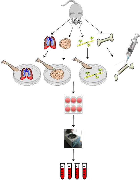
Figure 1. Overview Schematic of the Process of Organ Isolation and Generation of Conditioned Media. Tissues (lungs, brain, femur, tibia and lymph nodes) are harvested from the mouse. Lungs, brain and lymph nodes are minced with a scalpel. Bone marrow is harvested from the medullary cavity of the bones by pushing PBS through the cavity with a 27½ G needle. Lung and brain tissue fragments are incubated for a period of 24 hr before diluting with three equivalent volumes of fresh basal media and collected to make lung- and brain-conditioned media. Bone marrow and lymph node stromal cells are passaged 2 – 3 times before incubating with basal media to make bone and lymph node-conditioned media. Harvested media can be pooled and stored at -80 °C until ready for use in downstream assays such as protein arrays, cellular growth assays, cellular migration assays, tumorsphere formation, and RT-PCR. Please click here to view a larger version of this figure.
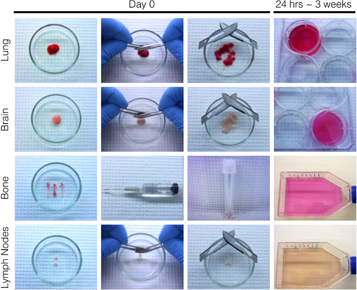
Figure 2. Representative Photographic Images of the Procedure for Organ Isolation and Generation of Conditioned Media. At Day 0, the lungs, brain, femur, tibia and lymph nodes are collected from the mouse. The lungs, brain and lymph nodes are placed in a 60 mm2 glass petri dish and minced with two scalpel blades to approximately 1 mm3 in size. The bone marrow is harvested from the medullary cavity of the bones by pushing PBS through the cavity with a 27 ½ G needle into a fresh tube of PBS. The lung and brain tissue fragments are incubated for a period of 24 hr at 37 °C and 5% CO2 before diluting with three equivalent volumes of fresh media and collected to make lung- and brain-conditioned media after 24 hr. The bone marrow and lymph node stromal cells are incubated at 37 °C and 5% CO2 and passaged 2 – 3 times (~ 3 weeks total) before replacing media with serum-free basal media to make bone and lymph node-conditioned media. Please click here to view a larger version of this figure.
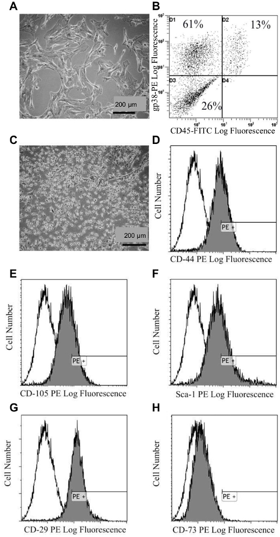
Figure 3. Characterization of Isolated Lymph Node and Bone Marrow Stromal Cells. (A) Bright-field microscopy image showing the morphology of lymph node stromal cells (LNSCs). (B) Representative flow cytometry analysis of LNSCs using a phycoerythrin (PE)-conjugated gp38 antibody and a fluorescein isothiocyanate (FITC)-conjugated CD45 antibody. (C) Bright field microscopy image showing the morphology of bone marrow stromal cells (BMSCs). (D-H) Representative flow cytometry analysis of BMSCs using PE conjugated (black profiles) antibodies against (D) CD44, (E) CD106, (F) Sca-1, (G) CD29, (H) CD73 antibodies relative to the isotype control (white profiles). A minimum of 10,000 viable events were collected per sample. This figure has been modified from 13. Please click here to view a larger version of this figure.
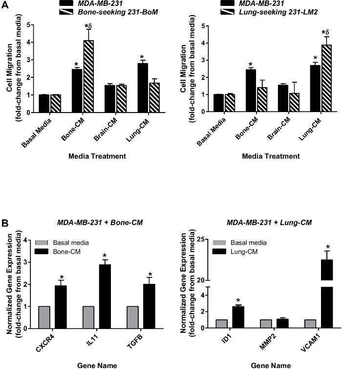
Figure 4. Organ-appropriate Migration Patterns and Gene Expression Changes in Response to Organ-conditioned Media. (A) Bone-seeking 231-BoM variants (left, striped bars) or lung-seeking 231-LM2 variants (right, striped bars) of the MDA-MB-231 human breast cancer cell line as well as parental MDA-MB-231 cells (both panels, solid bars) were plated onto gelatin-coated inserts (with 8 µm pores) prior to placement in basal media (DMEM:F12+ concentrated mitogen supplement) or organ-CM. Plates were incubated at 37 °C and 5% CO2 for 18 hr. (B) Whole population parental MDA-MB-231 cells were exposed to either bone-CM (left) or lung-CM (right) for 48h. RNA was isolated and quantified using RT-qPCR. Gene expression changes in response to organ-CM (black bars) were normalized to GAPDH and expressed relative to baseline expression in basal media (grey bars). Data are presented as mean ± SEM (N = 3; fold-change from respective negative control of basal media). * = significantly different from basal media; δ = significantly different from parental cell line treated with the same media conditions; P <0.05, ANOVA. Please click here to view a larger version of this figure.
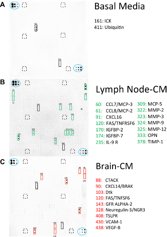
Figure 5. Organ-conditioned Media Contains Potential Soluble Mediators of Metastatic Behavior. A biotin label-based mouse antibody array was used to identify soluble factors present in lymph node-CM and brain-CM. Membranes were incubated with biotinylated samples of (A) basal media (DMEM:F12+ concentrated mitogen supplement), (B) lymph node-CM or (C) brain-CM at 4 oC O/N and visualized using chemiluminescence. Blue ovals indicate internal positive controls, lined boxes indicate internal negative controls and solid boxes indicate soluble factors of interest that are potentially involved in the metastatic behavior of breast cancer cells to the lymph node or brain. Please click here to view a larger version of this figure.
Metastasis is a complex process by which a series of cellular events are ultimately responsible for tissue invasion and distant tumor establishment4,30,31. The ex vivo model system presented here can be utilized to study two important aspects of metastatic progression: cancer cell homing or migration to a specific organ ("getting there") and growth in that organ ("growing there"). Many studies have previously focused on identifying key molecular characteristics associated with the cancer cells themselves that contribute to the metastasis process. For example, work done by Joan Massagué's group has identified distinct gene signatures associated with lung-specific, bone-specific, and brain-specific patterns of spread10-12,32. While this work provides important insights with regards to the contribution of cancer cells to this process, much less is known about the role of organ-specific factors contributed by the secondary microenvironment, which has remained relatively challenging to study13.
This ex vivo model system has previously been validated as an accurate representation of the secondary organ microenvironment by demonstrating that breast cancer cell line-specific migration and growth in organ-CM mirrors these cells lines' in vivo pattern of metastatic spread. A study by Chu and colleagues used four different human breast cancer cell lines with varying metastatic potential in vivo, including MDA-MB-231 and SUM-159 (highly metastatic, metastasize to the lymph node, lung, liver, bone and brain in vivo) and the SUM-149 and MDA-MB-468 (moderately metastatic, metastasize to the lymph node and lung in vivo) in. Both the MDA-MB-231 and SUM-159 cell lines displayed enhanced migration toward bone marrow-, lymph node- and lung-CM while the SUM-149 and MDA-MB-468 cell lines preferentially migrated toward lung-CM13. The current study demonstrates that bone- and lung-seeking MDA-MB-231 variants both prefer to migrate to bone marrow- and lung-CM respectively and that exposure to organ-CM can promote the expression of bone- and lung-associated metastatic genes previously identified by Massagué and colleagues11,12. Taken together, these results demonstrate that organ-CM provides an ex vivo platform representative of organ-specific soluble factors found in vivo that influence the phenotype and behavior of breast cancer cells.
Identification of soluble factors present within specific organs and their relative levels of expression provides a starting point for determining their potential influence on cancer cell behavior. Furthermore, using techniques such as immunodepletion can allow the user to effectively deplete a protein of interest from organ-CM to determine its effect on in vitro cellular behavior. For example, many soluble factors present within specific organ microenvironments function as chemoattractants and have the potential to induce cellular migration and proliferation. Previous work done using lung-CM has led to the identification of many soluble factors present in lung-CM that may act as chemoattractants for breast cancer cells, including osteopontin (OPN) and L-selectin (SELL)13. By immunodepleting these factors from lung-CM, Chu and colleagues were able to demonstrate reduced cellular migration and/or growth of highly metastatic MDA-MB-231 breast cancer cells13. These results show that conditioned media generated from whole organs provides a valuable resource for studying the effects of specific soluble factors on cancer cell behavior, through the use of traditional in vitro techniques.
Although this is a fairly straightforward model system, there are some steps in the experimental protocol that are critical for successful generation of organ-conditioned media. First, the use of mice between the defined ages of 6-12 weeks for harvesting of organs is important in order to control for age-related effects, either related to development (before 6 weeks) or related to aging in older mice. Secondly, sterility is of utmost importance throughout the protocol and can be best ensured through the use of rigorous aseptic technique and work in a sterile tissue culture hood, using sterilized tissue culture consumables and reagents as well as mild antibiotics in all culture media and supplements (i.e., penicillin/streptomycin). One of the challenges with using in vivo or ex vivo model systems is the inherent animal-to-animal variability. If this observed, troubleshooting steps include pooling of conditioned media obtained from multiple batches of mice before use in downstream experiments, as well as ensuring that lung- and brain-conditioned medias are accurately weight-normalized in each media collection experiment by resuspension in a 4:1 media:tissue ratio (volume/weight). In addition, the primary lymph node and bone marrow stromal cells should not be allowed to exceed 70% confluency at any given passage or be grown beyond 5 total passages, otherwise they will terminally differentiate.
Modifications to the protocol presented in this article could be envisioned to allow application to different fields of research. Although this model has so far been used exclusively for breast cancer research, it can potentially be used to study the role of organ-derived soluble factors in other types of cancer as well as additional normal physiological and disease states. In addition, while this method has been used here with immunocompromised nude mouse models, this technique is theoretically not limited to this species of mice and even could potentially be used with other types of animal models.
While this model provides important insight as to the contribution of specific organ-derived soluble factors on cancer cell behavior, it is not without its limitations. First this model focuses solely on the soluble component of organ microenvironments, which neglects the importance of the extracellular matrix (ECM). The ECM is the non-cellular component present within all tissue types and functions to provide both a physical scaffold for cells, as well as the ability to induce crucial biochemical and biomechanical cues required for various cellular processes, including morphogenesis, cellular differentiation and homeostasis33-35. Importantly, the physical and biochemical composition of the ECM is widely heterogeneous and can vary greatly between tissues types. Therefore, the tissue-specific composition of the ECM may also have an influence on organ tropism during cancer metastasis that may be overlooked with this model. The ECM may also act to concentrate specific soluble factors in a way that appropriately presents them to certain cells, and without this fine tuning of the ECM, the induction of cellular signaling may be diminished or altered36. Although the model mentioned in this paper lacks an appropriate ECM component, many studies could be strengthened by using a complimentary approach utilizing both the model mentioned here along with a suitable ECM component. For example, commercially available primary organ fibroblasts that synthesize endogenous ECM or recombinant ECM components could be used in addition to generated organ-CM to determine the full extent of the whole organ microenvironment on cellular behavior.
It is possible that the soluble microenvironment generated from organ-CM may not appropriately mimic what is present physiologically during the process of metastasis. Cancer cell survival, growth and progression are highly dependent upon cancer cell-host interactions4,30,31. Generation of whole organ-CM may result in the presence of soluble factors not normally expressed by cell types that interact with cancer cells during metastatic progression. Furthermore, it has previously been demonstrated that the presence of a primary tumor can modulate the microenvironment of distant metastatic sites, thereby "priming" them for metastasis37. Since the current protocol uses organs derived from healthy, non tumor-bearing mice, it would be worthwhile to compare the soluble factors derived from organs from mice bearing a primary breast tumor.
Due to the mechanical separation of whole organs during the generation of various organ-CM, intracellular factors may be released into the surrounding media which may not normally be encountered physiologically by cancer cells. Additionally, bone- and lymph node-derived conditioned media requires the culturing of stromal cells isolated from these organs prior to their collection. The cell types present during ex vivo culturing may not accurately represent the breadth of cellular types encountered physiologically by cancer cells. While stromal cells principally secrete many factors associated with various organ microenvironments, using these methods may not account for the influence of other cell types present within tissues and organs38. Finally, we included a mitogen supplement in the base media used to generate organ-conditioned media, because we found that this was required to maintain the viability of both the cultured organs (lung and brain) and the isolated BMSC and LNSC. However, we recognize the caveat that the presence of this mitogen supplement could have stimulated the organs to artificially produce soluble factors that they might not normally make,
In summary, many studies have indicated that specific organ microenvironments can profoundly influence tumor cell biology. Despite the discussed limitations, the ex vivo model system presented here represents a comprehensive technique for studying the influence of the soluble microenvironment of many solid organs on cellular behavior. In the laboratory of the authors, this model system has been and continues to be used as a means of studying many of the cellular events associated with metastasis, including invasion, migration, proliferation, stem-like behavior, and gene expression changes. The data gained from these studies could in turn inform the potential clinical application of this model system to primary breast cancer cells isolated from patient tumors in order to determine their response to various microenvironments representing potential metastatic sites. Taken together, it is hoped that further understanding of the interaction between the microenvironment and tumor cells during the process of metastatic organ tropism will lead to improvement of cancer treatment in the future.
The authors have nothing to disclose.
This work was supported by grants from the Canadian Breast Cancer Foundation-Ontario Region, the Canada Foundation for Innovation (No. 13199), and donor support from John and Donna Bristol through the London Health Sciences Foundation (to A.L.A.). Studentship and fellowship support were provided by the Ontario Graduate Scholarship program (Province of Ontario, to G.M.P. and J.E.C.), the Canada Graduate Scholarship-Master’s program (to M.M.P), the Canadian Institutes of Health Research (CIHR)-Strategic Training Program (to M.M.P., G.M.P and J.E.C.) and the Pamela Greenaway-Kohlmeier Translational Breast Cancer Research Unit at the London Regional Cancer Program (to M.M.P., G.M.P., J.E.C. and Y.X.). A.L.A. is supported by a CIHR New Investigator Award and an Early Researcher Award from the Ontario Ministry of Research and Innovation.
50 ml conical tubes | Thermo Scientific (Nunc) | 339652 | Keep sterile |
1X Phosphate-buffered saline | ThermoFisher Scientific | 10010-023 | Keep sterile |
Nude mice | Harlan Laboratories | Hsd:Athymic Nude-Foxn1nu | Use at 6-12 weeks of age |
Polystyrene foam pad | N/A | N/A | The discarded lid (~4 x 8 inches or larger) of a polystyrene foam shipping container can be used for this purpose. Sterilize by wiping with ethanol. |
Forceps | Fine Science Tools | 11050-10 | Keep sterile |
Scissors | Fine Science Tools | 14058-11 | Keep sterile |
Gauze pads | Fisher Scientific | 22-246069 | Keep sterile |
60 mm2 glass petri dishes | Sigma-Aldrich | CLS7016560 | Keep sterile |
Scalpel blades | Fisher Scientific | S95937A | Keep sterile |
DMEM:F12 | Life Technologies | 21331-020 | Warm in 37 °C water bath before use, keep sterile |
1 x Mito+ Serum Extender | BD Biosciences | 355006 | Referred to as "concentrated mitogen supplement" in the manuscript. Keep sterile |
Penicillin-Streptomycin (10,000 U/mL) | Life Technologies | 15140-122 | Keep sterile |
Rosewell Park Memorial Institute 1640 (RPMI 1640) | Life Technologies | 11875-093 | Warm in 37 °C water bath before use, keep sterile |
Fetal Bovine Serum | Sigma-Aldrich | F1051-500ML | Keep sterile |
Trypsin/EDTA solution | ThermoFisher Scientific | R-001-100 | Warm in 37 °C water bath before use, keep sterile |
6-well tissue culture plates | Thermo Scientific (Nunc) | 140675 | Keep sterile |
0.22 μm syringe filters | Sigma-Aldrich | Z359904 | Keep sterile |
T75 tissue culture flasks | Thermo Scientific (Nunc) | 178905 | Keep sterile |
Transwells | Sigma-Aldrich | CLS3464 | Keep sterile, use for migration assays |
Anti-mouse Sca-1 | R&D Systems | FAB1226P | use at 10 µl/106 cells |
Anti-mouse CD105 | R&D Systems | FAB1320P | use at 10 µl/106 cells |
Anti-mouse CD29 | R&D Systems | FAB2405P-025 | use at 10 µl/106 cells |
Anti-mouse CD73 | R&D Systems | FAB4488P | use at 10 µl/106 cells |
Anti-mouse CD44 | R&D Systems | MAB6127-SP | use at 0.25 µg/106 cells |
Anti-mouse CD45 | eBioscience | 11-0451-81 | use at 5 µl/106 cells |
Anti-mouse gp38 | eBioscience | 12-5381-80 | use at 10 µl/106 cells |
β-mercaptoethanol | Sigma-Aldrich | M6250 | Keep sterile |
Protein arrays | RayBiotech Inc. | AAM-BLM-1-2 | Use 1 array per media condition (including negative control), in triplicate |