MicroRNAs play an important regulatory role and are emerging as novel therapeutic targets for various human diseases. It has been shown that miRNAs are carried in high density lipoproteins. We have developed a simplified method to rapidly isolate purified HDL suitable for miRNA analysis from human plasma.
Small non-coding RNAs (miRNAs) have been implicated in a variety of human diseases including metabolic syndromes. They may be utilized as biomarkers for diagnosis and prognosis or may serve as targets for drug development, respectively. Recently it has been shown that miRNAs are carried in lipoproteins, particularly high density lipoproteins (HDL) and are delivered to recipient cells for uptake. This raises the possibility that miRNAs play a critical and pivotal role in cellular and organ function via regulation of gene expression as well as messenger for cell-cell communications and crosstalk between organs. Current methods for miRNA isolation from purified HDL are impractical when utilizing small samples on a large scale. This is largely due to the time consuming and laborious methods used for lipoprotein isolation. We have developed a simplified approach to rapidly isolate purified HDL suitable for miRNA analysis from plasma samples. This method should facilitate investigations into the role of miRNAs in health and disease and in particular provide new insights into the variety of biological functions, outside of the reverse cholesterol transport, that have been ascribed to HDL. Also, the miRNA species which are present in HDL can provide valuable information of clinical biomarkers for diagnosis of various diseases.
MicroRNAs are endogenous non-coding tiny RNA species that are highly conserved and are considered key players in the regulation of various biological processes by degrading or repressing specific target messenger RNAs1. Because miRNAs act intracellularly they have been explored as tissue-derived biomarkers which led to the discovery of tissue-specific functions of these miRNA. However, miRNAs are also found extracellularly either associated with proteins or in exosomes/micro vesicles that effectively can shield them from degradation by extracellular RNases2. More recent studies have shown that the protective effect of HDL may not be closely linked to its capability to promote cholesterol efflux but rather to its non-cholesterol cargo, in particularly as a circulating miRNAs carrier 3, 4. These miRNAs may not only modulate lipid metabolism but are also associated with anti-inflammatory, antioxidant and antithrombotic effects of the HDL-miRNA complex 5, 6.
To further explore the role of miRNAs carried in HDL particles, a simple and easy protocol needs to be established for miRNA extraction from isolated highly purified HDL for use in clinical routine. Numerous methods have been described to isolate HDL. These methods are either very time consuming or require large volume of plasma that may require sample pooling, extensive dialysis for desalting isolated lipoproteins and they do not completely remove exosomes as a source of miRNAs3, respectively. Here we describe a simple and rapid method that can isolate miRNA from highly purified HDL utilizing small volume of blood samples on a larger scale. We believe that this method may serve as good reference to promote research into the role of circulating miRNAs and in particular the role of HDL in facilitating communication between various cells and organs.
1. Collection of Blood Samples
2. Exosome Removal from Plasma
3. Density Gradient Ultracentrifugation (Figure 1).
4. Isolation of VLDL
5. Isolation of LDL
6. Isolation of HDL
7. Desalting and Concentration of Lipoprotein Fractions
8. Agarose Gel Electrophoresis
9. RNA Extraction and Purification
10. Reverse Transcription (RT-PCR)
11. Real-time PCR (qRT-PCR)
Isolation of High Density Lipoprotein After Removal of Exosomes
To obtain miRNA from highly purified HDL it is necessary to remove exosomes that represent a source of miRNA contamination7. This was done prior to density gradient ultracentrifugation with a commercially available kit. For practical purposes a three step standard density gradient ultracentrifugation protocol developed by commercial company was modified (Figure 1). This protocol requires centrifugation with a fixed-angle rotor at a speed of 140,000 rpm which is substantially faster than commonly used protocols with centrifugation forces up to 54,000 rpm3, 8, 9, and 10. Employing a widely available T-1270 rotor with a maximum force of 70,000 rpm, centrifugation was initially carried out with polyallomer tubes which have failed to resist the centrifugation forces. To avoid collapse of tubes, polycarbonate tubes were successfully used. Centrifugation time is critical for lipoprotein oxidation and potentially miRNA degradation11. Therefore several different centrifugation times, ranging from a total of 8 to 96 hr were tested. Furthermore temperature at which centrifugation was carried out was adjusted based on centrifugation time and force, respectively.
Purity of the High Density Lipoprotein Fractions
Purity of isolated high density lipoprotein fractions were checked with agarose gel electrophoresis. Figure 2. illustrates a typical electrophoresis result for the HDL subtraction isolated by gradient ultracentrifugation. It clearly showed that HDL was devoid of any contamination of VLDL and exhibits typical α-mobility. The absence of any α-migrating lipoproteins in the VLDL and LDL fractions demonstrates the complete recovery from HDL during the ultracentrifugation step. The HDL fraction, however, also showed some trace of β-mobility, a known electrophoretic banding pattern due to contamination with lipoproteins (Lp(a))12.
Elimination of Lp(a) from Isolated HDL
Studying miRNAs carried in HDL requires isolation of HDL of highest purity. Interference of Lp(a) with HDL potentially contributes to cross-contamination of HDL with miRNAs carried in LDL. To minimize this contamination of HDL, 15 μl of β-mercaptoethanol 10 was added to solution C during the last centrifugation step (Figure 1). Addition of β-mercaptoethanol has been shown not to change HDL density properties10. As displayed in Figure 3., addition of β-mercaptoethanol resulted in the absence of any b-migrating lipoproteins consistent with very effective removal of Lp (a).
Purification of miRNA from HDL
Isolation of miRNA was initially attempted employing lysis reagent which is commonly used for RNA extraction from blood. Although this method resulted in a very good RNA yield (91.45 ng/µl) but after spectrophotometric analysis showed unsatisfactory RNA purity. Additional purification of the isolated RNA by removing phenol improved the purity but the RNA yield was now significantly low. Next, another kit was tested which showed acceptable RNA purity (260/280 nm ratio of 1.7) but the RNA yield was 26-fold lower compared with the lysis reagent (3.45 vs. 91.45 ng/µl). The best results for acceptable RNA purity with a good yield were obtained with the miRNeasy serum/plasma kit (48.2 ng/µl; 260/280 nm ratio of 1.6) and therefore this extraction procedure was subsequently employed for the detection of miRNA from the isolated HDL lipoprotein fraction.
To demonstrate the feasibility to quantify miRNAs carried in human HDL purified with this method, we chose to amplify miR-223. This miRNA was chosen because miR-223 was identified in cargo of HDL3 and was shown to repress HDL cholesterol uptake5. The Figure 4A. shows a typical log plot of amplification curves comparing baseline threshold and threshold cycle values after optimization of the PCR reaction for the miR-223 and the spiked-in Cel-mir-39. This synthetic gene was used as internal control as there are no reports of known normalization control for miRNA in plasma. In addition, several potential established endogenous housekeeping genes like RNU6-2, RNU-48, HY3 could not be detected and SNORD95 could be detected in purified HDL, but, the difference in Ct values of miR-223 and SNORD95 is less than five (data not shown). Melting curve analysis illustrated in Figure 5. clearly shows a distinct single peak consistent with amplification of a selective miRNA in the preceding PCR. As illustrated in Figure 4B., both miR-223 and the reference Cel-mir-39 could consistently be detected in all six pro bands. Relatively small variations among all individuals were observed for Cel-mir-39 compared with miR-223. These findings support isolation of HDL at high purity to allow detection of its miRNA cargo.
Detection of miR-223 in Purified HDL
Real time quantitative PCR method was used to quantify double hairpin-structure miRNA precursors to single strand miRNA. This method needs a forward/reverse gene-specific primers and a thermostable reverse transcriptase to convert the hairpin structure of the miRNA to cDNA. The cDNA was subsequently amplified and quantified using real-time qPCR with the help of SYBR green detection. MicroRNA from serum, plasma and purified HDL plasma can be accurately profiled using the miRNA RT PCR system.
Our experimental product of mean miR-223 gene expression showed a Ct value of 30.9 in purified HDL and its corresponding negative controls were above cut-off 35 (NTC = 38.1 Ct value, NRT and NAC were not amplified). In this experiment, we saw in the purified HDL samples the formation of Primer-dimer with low melting temperature (73 °C in miR-223 and 75.5 °C in Cel-miR-39) and higher Ct value than the specific miRNA assays (Table 1). The Primer-dimers in Table 1. occur probably because of high concentration of primers in the solution. This occurs mostly when primer molecules attach to each other after 30 PCR cycles 13. This allows them to anneal to other primer molecules and in effect, become linear mini-templates and it will cause higher background and may lead to a generation of Ct value < 40 for NTC (No template control) samples.
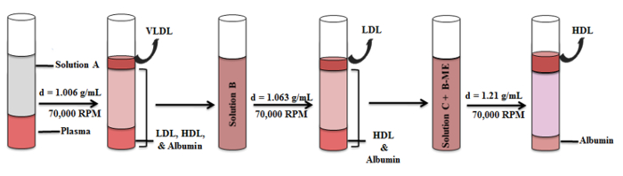
Figure 1. Schematic Representation of the HDL Isolation Procedure. HDL was prepared by density gradient ultracentrifugation in a series of three centrifugation steps. Distribution of lipoprotein bands and intermediate fractions in the density gradient are illustrated. Please click here to view a larger version of this figure.
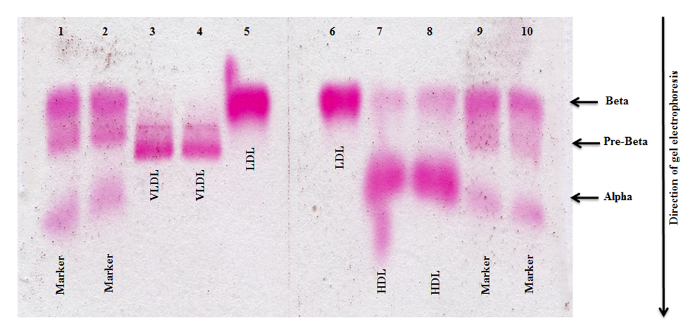
Figure 2. Agarose Gel Electrophoresis of Lipoproteins Isolated Without β-mercaptoethanol. VLDL, LDL and HDL fractions isolated by density gradient ultracentrifugation without adding β-mercaptoethanol to solution C demonstrate the presence of Lp (a) in the HDL fraction. Lanes 1, 2, 9, and 10 each represent the size marker; Lanes 3/4, 5/6 and 7/8 represent VLDL, LDL and HDL respectively. Please click here to view a larger version of this figure.
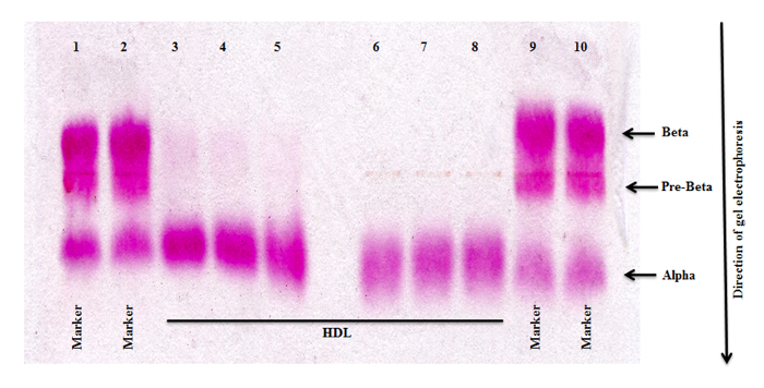
Figure 3. Agarose Gel Electrophoresis of HDL Isolated with β-mercaptoethanol. Adding β-mercaptoethanol to solution C before the last centrifugation step resulted in the removal of Lp(a) from the HDL fraction. Lanes 1, 2, 9 and 10 each represent the size marker; Lanes 3-8 represent HDL. Please click here to view a larger version of this figure.
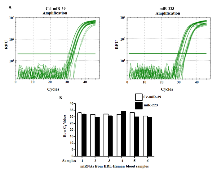
Figure 4. Amplification Plot of Two Different miRNAs and Expression of Cel-miR-39 and miR-223. (A) Real-time quantitative PCR employing isolated HDL was carried out as described. The PCR was run for 45 cycles and the point at which the curve intersects the threshold is the CT-Value. The data show the expression of Cel-miR-39 (left panel) and miR-223 (right panel), respectively. The horizontal line represents the detection threshold. A cycle number of 35 was set as cutoff for positive amplification. (B) Representative real-time PCR data from isolated HDL obtained from 6 samples are shown. Raw mean Ct values are shown for Cel-miR-39 and miR-223, whose expression levels vary less than 2 Ct value in purified HDL plasma fraction between 6 samples, each from a different donor. Expression profiling was performed using the PCR System and the Human Serum & Plasma miRNA qRT-PCR. Please click here to view a larger version of this figure.
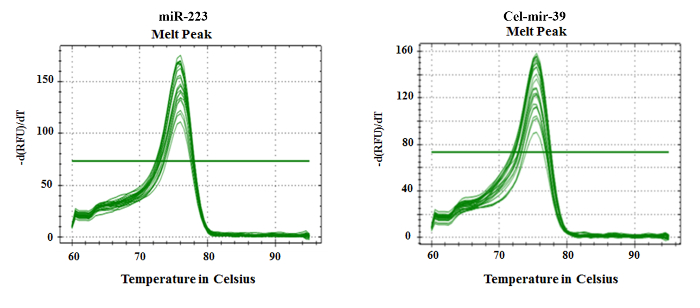
Figure 5. Melting Curves for Cel-miR-39 and miR-223. As illustrated, the presence of a single peak indicates specific amplification of Cel-miR-39 (right panel) and miR-223 (left panel), respectively. Please click here to view a larger version of this figure.
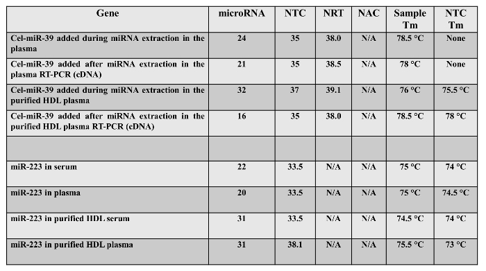
Table 1. Row Ct value of negative and positive control of synthetic Cel-miR-39 and miR-223 characterized in serum, plasma, cDNA, and purified HDL fraction. NTC (No template control), NRT (No reverse transcription enzyme), NAC (No amplification control, only water and reagents of qRT-PCR).
Identification of novel biomarkers from blood will aid in the clinical diagnosis and prognosis of various diseases. MicroRNAs have known to possess all the qualities of biomarkers and have been shown in various studies 14-17. In this study we have demonstrated rapid and simple easy method to isolate miRNA from plasma HDL. Conventional density gradient ultra-centrifugation method of isolation of VLDL, LDL and HDL depends on accurate sampling of plasma, precise preparation of the buffer solution, measurement of density and quantitative transfer of bottom lipoprotein fractions8. There are numerous methods that have been described to isolate HDL8 -11. These methods are often either very laborious or require large volume of plasma and extensive dialysis for desalting isolated lipoproteins and do not completely remove exosomes and lipoproteins as a source of miRNAs 3. To address these shortcomings we have established this simple and fast method of isolation of miRNA from plasma HDL.
The key goal and primary purpose of this study was to separate and isolate HDL-miRNA complex by ultracentrifugation without RBCs, buffy coat, exosomes, secretory vesicles, VLDL, LDL and Lipoproteins (a) interference, then isolate miRNAs from purified HDL. Contamination and interference of any of these components with HDL will alter the expected experimental results and miRNA profiling data. Isolation of DNA, RNA and miRNAs from blood is a very difficult task due to its complex nature. It has always been posed with lot of technical challenges to nucleic acids (including miRNAs) extraction and purification compared to cells, tissues and organ samples18. Also there are relatively less miRNA genes present in circulating blood cells. Blood is a known carrier for exosomes, secretory vesicles and HDLs along with free circulating miRNAs, which are relatively less18. Therefore, larger amount of blood plasma sample volume is required for processing and isolation of HDL-miRNA complex. In addition, the short nature of the miRNAs and their target sequences, makes it even difficult to achieve sufficient specificity with standard PCR oligonucleotide technologies. The cellular components of blood are always know to secrete miRNA and mRNA in response to change in external environment including temperature, microorganisms and toxins or stress. Also presence of nucleases, RNases, circulating proteins and other enzymes in blood will affect these miRNA isolation.
The circulating miRNA amplification also lacks a well-established known housekeeping genes like β-actin or GAPDH for data normalization while using the ΔΔCt method of qRT-PCR. This again poses one of the major hurdle for miRNA profiling from circulating blood serum or plasma19. Commonly used short non coding snoRNAs and snRNAs are rarely expressed in blood and this makes it further difficult in utilizing them as internal control genes. To solve these demerits of snoRNAs and snRNAs and to avoid any technical difficulties we used Serum/Plasma synthetic Spike-In control in the assays as internal control gene as followed according to the manufacturer protocol. It helps in normalization for any nonspecific amplification and changes occurred during miRNA purification and PCR reaction18. Even in the case of degradation of miRNA or absence of any specific miRNA amplification signals, the spike-in control should amplify in qRT-PCR reactions and give an expected signal with a reasonably constant and uniform Ct value. Based on the spike in control amplification along with three negative controls and positive controls we got good optimal data validation for miR-223 gene. This confirmed that we have got good yield of miR-223 from circulating plasma HDL from this simple method.
In conclusion, we have established a method of density gradient ultracentrifugation (DGUC) with the addition of β-mercaptoethanol for purification of miRNA from plasma HDL. The method has several advantages: (a) VLDL, LDL and HDL are separated within a short period of time by floor ultracentrifuge compare to other methods that needs 24 – 96 hr 8-11; (b) LDL and HDL dialysis, desalting/concentrating, take place within 1 hr compare with other methods that took 24 or more hours8,9; (c) Contamination of Lipoproteins is removed by β-mercaptoethanol in HDL fraction, but is not intended to be used in the LDL fraction; (d) The sample volume required is only 1 ml for all stepwise VLDL, LDL and HDL isolations compare with the other methods that needs more specimen volume; (e) It minimized oxidative damage to HDL during isolation11; (f) A maximum of 12 samples can be analyzed in a single lipoprotein separation analytical run; (g) The 250 µl sample volume of extracted HDL is enough to analyse miRNA concentration/purity, agarose gel electrophoresis, protein concentration, microarray and RT-qPCR procedures. The limitations of our method is that small percentage of HDL-miRNA may lose during the exosome precipitation step and the required plasma sample volume should be more than 200 µl to isolate HDL-miRNA.
Finally, the plasma microRNAs are not only derived from damaged or renegade blood cells in the circulation but also from healthy normal blood cells and cells from other parts of the body which includes various healthy and diseased tissues and organs affected by ongoing health status of the body 20. This present study has systematically purified mature miR-223 from isolated HDL of human plasma. This study clearly demonstrates that levels of mature miR-223 in the highly purified HDL plasma are detectable, stable, reproducible and consistent among individuals sample, thereby greatly facilitating clinical use of futurity tests for lipoproteins, liver, cardiovascular and other metabolic syndromes. Surprisingly, mature miRNAs, particularly miR-223 from HDL plasma are probably resistant to nuclease digestion and other harsh conditions which potentially explain the stability of miR-223 in purified HDL. The mechanism of resistance of miR-223 to RNases requires further study. Obviously, studying miRNA expression profiles in these purified HDL plasma would shed light on future miRNA markers in studying various maladies. These HDL associated microRNAs in the plasma can be used as a potential clinical diagnostic biomarkers and personalized medicine in the future.
The authors have nothing to disclose.
This work was supported, in whole or in part, by NIH Grants R01 AA 020758-04, U01DK 061731-13 and T32 DK 007150-38 to AJS and T32 DK 007150-38 to AA. This is original work and is not under consideration elsewhere for publication.
Plastic Vacutainer Lavender K2EDTA tubes | Becton, Dickinson and Company | 366643 | |
Centrifuge | Thermo Scientific, Sorvall Legend X1R | 75004261 | |
Densito 30PX densitometer | Mettler Toledo | MT51324450 | |
ExoQuick solution | Invitrogen | 4484451 | |
Polycarbonate thick-walled ultracentrifuge tube | Thermo Scientific | O3237 | |
Sorvall WX100 ultracentrifuge | Thermo Scientific | 46902 | |
Fat Red 7B | Sigma-Aldrich | 201618 | |
β-mercaptoethanol | Sigma-Aldrich | ||
Amicon Ultra-15 Centrifugal filter devices 10K | Millipore | UFC901008 | |
Amicon Ultra-centrifugal filter devices 3K | Millipore | UFC800308 | |
QuickGel Lipo kit | Helena Laboratories | 3344,3544T | |
Human lipoprotein standards for VLDL, LDL and HDL | LipoTrol; Helena Laboratories | 5069 | |
Rep Prep buffer | Helena Laboratories | 3100 | |
RNeasy MinElute spin columns | Qiagen | ||
NanoDrop 1000 analyzer | Thermo Scientific | ||
miScript II RT Kit | Qiagen | 218161 | |
CFX96 Touch real-time PCR detection system | BioRad | ||
miRNeasy Serum/Plasma Kit | QIAGEN | 217184 | |
miScript Primer Assays | QIAGEN | 141078139 | |
miScript SYBR Green PCR Kit | QIAGEN | 218073 | |
miRNeasy Serum/Plasma Spike-In Control | QIAGEN | 219610 |