This manuscript describes a soft lithography-based technique to engineer uniform arrays of three-dimensional (3D) epithelial tissues of defined geometry surrounded by extracellular matrix. This method is amenable to a wide variety of cell types and experimental contexts and allows for high-throughput screening of identical replicates.
The architecture of branched organs such as the lungs, kidneys, and mammary glands arises through the developmental process of branching morphogenesis, which is regulated by a variety of soluble and physical signals in the microenvironment. Described here is a method created to study the process of branching morphogenesis by forming engineered three-dimensional (3D) epithelial tissues of defined shape and size that are completely embedded within an extracellular matrix (ECM). This method enables the formation of arrays of identical tissues and enables the control of a variety of environmental factors, including tissue geometry, spacing, and ECM composition. This method can also be combined with widely used techniques such as traction force microscopy (TFM) to gain more information about the interactions between cells and their surrounding ECM. The protocol can be used to investigate a variety of cell and tissue processes beyond branching morphogenesis, including cancer invasion.
The development of branched epithelial tissues, known as branching morphogenesis, is regulated by cell-derived, physical, and environmental factors. In the mammary gland, branching morphogenesis is an iterative process through which guided collective cell migration creates a tree-like architecture. The first step is primary bud formation from the milk ducts, followed by branch initiation and elongation1,2. Invasion of branches into the surrounding stroma is induced by the systemic release of steroid hormones at puberty. New primary buds then initiate from the ends of existing branches, and this process continues to create an epithelial tree3. Although many important biochemical signals have been identified, a comprehensive understanding of the cell biological mechanisms that guide this complex developmental process is currently lacking. Moreover, mechanistic studies on the influences of specific cues are difficult to deconstruct from experiments in vivo, as precise spatiotemporal perturbations and measurements are often not possible.
Three-dimensional (3D) culture techniques, such as whole organ culture, primary organoids, and cell culture models, are useful tools for systematically investigating the mechanisms underlying tissue morphogenesis4-6. These can be particularly useful for determining the influences of specific factors individually, such as mechanical forces and biochemical signals, on a variety of cell behaviors, including migration, proliferation, and differentiation.6 Engineered cell culture models, in particular, readily enable the perturbation of individual cells and their microenvironment.
One such culture model uses a microfabrication-based approach to engineer model mammary epithelial tissues with controlled 3D structure that consistently and reproducibly form branches that migrate collectively when induced with the appropriate growth factors. The major advantage of the model is the ability to precisely manipulate and measure the effects of physical and biochemical factors, such as patterns of mechanical stress, with high statistical confidence. This technique, together with computational modeling, has already been used to determine the relative contributions of physical and biochemical signals in the guidance of the normal development of mammary epithelial tissues and other branched epithelia7-11. Presented here is a detailed protocol for building these model tissues, which can be readily extended to other types of cells and extracellular matrix (ECM) gels, and which serves as a potential tool for the testing of therapeutics.
1. Preparation of Solutions
2. Preparation of Elastomeric Stamps for 3D Micropatterning
Note: Elastomeric stamps are made with PDMS.
3. Preparation of 3D Epithelial Tissues
4. Immunofluorescence and Image Analysis
General schematic of mammary epithelial tissue microfabrication
A general schematic of the microfabrication procedure outlining the experimental work flow is shown in Figure 1. The end result is an array of epithelial tissues of identical geometry and spacing that are completely embedded within an ECM gel. A representative experiment uses EpH4 mouse mammary epithelial cells cultured in a gel of bovine type I collagen at a concentration of 4 mg/ml. To ensure the highest quality of engineered tissues, the techniques outlined in the protocol should be followed closely. Figures 2A and 2B show low and high magnification views of arrays of rectangular wells that have been molded into a type I collagen gel prior to cell seeding. The shape of the wells is determined by the shape of the features on the silicon master. It is important to lift the PDMS mold straight up from the collagen so as not to distort the cavity geometry. Figure 2C shows rectangular wells in a type I collagen gel that have been filled with mammary epithelial cells (excess cells have been washed off the surface of the collagen). In this example, each 200 μm x 50 μm rectangular well contains approximately 80-100 cells.
Addition of growth factors induces morphogenesis
24 hours after seeding, the tissues may be treated with growth factors such as HGF or EGF and cultured over several days to model branching morphogenesis. Typically, branches begin to form as early as 4 hours after growth factor stimulation. Figure 3A shows representative results from rectangular mammary epithelial tissues within a type I collagen gel 24 hours after the microfabrication procedure, after which cells have adhered to the collagen and to each other. No branches are observed prior to growth factor addition. Figure 3B shows a representative rectangular tissue that has undergone branching 24 hours after the addition of HGF at 10 ng/ml. In this case, branches occur at the ends of the tissues (as opposed to the middle), where the cells experience the highest mechanical stress9. Multiple tissues of identical initial geometry in the same gel can then be imaged to determine population averages of branch location and branch length, enabling high-throughput analysis.
Immunofluorescence staining to visualize protein localization
Immunofluorescence staining of the tissue arrays in the culture model allows us to determine protein localization within a tissue with high statistical confidence. Figure 4A shows representative results from a branching rectangular mammary epithelial tissue stained for focal adhesion kinase (FAK). Creating frequency maps of tissues of identical geometry can be used to visualize the average spatial localization of proteins of interest within the tissues, which can be compared to the localization of other proteins as well as branching activity. Figure 4B shows a frequency map of average FAK staining for 50 tissues showing FAK enrichment at the short ends of rectangular tissues, where branching typically occurs.
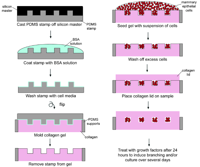
Figure 1. Schematic outlining the microfabrication procedure. Please click here to view a larger version of this figure.
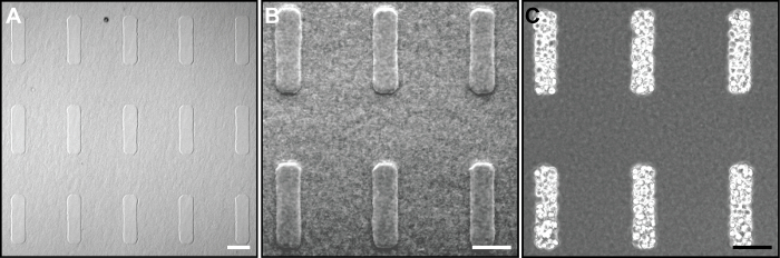
Figure 2. Images taken during the microfabrication process. (A) Low and (B) and high magnification phase-contrast images of rectangular cavities in type I collagen created using an elastomeric PDMS mold. (C) Cavities from (A) and (B) are filled with mammary epithelial cells. Scale bars, 100 μm. Please click here to view a larger version of this figure.
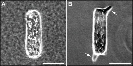
Figure 3. Microfabricated tissues undergo branching morphogenesis. (A) Phase-contrast image of a representative rectangular tissue 24 hr after microfabrication. (B) Phase-contrast image of a representative rectangular tissue that has started to undergo branching 24 hr after the addition of HGF at 10 ng/ml. White arrows indicate newly formed branches. Scale bars, 100 μm. Please click here to view a larger version of this figure.
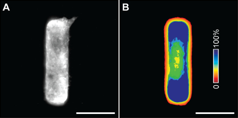
Figure 4. Immunofluorescence staining of microfabricated tissues. (A) Immunofluorescence staining for FAK in a mammary epithelial tissue after branch initiation. (B) A frequency map of average FAK staining in 50 tissues. Scale bars, 100 μm. Please click here to view a larger version of this figure.
The protocol described above outlines a method to produce identical epithelial tissues of pre-defined shape, enabling spatial control of the mechanical stress experienced by cells in the tissue. An elastomeric mold is used to create cavities in type I collagen that are then filled with epithelial cells and covered with an additional collagen layer such that cells are completely encapsulated in a 3D collagen matrix environment. Further culture of these tissues and treatment with growth factors to induce branching from the initial architecture make this system amenable for the study of branching morphogenesis of epithelial cells. There are several critical steps in the protocol. The first is lifting the PDMS molds straight upwards from the gelled collagen prior to cell seeding. Any horizontal movement during this step will distort the cavities, and they will no longer retain the desired geometry. Then, after the cavities have been seeded with cells, it is important to carefully wash off any excess cells that remain on the surface, as these may interfere with the behavior of the patterned tissues, particularly if they are very close to the tissues. Washing too rigorously, however, may result in cells being washed out of the cavities. Lastly, in the final micropatterning step when the cell-containing collagen gels are being immersed in culture medium, it is important to add the medium slowly and directly on top of the coverslip. Adding medium to the side of the dish away from the coverslip may result in detachment of the coverslip and the collagen lid. Each of these steps needs to be performed with care to achieve the best results.
Over the past few decades, several culture models have emerged for the study of epithelial morphogenesis, many of which involve the ex vivo culture of intact organs or explants of organs. Major advantages of culturing tissue taken directly from an animal are that tissue architecture remains intact and that cell-cell interactions are maintained. However, these models are not readily amenable to studies investigating the specific effects of a particular cell type or physical microenvironment. Additionally, organs and organ explants can be fragile and difficult to culture ex vivo. Others used primary mammary epithelial cells12, in which gene expression can be more readily manipulated, though this method does not conserve in vivo tissue architecture. Moreover, like intact organs, these cells can also be difficult to culture for long periods of time5. There is a need, therefore, for more robust culture models of epithelial morphogenesis that can be more precisely controlled and readily perturbed to gather reproducible data.
A widely used 3D cell culture technique is the formation of epithelial cell clusters embedded in collagen or other ECM proteins11,13. Disadvantages of this technique, however, include the inability to precisely control or predict the location of clusters within the gel, cluster size and shape, and branching sites. In addition, it is difficult to manipulate or measure the mechanical cues experienced by cells in different regions of the tissue clusters. Engineered tissues enable better control of these parameters.
The engineered tissue protocol described here is an easy to use, versatile, and reproducible 3D cell culture method that eliminates the heterogeneity commonly found in ex vivo culture systems and 3D clusters. Such engineered cell culture models provide a platform for more precise control of physical and biochemical signals, and results can be easily combined with computational models to predict the effects of perturbations or measure cellular forces using techniques such as traction force microscopy (TFM)9,10. A caveat of the model system, however, is that it does not completely replicate the cellular, chemical, and physical microenvironment within the mammary gland in vivo. Nonetheless, essentially all parts of the system are modifiable: including the cells used (in which gene expression can also be altered by transfection), tissue geometry, ECM composition, and other biochemical factors (ex. growth factors). Primary mammary epithelial cells can also be cultured in this system; they form a lumen and undergo branching morphogenesis from the same locations as immortalized cell lines7. Moreover, the model can be modified to incorporate other cell types to more appropriately represent the native mammary gland microenvironment14, or to investigate the underlying mechanisms that give rise to any branched organ.
The authors have nothing to disclose.
This work was supported in part by grants from the NIH (HL118532, HL120142, CA187692), the David & Lucile Packard Foundation, the Camille & Henry Dreyfus Foundation, and the Burroughs Welcome Fund. A.S.P. was supported in part by a Charlotte Elizabeth Procter Honorific Fellowship.
Polydimethylsiloxane (PDMS) | Ellsworth Adhesives | Sylgard 184 | |
PDMS curing agent | Ellsworth Adhesives | Sylgard 184 | |
Lithographically patterned silicon master | self-made | N/A | |
Plastic weigh boat | Fisher Scientific | 08-732-115 | |
100-mm-diameter Petri dishes | BioExpress | D-2550-2 | |
Ethyl Alcohol 200 Proof | Pharmco-Aaper | 111000200 | Make a 70% EtOH (v:v) solution by mixing with dH2O |
Razor blade | American Safety Razor | 620179 | |
1:1 Dulbecco’s Modified Eagle’s Medium : Ham’s F12 Nutrient Mixture (DMEM/F12) (1:1) | Hyclone | SH30023FS | |
Fetal Bovine Serum (FBS) | Atlanta Biologicals | S11150H | |
10x Hank’s balanced salt solution (HBSS) | Life Technologies | 14185-052 | |
Insulin | Sigma Aldrich | I6634-500MG | |
Gentamicin | Life Technologies | 15750-060 | |
10X Phosphate-buffered saline (PBS) | Fisher Scientific | BP399-500 | |
Sodium hydroxide (NaOH) | Sigma Aldrich | 221465-500G | |
Bovine type I collagen (non-pepsinized) | Koken | IAC-50 | |
Albumin from bovine serum (BSA) | Sigma Aldrich | A-7906 | |
Curved stainless steel tweezers | Dumont | 7 | |
35-mm-diameter tissue culture dishes | BioExpress | T-2881-6 | |
15 mL conical tubes | BioExpress | C-3394-2 | |
1.5 mL Eppendorf Safe-Lock Tube | USA Scientific | 1615-5500 | |
Circular #1 glass coverslips, 15-mm in diameter | Bellco Glass Inc. | Special order | |
0.05% 1X Trypsin-EDTA | Life Technologies | 25300-054 | |
Paraformaldehyde | VWR | 100503-916 | |
Triton X-100 | Perkin Elmer | N9300260 | Detergent |
HGF | Sigma Aldrich | H 9661 | Resuspended in dH2O at 50 mg/mL |
Rabbit anti-mouse FAK antibody | Life Technologies | AMO0672 | |
Goat anti-rabbit Alexa 488 antibody | Life Technologies | A-11034 | |
Adobe Photoshop | Adobe | N/A | Used for color-coding pixel frequency maps. |
FIJI (ImageJ) | NIH | N/A | Free image analysis software used for thresholding, registering, and overlaying images to create a pixel frequency map. The StackReg plugin was used for registering binary images. |