1. Chip Design and Fabrication
- Draw patterns to be ablated on PMMA substrates and double-side tapes using commercial software 24.
- To study the effects of chemical concentrations and shear stresses, draw a "Christmas tree" pattern with a varying width at its end in each of the five culture areas (Figure 1A and 1B).
- To study the effects of chemical concentrations and electric fields, draw a "Christmas tree" pattern with two more fluidic channels for salt bridges (Figure 2A and 2B).
- Scribe an individual pattern on a PMMA sheet or a double-side tape by loading the corresponding file into the CO2 laser scriber.
- Turn on the laser scriber, and check its connection to the computer. Open the pattern to be ablated using the commercial software.
- Place a PMMA sheet or a double-sided tape on top of the stage of the scriber. Adjust the focus of the CO2 laser if necessary using the calibration bar and the visible He-Ne laser.
- Load the pattern into the scriber for ablation on the PMMA sheet or the tape.
- Pick up the patterned sheet or tape, remove unwanted pieces, and clean the surface with nitrogen blowing.
NOTE: The thickness of the PMMA sheet is 1 mm, and that of the double-sided tape is 0.07 mm or 0.22 mm.
2. Chip Assembly
- With super glue, attach acrylic adaptors (length x width x height = 10 mm x 10 mm x 5 mm, with screw threads in the middle) to the top-most layer of the chip by aligning the screw threads and the holes on the top-most layer. These adaptors serve as medium inlets/outlets and salt bridge connectors.
- Assemble the microfluidic chip inside a laminar flow hood.
- Assemble the chemical-shear stress microfluidic chip (CSS chip).
- Attach 3 sheets of scribed PMMA sheets using 2 pieces of scribed double-sided tape. (Figure 1A and 1B).
- Add one more piece of the 0.07 mm thick double sided tape to the bottom of the chip and attach the chip to a 10 cm diameter Petri dish.
- Put the assembly chip in the vacuum chamber for overnight.
- To assemble the chemical-electric field chip (CEF chip), attach the PMMA sheet to a double-sided tape of thickness = 0.22 mm (Figure 2A and 2B).
- Attach the scribed PMMA sheet and double-sided tape to a 10 cm diameter Petri dish using this same piece of tape.
- Leave the assembled chip inside the hood and expose it to UV for 30 min for sterilization.
- Connect the inlets to two 3-ml syringes via plastic tubes and finger-tight nuts. Connect the outlet to a waste tube via a plastic tube and a finger-tight nut.
NOTE: Autoclave all tubes and nuts at 121 °C for 15 min prior to usage. - Slowly depress the plunger on the syringe to prime the fluidic channels with PBS. Push syringes back and forth to remove bubbles.
- Put the chips inside an incubator overnight at 37 °C under 5% CO2.
NOTE: These two steps are aimed to wash the chip and remove any remaining bubbles within the chip. - Take the chip out of the incubator.
- Prepare agar salt bridges for generating electric fields in the CEF chip.
- Dissolve 3% low melting point agarose in 1x phosphate buffered saline (PBS) buffer using a microwave.
- Inject solution-phased agarose into the salt bridge channel and insert the electrodes before solution solidifies.
- Place silver/silver-chloride electrodes into the tubular nuts.
- Connect the syringes to the syringe pumps, and continuously flow the culture medium (Dulbecco’s Modified Eagle’s medium, DMEM) into the fluidic channel for 10 min at a flow rate of 20 μl/min to replace PBS.
3. Cell Preparation and Experimental Setup
NOTE: Pre-warm 1x PBS, culture medium (DMEM plus FBS), and trypsin in a 37 °C water bath before usage.
- Plate 2 x 10⁵ lung cancer CL1-5 cells25,26 in a 10 cm Petri dish supplied with DMEM plus 10% fetal bovine serum (FBS). Incubate the cells inside an incubator under 5% CO2 at 37 °C until 90% confluence.
- Aspirate the medium and wash the cells once with pre-warmed 1x PBS. Add 2 ml of 0.05% trypsin buffer to the cells and wait for 2 to 3 min at 37 °C to detach the cells.
- Transfer the cells to a 15 ml sterile centrifugation tube and add 6 ml of culture medium into the tube. Gently invert the tube for mixing and take out 5 µl of the cell-containing medium for counting the cell number in a hemocytometer.
- Centrifuge the tube at 300 x g for 5 min. Suspend 106 cells in 1 ml of culture medium and place the cell-containing medium in a 1 ml syringe.
- Infuse the cell-containing medium into the microfluidic chip from the outlet and make sure that the solution distributes all through the five culture areas. Incubate the chip inside an incubator under 5% CO2 at 37 °C for 2 hr.
4. Experimental Setup
- Take the chip out of the incubator and place it on top of a transparent indium tin oxide (ITO) glass heater.
NOTE: The ITO glass is connected to a proportional-integral-derivative (PID) controller for maintaining the temperature at 37 ± 0.5 °C via feedback from a thermal coupler clamped tightly between the ITO heater and the chip. - Put the chip-heater assembly on top of a motorized XY stage of an inverted microscope for tracking cell migration or a fixed XY stage of an inverted fluorescent microscope for measuring the production of ROS.
- To generate different chemical concentrations, fill two syringes with 5 ml of chemical solutions of relative concentrations 0 and 1 (dissolved in culture medium) and pump them into the chip at desired flow rates: in the CSS chip, at a flow rate of 0.3 ml/min for 1 hr; in the CEF chip, at a flow rate of 20 µl/min for the first 20 min and a flow rate of 20 µl/hr for another 120 min (total time = 2 hr).
- In the CEF chip, for tracking cell migration, program the XY stage of the microscope to repeat taking photos, via a digital single-lens reflex (DSLR) camera, of certain field of views (FOVs) within culture areas every 15 min for 2 hr.
- For measuring the production of ROS, use the fluorescence-based indicator 2′-7′-dichlorodihydrofluoresce diacetate (2′-7′-DCFDA).
- Prepare the stock of 2′-7′-DCFDA at 10 mM in molecular biology grade dimethyl sulfoxide (DMSO). Dilute DCFDA in DMEM only without serum (5 µM in DMEM). The potential deacetylase could increase the background signals and decrease the signals in cells.
- After 1 hr of shear stress stimulus in the CSS chip or after 2 hr of EF stimulus in the CEF chip, pump 2′-7′-DCFDA (5 µM in DMEM) into the chip at a flow rate of 20 µl/min for the first 20 min and a flow rate of 20 µl/hr for another 20 min. For washing, pump DMEM into the chip at a flow rate of 20 µl/hr for 20 min.
- Take photos, via a charge-coupled device (CCD) camera, of certain FOVs within culture areas for analyzing the fluorescent intensities.
5. Calculations of Chemical Concentrations, Shear Stresses, and Electric Fields
- In both the CSS chip and the CEF chip, calculate the chemical concentrations in the five culture areas. For example, by injecting H2O2 of concentrations 0 and 200 µM from the two inlets, concentrations of 0, 25, 100, 175, and 200 µM are generated.
NOTE: By assuming that all liquids split-flow smoothly and equally around the fork, the relative concentrations in the five culture areas are 0, 1/8, 1/2, 7/8, and 1, respectively. - In the CSS chip, calculate the shear stress (τ) within each of the culture areas using
 27, where Q is the volume flow rate, η is the fluidic viscosity, h is the height of the channel, and w is the width of the channel.
NOTE: By setting Q = 0.3 ml/min in each inlet (Q = 0.12 ml/min in each culture area), η = 0.0008 Pa·s for culture medium, h = 1 mm, and w = 1 ~ 4 mm, the shear stress is calculated to range from 0.0048 Pa (4 mm wide region) to 0.0192 Pa (1 mm wide region). - In the CEF chip, calculate the EF strength within each of the culture areas using E = I/(σAeff) (Ohm's law), where I is the electric current flowing across the fluidic channel, σ is the electrical conductivity of the culture medium, and Aeff is the effective cross-sectional area of the channel.
NOTE: Using σ = 1.38 Ω-1m-1 for culture medium and Aeff = 0.22 mm2 (width = 1 mm and height = 0.22 mm), the EF strength is calculated to be E (mV/mm) = I (A) × 3.3 × 10⁶.- As shown in Figure 2D, treat the equivalent circuit as five C-section circuits with four (8 + 35 + 8) segments and one (5 + 35 + 5) segment.
NOTE: By analyzing this parallel circuit according to Kirchhoff's voltage law and Ohm's law, currents flowing across five culture areas, I1 through I5 from bottom to top, are calculated to be around 0.49I (area 1), 0.25I (area 2), 0.13I (area 3), 0.08I (area 4), and 0.05I (area 5), respectively, where I is the total direct current (dc). With an applied dc of 0.157 mA, EFs of 254, 130, 67, 41, and 26 mV/mm are generated within the five culture areas.
NOTE: For a simplified electrical analysis of the microfluidic network, all fluidic segments are considered as resistors with resistance proportional to their lengths.
- As shown in Figure 2D, treat the equivalent circuit as five C-section circuits with four (8 + 35 + 8) segments and one (5 + 35 + 5) segment.
6. Data Analysis
Note: Data analysis is performed using the ImageJ software.
- Analyze the Production of ROS.
- Run the ImageJ software. Go to "File" → Open to load a fluorescent image to be analyzed.
- Go to "Image" → "Type" → "16-bit" to change the image to a gray scale.
- Draw a polygon to enclose a cell. Go to "Analyze" → "Measure" to measure the mean fluorescent intensity of the cell.
- Repeat 6.1.3 to collect intensities from at least 50 cells of three independent experiments, and calculate the mean intensity with standard error of mean (SEM).
- Repeat 6.1.1-6.1.4 for each experimental condition.
- Analyze Cell Migration.
- Run the ImageJ software. Go to "File" → Open to load an image taken at time = 0 to be analyzed.
- Draw a polygon to enclose a cell. Go to "Analyze" → "Measure" to measure the center of mass of the cell as (x1, y1).
- Repeat 6.2.1- 6.2.2 to measure the center of mass of the same cell as (x2, y2) from another image take at time = t.
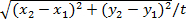
- Calculate the migration rate (in µm/hr) of this cell as
 .
. - Repeat 6.2.1-6.2.4 to collect migration rates from at least 50 cells of three independent experiments, and calculate the mean migration rate with standard error of mean (SEM).
- From 6.2.2 and 6.2.3, calculate the migration directedness of this cell as cosine θ or
 , where θ is the angle between the vector of the applied EF (from positive to negative) and the vector from the start to the end position of the cell (Figure 2G).
 - Repeat 6.2.6 to collect migration directedness from at least 50 cells of three independent experiments, and calculate the mean migration directedness with standard error of mean (SEM).
- Calculate the mean migration rate with SEM and the mean migration directedness with SEM for each experimental condition.
NOTE: A directedness of +1 indicates that all cells migrate toward the cathode, and a -1 value indicates that all cells migrate toward the anode. The directedness of a group of randomly moving cells is close to 0.
- Analyze Cell Alignment.
- Run the ImageJ software. Go to "File" → "Open" to load an image to be analyzed.
- Treat the cell as an ellipse and draw a line to indicate the long axis of a cell. Go to "Analyze" → "Measure" to measure the angle β between the line and the horizontal EF direction.
- Repeat 6.3.2 to collect β from at least 50 cells of three independent experiments, and calculate mean cosine β with standard error of mean (SEM).
- Repeat 6.3.1-6.3.3 for each experimental condition.
NOTE: A cosβ of +1 indicates that all cells align in parallel to the applied EF, and a 0 value indicates that all cells align perpendicularly to the applied EF.
The Chemical-shear Stress (CSS) Chip
The CSS chip is made of three PMMA sheets, each of thickness 1 mm, attached together via two double-sided tapes, each of thickness 0.07 mm (Figure 1A and 1B). The "Christmas tree" structure generates five relative concentrations of 0, 1/8, 1/2, 7/8, and 1 in the five culture areas. By designing the culture area as a triangle, a shear stress gradient, with a magnitude related the volume flow rate, the fluidic viscosity, and the dimension of the fluidic channel, is created within each of the areas. This chip is then attached, via another 0.07 mm thick double-sided tape, to a Petri dish for culturing lung cancer CL1-5 cells and the production of ROS was observed in response to different chemical concentrations and shear stresses. The production of ROS in lung cancer cells is highly related to lung cancer metastasis and development, and certain chemicals and shear stress were demonstrated to be involved in ROS generation 23.
First, to study the effect of H2O2, a chemical stimulus, on the production of ROS, cells were incubated with continuous flowing of H2O2 solutions at 0, 25, 100, 175, and 200 µM. As shown in Figure 1C, the fluorescent intensity increased as the concentration of H2O2 increased, indicating that H2O2 stimulated the production of ROS. Next, to investigate the effect of shear stress on the production of ROS, cells were exposed to a shear stress gradient of 0.0048 Pa to 0.0192 Pa. Figure 1D shows that the fluorescent intensity increased as the shear stress increased (the shear stresses were the highest and the lowest in the Front and Back areas, respectively), suggesting that higher shear stress induced more ROS production. Also, this CSS chip was used to study the production of ROS in response to different concentrations of α-tocopherol, an antioxidant of a form of vitamin E. Cells were stimulated by shear stress plus α-tocopherol of 0, 3.2, 12.5, 21.9, and 25 µg/ml. As shown in Figure 1E, for α-tocopherol concentrations lower than 21.9 µg/ml, the fluorescent intensity decreased as the concentration increased, indicating the effect of α-tocopherol in reducing the production of ROS. However, as the concentration increased to 25 µg/ml, the mean intensity also increased, suggesting that this high concentration of α-tocopherol did not eliminate much ROS compared to lower concentrations.
The Chemical-electric Field (CEF) Chip
The CEF chip is made of a 1 mm-thick PMMA and a 0.22-mm double-sided tape with fluidic channels patterned on it (Figure 2A and 2B). Similarly, the "Christmas tree" structure creates five relative concentrations of 0, 1/8, 1/2, 7/8, and 1 in the five culture areas. In addition, these five parallel areas are connected perpendicularly to form a fluidic path analogous to an electric circuit. The electric field generated within a culture area is related to the conductivity of the fluid, the cross-sectional area of the channel, and the direct current (dc) passing through the area. According to Kirchhoff's voltage law and Ohm's law, the currents in the five culture areas are 0.49I, 0.25I, 0.13I, 0.08I, and 0.05I, respectively, where I is the applied dc (Figure 2C). This chip is attached, via the 0.22 mm thick double-sided tape, to a Petri dish for culturing lung cancer CL1-5 cells to observe cell migration and the production of ROS in response to different chemical concentrations and electric fields.
First, this chip was used to study the production of ROS in response to different concentrations of honokiol, different strengths of EFs, and combined treatments of both. Honokiol, a small-molecule polyphenol isolated from the genus Magnolia, was found to have antiangiogenic, anti-inflammatory, and antitumor properties in preclinical studies 28. As shown in Figure 2D, the ROS produced was almost the same for EFs lower than 67 mV/mm, but was increased with increasing EFs above this value. Figure 2E shows that under combined treatments of electric fields and honokiol, the ROS level stayed almost the same, indicating that honokiol inhibited the production of exogenous ROS (i.e., ROS related to EF stimulus), especially under higher EFs. The cell migration under single or coexisting chemical/electrical stimuli was also investigated using this CEF chip. As seen in Figure 2F, without the addition of honokiol, the migration rate increased as the EF strength increased. After adding honokiol of different concentrations, the migration rate decreased in general, suggesting that honokiol reduced cell migration possibly via inhibiting the production of ROS. The migration directedness is shown in Figure 2G. In the presence of EF only, lung cancer cells showed prominent directional migration toward the anode (with negative values). After adding honokiol of different concentrations, there was a slight decrease in migration directedness for all EF-stimulated areas.
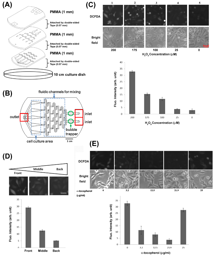
Figure 1: The Chemical-shear Stress (CSS) Chip Used to Study the Effects of Chemicals and Shear Stresses on Lung Cancer Cells.23
(A) Three layers of PMMA substrates are bound together via two double-sided tapes to form the CSS chip. (B) The integrated CSS chip. (C) Top: Fluorescent and bright-field images of CL 1-5 cells after being treated with different concentrations of H2O2. Scale bar = 50 µm. Bottom: Mean fluorescent intensity with SEM plotted at different concentrations of H2O2. (D) Top: Fluorescent images of CL 1-5 cells under different shear stresses. Scale bar = 50 µm. Bottom: Mean fluorescent intensity with SEM plotted at different shear stresses. (E) Top: Fluorescent and bright-field images of CL 1-5 cells after being stimulated with shear stress with different concentrations of α-tocopherol. Scale bar = 50 µm. Bottom: Mean fluorescent intensity with SEM plotted at different concentrations of α-tocopherol. Data presented here was originally published in reference 23. Please click here to view a larger version of this figure.
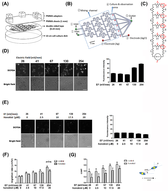
Figure 2: The Chemical-electric field (CEF) Chip Used to Study the Effects of Chemicals and Electric Fields on Lung Cancer Cells 22.
(A) One layer of PMMA is bound onto a culture dish via a double-sided tape to form the CEF chip. (B) The fluidic pattern of the CSS chip. (C) The equivalent electrical circuit of the microfluidic chip. (D) Left: Fluorescent and bright-field images of CL 1-5 cells after being treated with different strengths of EFs. Scale bar = 50 µm. Right: Mean fluorescent intensity with SEM plotted at different strengths of EFs. (E) Left: Fluorescent and bright-field images of CL 1-5 cells after being treated with combined honokiol and EFs. Scale bar = 50 µm. Right: Mean fluorescent intensity with SEM plotted at combined honokiol and EFs. (F) The migration rates of CL1-5 cells after being treated with EFs only (marked as control) and combined honokiol and EFs (marked as honokiol). (G) The migration directedness of CL1-5 cells after being treated with EFs only (marked as control) and combined honokiol and EFs (marked as honokiol). Data presented here was originally published in reference 22. Please click here to view a larger version of this figure.
