In these studies, we provide methodology for novel, neonatal, murine cardiac scaffolds for use in regenerative studies.
The only definitive therapy for end stage heart failure is orthotopic heart transplantation. Each year, it is estimated that more than 100,000 donor hearts are needed for cardiac transplantation procedures in the United States1-2. Due to the limited numbers of donors, only approximately 2,400 transplants are performed each year in the U.S.2. Numerous approaches, from cell therapy studies to implantation of mechanical assist devices, have been undertaken, either alone or in combination, in an attempt to coax the heart to repair itself or to rest the failing heart3. In spite of these efforts, ventricular assist devices are still largely used for the purpose of bridging to transplantation and the utility of cell therapies, while they hold some curative promise, is still limited to clinical trials. Additionally, direct xenotransplantation has been attempted but success has been limited due to immune rejection. Clearly, another strategy is required to produce additional organs for transplantation and, ideally, these organs would be autologous so as to avoid the complications associated with rejection and lifetime immunosuppression. Decellularization is a process of removing resident cells from tissues to expose the native extracellular matrix (ECM) or scaffold. Perfusion decellularization offers complete preservation of the three dimensional structure of the tissue, while leaving the bulk of the mechanical properties of the tissue intact4. These scaffolds can be utilized for repopulation with healthy cells to generate research models and, possibly, much needed organs for transplantation. We have exposed the scaffolds from neonatal mice (P3), known to retain remarkable cardiac regenerative capabilities,5-8 to detergent mediated decellularization and we repopulated these scaffolds with murine cardiac cells. These studies support the feasibility of engineering a neonatal heart construct. They further allow for the investigation as to whether the ECM of early postnatal hearts may harbor cues that will result in improved recellularization strategies.
Heart failure is common and deadly. It is a progressive disease that results in decreased contractility of the heart, which impairs blood flow to organs and leaves the metabolic demands of the body unmet. It is estimated that 5.7 million Americans have heart failure and it is the primary cause of hospitalization in the United States9. The collective cost of treating patients in heart failure in the United States exceeds $300 billion dollars per year 9-10. The only definitive therapy for end stage heart failure is orthotopic heart transplantation. Each year, it is estimated that more than 100,000 donor hearts are needed for cardiac transplantation procedures in the United States1-2. Due to the limited numbers of donors, only approximately 2,400 transplants are performed each year in the U.S.2. Clearly, this organ shortage needs to be addressed as other strategies are required to produce additional organs for transplantation and, ideally, these organs would be autologous so as to avoid the complications associated with rejection and lifetime immunosuppression.
Mammalian adult cardiomyocytes demonstrate a limited regenerative capacity upon injury but recent evidence suggests that mammalian neonatal hearts maintain a remarkable regenerative capacity following injury5-8. Specifically, following partial surgical resection, a regenerative window has been discovered between birth and postnatal day 7. This regenerative period is characterized by a lack of fibrotic scar, formation of neovascularization, release of angiogenic factors from the epicardium, and cardiomyocyte proliferation 5-8,11. This regenerative window of time provides the potential for using the neonatal heart as a novel source of material for the development of a bioartificial heart.
The extracellular matrix is known to provide important cues to promote cardiomyocyte proliferation and growth. Distinct differences in the availability of molecules in the neonatal and adult matrices12 and their ability to promote regeneration have been explored13. Decellularized adult matrices have been used in several studies to provide an ECM scaffold for cellular repopulation and the generation of a bioartificial heart. While these studies, and new discoveries in stem cell technologies, are advancing rapidly, several hurdles have yet to be met. For example, limitations in preserving native structure of the matrix, cellular integration into the matrix wall, and ability to support proliferation and growth all limit the success of this approach. While superior regenerative attributes have been ascribed to the neonatal heart, the practical aspects of using such a tissue have limited its exploration.
Based on the demonstrated regenerative capacity of the neonatal heart, we have developed novel matrices by developing a technique of decellularization for the P3 mouse heart. The P3 heart was chosen for these studies as it is within the window of cardiac regeneration as previously determined6 but the heart is large enough to harvest, decellularize and recellularize. The goal of this study is to demonstrate the feasibility of creating a matrix from a neonatal mouse heart. Our studies provide evidence for the feasibility of decellularizing a minute, neonatal heart while maintaining the structural and proteinaceous integrity of the ECM. We also demonstrate the ability to recellularize this cardiac ECM with mCherry expressing cardiomyocytes and we have examined these cardiomyocytes for expression of various cardiac markers following recellularization. This technology will allow for the testing of the superiority of a neonatal matrix for the development of a bioartificial heart.
All mouse experiments were performed in accordance with US Animal Welfare Act and were approved by the Institutional Animal Care and Use Committee at the University of Minnesota.
1. Method for Mouse Heart Isolation
2. Method for Decellularization by Langendorff Perfusion
3. DNA Determination
4. Fixation and Sectioning of Tissue
5. P1 Neonatal Murine Ventricular Cardiomyocytes for Recellularization
6. Bioreactor Recellularization of P3 Heart Matrix
Decellularization
On average, the time to decellularization of a P3 heart using this protocol is approximately 14 hr. given an average heart weight of 23 mg for the P3 neonate.
Acellularity
Figure 3a demonstrates a fully intact P3 neonatal heart (whole mount). Figure 3b shows the same heart following decellularization. Figures 4a and 4b show the hematoxylin and eosin staining of intact and decellularized hearts, respectively. Note the absence of hematoxylin positive nuclei and the diminution of eosinophilic structures in the decellularized heart. In addition, the DNA content of the decellularized heart is significantly reduced from 68.08 ± 2.25 µg in the intact heart (n=6) to 4.73 ± 2.27 µg in the decellularized heart (n=5).
Collagen Immunoreactivity
The maintenance of the extracellular matrix (ECM) following decellularization is essential to repopulation with exogenous cells and to the functionality of the matrix. To evaluate the content of the neonatal ECM in intact and decellularized ECM, immuno-staining for collagen IV was performed. Figure 4c and d demonstrate that collagen IV is robustly expressed in both the intact and decellularized heart and that the localization of this protein is maintained following cell removal, while DAPI positive nuclei are effectively removed (Figure 4e-h).
DNA Content
The presence of DNA is used as an additional indication of cellularity. In Figure 5, DNA is demonstrated to be decreased by 93% in neonatal hearts following detergent based decellularization. This degree of DNA reduction is consistent with reports in the literature using detergent based decellularization in other tissues15-16.
Recellularization
We have performed immunohistochemistry to determine the expression of various cardiomyocyte markers in the recellularized heart. Figure 6 illustrates cells that have migrated into the wall of the left ventricle (Figure 6A and 6B). Figure 6C demonstrates the DAPI labeling of the recellularized heart. Figure 6D-G illustrates cells that are positive for NKX 2.5, mCherry, α-actinin and DAPI, respectively. NKX2.5 is known to label cardiac progenitor cells, whereas α-actinin is a sarcomeric protein that marks differentiated cardiomyocytes. We have observed a majority of cells which express all of these markers (pink; Figure 6H), indicating that these cells continue to express cardiomyocyte markers even after 23 days of perfusion.
Figure 1. Schematic of decellularization hardware. A. 60 cc syringe barrel reservoir for detergent solution. B. Catheter assembly with irrigation syringe as detailed. C. Detail of the drawn PE tubing catheter tip. D. Decellularization chamber and septum with drain. Please click here to view a larger version of this figure.
Figure 2. Schematic representation of the heart bioreactor. 1. Humidification of carbogen gas (Green lines represent gas flow). 2. Peristaltic pump drive for media perfusion and oxygenation (Purple lines represent media flow through oxygenator). 3. Thin wall oxygenator. 4. Sheet oxygenator and media reservoir. 5. Preload chamber and bubble trap. 6. Heart chamber (Red lines represent media flow to and from the heart). Please click here to view a larger version of this figure.
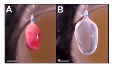
Figure 3. Whole mount of P3 neonatal mouse heart before (A) and after decellularization (B). This heart is decellularized using the Langendorff perfusion method as described in Method 2. Note that the heart becomes translucent and slightly enlarged following perfusion (B). Scale = 2mm. Please click here to view a larger version of this figure.
Figure 4. Histology of native and decellularized P3 neonatal mouse heart. Cryostat section (10 µm) were stained with H&E (A, B), Collagen IV (C, D, G, H) and DAPI (E, F, G, H). Merged images are represented in G and H. H&E staining shows an absence of cell nuclei and cytoplasm in decellularized tissue (B) when compared to the native heart (A). While the Collagen IV content remains following decellularization (D), DAPI staining (a marker of nuclei) is abolished. The merged images demonstrate the colocalization of Collagen IV and DAPI in the naïve heart (G) and the absence of this colocalization in the decellularized heart (H). These data indicate that cells no longer populate the collagen matrix of the heart. Scale = 500 µm. Please click here to view a larger version of this figure.
Figure 5. Evaluation of DNA content. Control (n=6) and decellularized (n=5) hearts assayed for DNA content by the pico-green method. Quantitation expressed as µg of DNA per heart ± standard deviation. Asterisk indicates p<0.01 compared to control. These data indicate that decellularization reduces DNA content significantly in the P3 neonatal heart. Please click here to view a larger version of this figure.
Figure 6. Histology of P3 heart matrix 23 days following recellularization with P1 mCherry expressing cardiomyocytes. Stained with H&E (A, scale = 250 µm, B, scale = 50 µm), DAPI (C, scale = 250 µm), NKX 2.5, mCherry, α-actinin, DAPI, and merged (D, E, F, G, H, scale = 50 µm). We have demonstrated the effective repopulation of the collagen matrix with P1 cardiomyocytes (A-C). Additionally, we observed that m-cherry positive cardiomyocytes express Nkx2.5 and α-actinin 23 days following introduction into the collagen matrix. These data indicate that these cells maintain their cardiomyocyte identity for extended periods of time. Please click here to view a larger version of this figure.
The dependence of this technique on repeated perfusions of the heart makes the avoidance of an embolism a critical component of a successful outcome. From the initial catheterization of the heart in Steps 2.2-2.6, to the changes of solution between Steps 2.8-2.14, there are manipulations that can allow introduction of air bubbles which compromise the flow of perfusate into the myocardium. Due to the diminutive size of the neonatal heart, even minute bubbles in the vasculature can cause a technical infarct, thus rendering the decellularization incomplete. Additionally, in the later wash steps, incomplete perfusion can result in detergent residue that negatively impacts biocompatibility. Moreover, rapid changes in temperature, such as when removing the matrix from 4 °C storage as suggested in Step 2.14, should be approached with care as this can also be a source of air bubble formation when dissolved gas moves out of solution with the change in temperature. When proceeding to recellularization, additional care should be applied, when preparing the syringe with the cell suspension, to ensure the infusion is bubble free (Step 6.4).
It may be necessary to manipulate the specifics of this protocol to accommodate other tissues types. There are a number of decellularization protocols reported4,9,11 which could provide guidance. The end goal(s) of any decellularization protocol should include the maintenance of the ECM protein structure and related biochemistry, and adequate removal of native cellular components as exemplified by residual DNA. In this setting, the application of excessive perfusion pressure leads to disruption of the ECM ultrastructure, which can be visualized histologically.
The size of the P3 mouse heart puts some constraints on cell administration compared to adult tissue. While adult hearts present a thick enough ventricular wall that makes transmural injection a possible cell delivery modality, the P3 heart is small enough that a needle, the size of which will not lyse a cell suspension, does substantial damage to the heart. Perfusion is a viable delivery strategy, but depends on cells of a certain size and shape to be delivered effectively. Neonatal murine myocytes work well in this regard. Other small circular cells have also been shown to serve this purpose11. The size scale of these hearts precludes the use of conventional physiologic functional analysis such as pressure volume catheters. Other approaches based on video capture may have to be considered.
These data support the feasibility of decellularization and recellularization of the neonatal mouse heart. Previous studies have demonstrated the use of adult hearts for the purpose of decellularization/recellularization studies. The matrices produced from these adult hearts have been repopulated with neonatal cardiomyocytes4 and human induced pluripotent stem cells (hiPSCs)17. In the case of the hiPSCs, the repopulated hearts displayed a decrease in pluripotency markers such as NANOG, SOX2, and OCT4 and formed muscle-like structures; all suggestive of maturation. The extracellular cues of the ECM, however, have been shown to play a critical role in the development of cells, tissues, and organs which prompted us to attempt to generate matrices from tissues known to harbor regenerative capacity. In our studies, we demonstrate that recellularized hearts still express cardiomyocyte markers, even after prolonged culture. These data indicate that, using novel matrices, cardiomyocytes can be maintained for extended periods of time without losing their identity as cardiomyocytes. Our data support the technique and feasibility for decellularizing neonatal mouse heart. The neonatal matrices hold the potential for providing novel constructs for repopulation studies as well as for the production of gels that have superior regenerative capacity. Using these neonatal ECMs, we are now addressing the superiority of these scaffolds to form functional tissues with a variety of cell types, including hiPSCs.
The authors have nothing to disclose.
The authors gratefully acknowledge Ms. Cynthia DeKay for the preparation of the figures.
1. Materials For Mouse Heart Isolation | |||
P1 mouse pups (as shown; B6;D2-Tg(Myh6*-mCherry)2Mik/J) | Jackson Laboratories | 21577 | or equivalent |
60 mm Culture dish | BD Falcon | 353004 | or equivalent |
Phosphate buffered saline pH 7.4 (sterile) | Hyclone | SH30256.01 | or equivalent |
Single Use Blade | Stanley | 28-510 | or equivalent |
Standard Scissors | Moria Bonn (Fine Science Tools) | 14381-43 | or equivalent |
Spring Scissors 10 cm | Fine Science Tools | 15024-10 | or equivalent |
Vannas Spring Scissors – 3mm Cutting Edge | Fine Science Tools | 15000-00 | or equivalent |
#5 Forceps | Dumnot (Fine Science Tools) | 11295-00 | or equivalent |
2. Materials For Decellularization | |||
Inlet adaptor | Chemglass | CG-1013 | autoclavable |
Septum | Chemglass | CG-3022-99 | autoclavable |
1/8 in. ID x 3/8 OD C-Flex tubing | Cole-Parmer | EW-06422-10 | autoclavable |
Male luer to 1/8" hose barb adaptor | McMaster-Carr | 51525K33 | autoclavable |
Female luer to 1/8" hose barb adaptor | McMaster-Carr | 51525K26 | autoclavable |
Prolene 7-0 surgical suture | Ethicon | 8648G | or equivalent |
Ring stand | Fisher Scientific | S47807 | or equivalent |
Clamp | Fisher Scientific | 05-769-6Q | or equivalent |
Clamp regular holder | Fisher Scientific | 05-754Q | or equivalent |
60 cc syringe barrel | Coviden | 1186000777T | or equivalent |
Beaker | Kimble | 14000250 | or equivalent |
22g x 1 Syringe Needle | BD | 305155 | or equivalent |
12 cc syringe | Coviden | 8881512878 | or equivalent |
3-way stop cock | Smith Medical | MX5311L | or equivalent |
22 x 1 g needle | BD | 305155 | or equivalent |
PE50 tubing | BD Clay Adams Intramedic | 427411 | Must be formable by heat. Polyethylene recommended |
1% SDS | Invitrogen | 15525-017 | Ultrapure grade recommended. Make up fresh solution and filter sterilize before use. |
1% Triton X-100 | Sigma-Aldrich | T8787 | Make up fresh solution from a 10% stock and filter sterilize before use. |
Sterile dH2O | Hyclone | SH30538.02 | Or MilliQ system purified water. |
1X Pen/Strep | Corning CellGro | 30-001-Cl | or equivalent |
3. Materials For DNA Quantitation | |||
Proteinase K | Fisher | BP1700 | >30U/mg activity |
KCl | Sigma-Aldrich | P9333 | or equivalent |
MgCl*6H2O | Mallinckrodt | 5958-04 | or equivalent |
Tween 20 | Sigma-Aldrich | P1379 | or equivalent |
Tris base/hydrochloride | Sigma-Aldrich | T1503/T5941 | or equivalent |
Pico-Green dsDNA assay kit | Life Technologies | P7589 | requires fluorimeter to read |
4. Method for fixation and sectioning of tissue. | |||
Paraformaldehyde | Sigma-Aldrich | P6148 | or equivalent |
Gelatin Type A from porcine skin | Sigma-Aldrich | G2500 | must be 300 bloom or greater |
5. Method for tissue histology | |||
Cryomolds 10 x 10 x 5mm | Tissue-Tek | 4565 | or equivalent |
Cryostat | Hacker/Bright | Model OTF | or equivalent |
Microscope Slides 25 x 75 x 1 mm | Fisher Scientific | 12-550-19 | or equivalent |
Hematoxylin 560 | Surgipath/Leica Selectech | 3801570 | or equivalent |
Ethanol | Decon Laboratories | 2701 | or equivalent |
Define | Surgipath/Leica Selectech | 3803590 | or equivalent |
Blue buffer | Surgipath/Leica Selectech | 3802915 | or equivalent |
Alcoholic Eosin Y 515 | Surgipath/Leica Selectech | 3801615 | or equivalent |
Formula 83 Xylene substitute | CBG Biotech | CH0104B | or equivalent |
Permount Mounting Medium | Fisher Chemical | SP15-500 | or equivalent |
Collagen IV Antibody | Rockland | 600-401-106.1 | or equivalent |
α-Actinin Antibody | Abcam | AB9465 | or equivalent |
mCherry Antibody | Abcam | AB205402 | or equivalent |
NKX2.5 Antibody | Santa Cruz Biotechnology | SC-8697 | or equivalent |
Donkey anti-mouse AF488 Antibody | Life Technology | A21202 | or equivalent |
Donkey anti-chicken AF594 Antibody | Jackson Immunoresearch | 703-585-155 | or equivalent |
Donkey anti-goat CY5 Antibody | Jackson Immunoresearch | 705-175-147 | or equivalent |
Fab Fragment Goat Anti-Rabbit IgG (H+L) AF594 | Jackson Immunoresearch | 111-587-003 | or equivalent |
Prolong Gold Antifade Mountant with DAPI | ThermoFisher | P36930 | or equivalent |
6. Isolation of neonatal ventricular cardiomyocytes using pre-plating. | |||
HBSS (Ca, Mg Free) | Hyclone | SH30031.02 | or equivalent |
HEPES (1M) | Corning CellGro | 25-060-Cl | or equivalent |
Cell Strainer | BD Falcon | 352340 | or equivalent |
50 mL tube | BD Falcon | 352070 | or equivalent |
Primeria 100 mm plates | Corning | 353803 | Primeria surface enhances fibroblast attachment promoting a higher myocyte purity |
Trypsin | Difco | 215240 | or equivalent |
DNase II | Sigma-Aldrich | D8764 | or equivalent |
DMEM (Delbecco's Minimal Essential Media) | Hyclone | SH30022.01 | or equivalent |
Vitamin B12 | Sigma-Aldrich | V6629 | or equivalent |
Fibronectin coated plates | BD Bioscience | 354501 | or equivalent |
Fetal bovine serum | Hyclone | SH30910.03 | or equivalent |
Heart bioreactor glassware | Radnoti Glass Technology | 120101BEZ | Must be sterilizable by autoclaving or gas. |