Novel isolation methods of primary endothelial cells from blood vessels are needed. This protocol describes a new technique that completely inverts blood vessels of interest, exposing only the endothelial side to enzymatic digestion. The resulting pure endothelial cell culture can be used to study cardiovascular diseases, disease modelling, and angiogenesis.
Cardiovascular disease is studied in both human and veterinary medicine. Endothelial cells have been used extensively as an in vitro model to study vasculogenesis, (tumor) angiogenesis, and atherosclerosis. The current standard for in vitro research on human endothelial cells (ECs) is the use of Human Umbilical Vein Endothelial Cells (HUVECs) and Human Umbilical Artery Endothelial Cells (HUAECs). For canine endothelial research, only one cell line (CnAOEC) is available, which is derived from canine aortic endothelium. Although currently not completely understood, there is a difference between ECs originating from either arteries or veins. For a more direct approach to in vitro functionality studies on ECs, we describe a new method for isolating Canine Primary Endothelial Cells (CaPECs) from a variety of vessels. This technique reduces the chance of contamination with fast-growing cells such as fibroblasts and smooth muscle cells, a problem that is common in standard isolation methods such as flushing the vessel with enzymatic solutions or mincing the vessel prior to digestion of the tissue containing all cells. The technique we describe was optimized for the canine model, but can easily be utilized in other species such as human.
Dogs are used as large animal model for cardiovascular disease research and can also suffer from inborn (genetic) vascular abnormalities1, 2. To study these diseases commercial endothelial cell lines are often used to assess endothelial cell (EC) functionality. For dogs there is one commercial endothelial cell line available (CnAOEC), derived from canine aorta. This cell line is mostly used in studies as control normal ECs3-5. In human cardiovascular research the most commonly used endothelial cell lines are Human Umbilical Vein Endothelial Cells (HUVECs) and Human Umbilical Artery Endothelial Cells (HUAECs) derived from human umbilical cord vein and artery, respectively. HUVECs have been used as the golden standard in vascular research since the 1980s6. They are considered to be the classic model system to study endothelial function and disease adaptation. Endothelial cells isolated from different blood vessels vary in appearance and functionality due to genetic background and exposure to the microenvironment7. In addition, HUVECs and HUAECs are derived from umbilical cord, a developmental vascular structure that might not fully mimic adult blood vessels with respect to the conditions they are exposed to and response to disease. Hence, translating results found in HUVECs and HUAECs to cardiovascular disease in general is inadequate.
When studying adaptation and behavior of adult ECs, primary ECs from the vessel of interest should be used as a more direct approach. To isolate these cells, several methods have been reported. A widely described method, which is also used for HUVECs, is flushing the vessel with an enzymatic digestion solution8. This often results in contamination with non-ECs such as smooth muscle cells and fibroblasts9. Another frequently used method for isolation is enzymatic digestion of minced vessel tissue followed by fluorescence-activated cell sorting (FACS) based on endothelial cell marker Cluster of Differentiation (CD)317, 8. FACS sorting and subsequent cell culture requires relatively large amounts of cells and is therefore not suitable for the isolation of endothelium from small blood vessels. We therefore aimed at developing a new robust method for isolating a pure endothelial cell population from various canine blood vessels with high purity. To test the efficiency of the new isolation method, we isolated and obtained pure Canine Primary Endothelial Cell (CaPEC) cultures from different canine arteries and veins, both large and small. This method also enables the culture of endothelial cells originating from diseased and/or aberrant vessels such as inborn intra- or extra-hepatic portosystemic shunts, a common disease in dogs2. The method allows the isolation of additional relevant cell types of the same vessel such as vascular smooth muscle cells since most of the vessel remains intact during the procedure.
Ethics statement: Blood vessels used in this study were harvested as surplus material obtained from fresh canine cadavers (n= 4) from healthy dogs euthanized for other unrelated research (University 3R policy). Aberrant blood vessels (intra- and extrahepatic portosystemic shunts, n= 1 each) were harvested post-mortem after informed consent of the owners from dogs presented to the University Clinic for Companion Animals of Utrecht University.
1. Isolation and Culture of Primary Canine Endothelial Cells
2. Characterization
Different blood vessels were successfully subjected to the described isolation protocol (Figure 2). It was possible to dissect and invert aorta, vena cava, vena porta, and coronary artery from healthy dogs (all vessels from each dog, n= 4). With the same approach ECs were isolated from two congenital portosystemic shunts (extrahepatic and intrahepatic, n= 1 each). Although aorta was easily inverted, thoracic aorta segments were more challenging than abdominal aorta. In thoracic segments the aorta has many intercostal arteries branching from it, which need to be ligated individually to ensure strictly endothelial exposure to the digestion solution. The dissected segment of caudal vena cava included the branching point of the renal veins, which needed to be ligated before inversion. For the portal vein the contributing branch of the vena gastroduodenalis was ligated before inversion of the blood vessel. The coronary artery in dogs is a much smaller blood vessel (approximately 1 – 2 mm diameter in a medium sized dog), from which we excised a segment of the circumflex branch. Because it has a small diameter, it proved to be easier to invert a rather short segment of 1 cm because the Mosquito forceps could not be inserted much further.
One day post isolation adhered cells were visible in the culture plate. In culture, CaPECs had a polygonal shape and displayed a tendency to grow in patches as shown in Figure 3. Numerous colonies of endothelial cells could be observed 3 – 6 days after isolation. After approximately 10 days in culture a confluency of 80% was reached and cells could be surpassed. On average the primary endothelial cultures could be maintained for a maximum of 8 passages (once weekly at a split rate of 1:4) at which point they stopped growing.
Isolated endothelial cells expressed endothelial cell marker CD31 as indicated by qPCR (Figure 4). The expression in endothelial cells derived from aorta, vena cava, and vena porta from four dogs was compared with a control culture of CnAOECs. The cultured primary cells had a comparable CD31 expression with the control ECs (Kruskall-Wallis, p=0.856).
CaPECs derived from aorta, vena cava and vena porta showed branching after 6 hr incubation on the angiogenesis slide as shown in Figure 5.
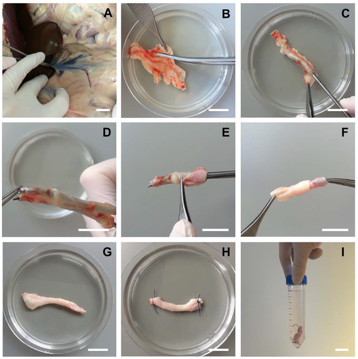
Figure 1: Dissecting and Inverting Blood Vessels for Endothelial Cell Isolation. A) The blood vessel of interest is aseptically removed from a fresh canine cadaver. B) Adherent tissue and/or fat surrounding the vessel should be removed carefully with surgical scissors without damaging the vessel itself (canine vena cava, abdominal segment of 5 cm). C) With a curved Halsted Mosquito forceps, the vessel can be entered without perforating the endothelial layer. D-G) Secure the forceps on the other end of the blood vessel and gently retract, thereby completely inverting the blood vessel. The endothelial layer is now on the outside of the vessel. H) Purse-string sutures are placed at the ends closing off the non-endothelial surface of the inverted vessel. I) The blood vessel is transferred to a 50 ml tube for washing and subsequent digestion. Scale bars indicate 2 cm. Please click here to view a larger version of this figure.
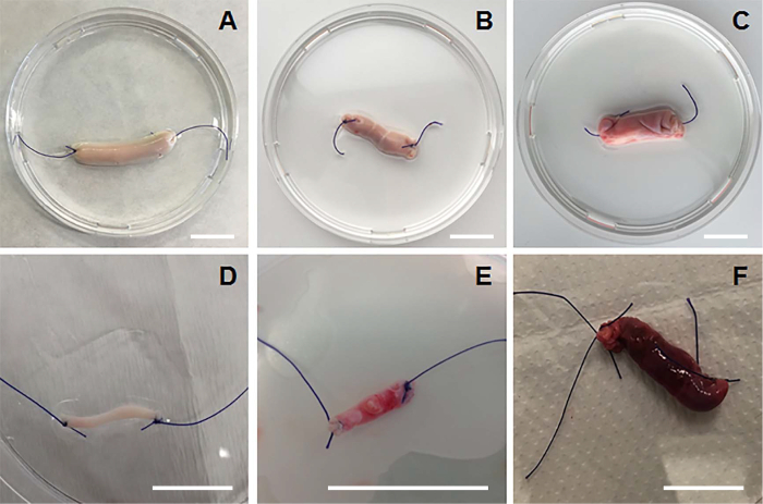
Figure 2: Inverted Vessels of Different Origin. A) An inverted aorta segment (healthy dog). B) An inverted vena porta (healthy dog). C) An inverted vena cava segment (healthy dog). D) An inverted coronary artery segment (healthy dog). E) An inverted extrahepatic portosystemic shunt derived from a Cairn terrier (age: 6 weeks old). F) An inverted intrahepatic portosystemic shunt derived from an Irish wolfhound (age: 8 weeks old). Scale bars indicate 2 cm. Please click here to view a larger version of this figure.
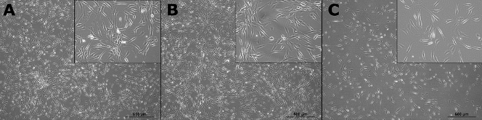
Figure 3: Cell Morphology. Pictures were taken two weeks after digestion of the vessels. A) ECs derived from canine aorta in passage 2. B) ECs derived from canine vena cava in passage 2. C) ECs derived from canine vena porta in passage 2. All pictures are taken with 4X original magnification. Scale bars indicate 500 µm. Insert shows 10X magnification. Please click here to view a larger version of this figure.
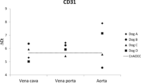
Figure 4: Gene Expression of CD31. Expression of CD31 in endothelial cells in passage 1 derived from aorta, vena cava, and vena porta (n= 4 dogs). No significant expression differences were observed between the CaPECs and the CnAOECs. Please click here to view a larger version of this figure.
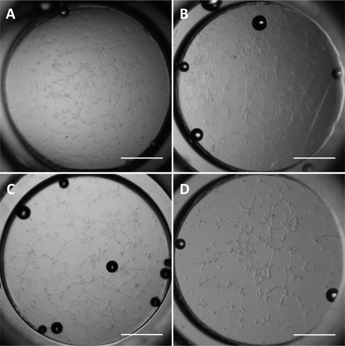
Figure 5: Angiogenesis. Photographs of CnAOECs and CaPECs from aorta, vena cava and vena porta after 6 hr incubation on an angiogenesis slide (20X magnification). Branch formation is visible after 6 hr in culture. A) CnAOECs. B) CaPECs derived from aorta. C) CaPECs derived from vena cava. D) CaPECs derived from vena porta. All cells were in passage 3. Scale bars indicate 1 mm. Please click here to view a larger version of this figure.
GOI | Direction | 5’-Sequence-3’ | Tm annealing | Product size (bp) | Genebank number | |
CD31 | Forward | GTTCTGCGTGTCAAGGTG | 59 °C | 85 | XM_005624261.1 | |
Reverse | TGTCCTTCCCAAACTCCA | |||||
beta-actin | Forward | GATATCGCTGCGCTTGTGGTC | 58 °C | 384 | NM_001195845 | |
Reverse | GGCTGGGGTGTTGAAAGTCTC | |||||
RPS19 | Forward | CCTTCCTCAAAAAGTCTGGG | 63 °C | 95 | XM_005616513 | |
Reverse | GTTCTCATCGTAGGGAGCAAG | |||||
B2MG | Forward | TCCTCATCCTCCTCGCT | 63 °C | 85 | AB745507 | |
Reverse | TTCTCTGCTGGGTGTCG |
Table 1: qPCR Primer Sets. qPCR primers for canine reference genes and CD31.
In studies focusing on canine ECs the CnAOEC primary line is used to model the endothelial lineages of the dog3, 12, 13. In human studies, the HUVEC culture is still considered the gold standard. Clearly, focusing merely on ECs derived from umbilical cord is a firm restriction in cardiovascular research. Endothelial cells have a specific gene expression pattern determining arteriovenous specification. In order to account for these differences in postnatal vessels we present this novel isolation method based on the specific anatomical location of the endothelial cells. The methods commonly used for primary EC isolation are flushing the vessel with an enzyme solution or mincing the vessel prior to digestion, two methods that both have a risk of contamination with non-ECs. We established a technique for the isolation of Canine Primary Endothelial Cells (CaPECs) with a lesser chance of contamination that can also be used on small vessels. This isolation method of endothelial cells is based on the inversion of the vessel, which avoids digestion of all other vessel cell types. It is important to remove the vessels aseptically, as illustrated in Figure 1A, to prevent bacterial or fungal contamination of the endothelial cultures. In case of bacterial growth an additional antimicrobial agent (e.g., gentamycin) can be added to the culture medium. In case of fungal infection treatment is often not successful in our experience.
In order for the inversion to succeed, it is critical to obtain a vessel that is approximately 5 cm in length. A second important step to facilitate inversion of the blood vessel is the removal of any adherent tissue and fat with surgical scissors. Inverting the vessel is done carefully by clamping the forceps at the opposite end and slowly inverting the vessel. When placing the purse-string sutures make sure to touch the endothelial layer as little as possible. Damage of the endothelium can result in poor yield of viable endothelial cells and access of the digestion media to underlying tissue. This can also happen if the vessel is not closed completely. The vessel can easily be handled by picking it up at the end of a ligature.
In specific cases a modification of the technique can be applied. In blood vessels with a small diameter it is sometimes impossible to insert a Mosquito forceps in order to invert the vessel. A solution is to place stay sutures on one end of the vessel and push their ligatures through the blood vessel using the forceps. At the other end they can then be secured with a Mosquito forceps and pulled, thereby inverting the vessel. Sometimes the vessel will tear as a consequence of the extra sutures, so this modification is not the preferred approach. Another problem that can arise is when many erythrocytes are present in the culture plates upon seeding of the primary cells. This can be the result of insufficient washing of the blood vessel prior to digestion. When this occurs, washing the wells on day 2 with warm HBSS is often sufficient to remove the majority of the erythrocytes. In any case, the erythrocytes will not persist in the culture and are lost upon passaging of the CaPECs.
The isolated CaPECs expressed CD31, indicating that the population of cells that was digested from the blood vessel is indeed of endothelial origin. As recently published, this marker is also expressed in endothelial cells from the canine mitral valve14. The formation of colonies in culture indicates the cultures start from either single cells or from small cell clusters. Rapid growth on endothelial specific media is also indicative of correct cell type. The primary cells can be cultured for eight passages, after which the cultures cease expansion. This is an indication of senescence and makes it less likely that stem/progenitor cells are cultured with this protocol. In an angiogenesis assay, CaPECs from aorta, vena cava and vena porta showed branching within 6 hr. This endothelial functionality provides solid evidence for their origin.
Based on availability only canine material was studied, but the isolation method has possible applications for isolation of human primary endothelial cells or endothelial cells from other organisms. The technique is also possible for very small vessels (1 cm length), but will yield less isolated cells. For this reason CaPECs obtained from small vessels will require an extra passage before they have reached a sufficient number for experiments.
The ability to perform the isolation method on small vessels is a great advantage for studies in vascular disease. ECs could be isolated from diseased or aberrant vessels like portosystemic shunts. These are vessels connecting the portal vein and the systemic circulation which causes blood to bypass the liver2. The shunt itself can be as small as 1 cm. Comparing the ECs from a shunt vessel with cells originating from a healthy caval vein and portal vein could give new information relevant for the shunt-development since the pathogenesis is still not completely understood. In addition, new angiogenic genes could be investigated on a functional level by performing genetic modifications such as siRNA mediated gene silencing. In conclusion, this novel isolation method can be a powerful model to study (congenital) cardiovascular diseases, disease modelling, and (tumor) angiogenesis15, 16.
The authors have nothing to disclose.
The authors would like to acknowledge Hans de Graaf and Tomas Veenendaal for their technical assistance in culturing the ECs.
Collagenase type II | Life Technologies | 17101-015 | |
Dispase | Life Technologies | 17105-041 | |
DMEM (1X) + GlutaMAX | Life Technologies | 31966-021 | |
Hank's Balanced Salt Solution | Life Technologies | 14025-050 | |
Canine Endothelial Cells Growth Medium | Cell Applications | Cn211-500 | |
CnAOECs | Cell Applications | Cn304-05 | |
Fetal Calf Serum (FCS) | GE Healthcare | 16000-044 | |
TrypLE Express | Life Technologies | 12604-013 | |
SPR | Bio-Rad | 170-8898 | |
iScript synthesis kit | Bio-Rad | 170-8891 | |
SYBR green super mix | Bio-Rad | 170-8886 | |
Recovery Cell Freezing Medium | Gibco/Life Technologies | 12648-010 | Keep on ice prior to use |
Freezing container, Nalgene Mr. Frosty | Sigma-Aldrich | C1562 | |
Gelatin | Sigma-Aldrich | G1890 | |
Surgical scissors (Mayo or Metzenbaum) | B. Braun Medical | BC555R | |
Mosquito forceps | B. Braun Medical | FB440R | |
Mosquito forceps curved | B. Braun Medical | FB441R | |
polyglactin 3-0 | Ethicon | VCP311H | |
Trypan blue | Bio-Rad | 145-0013 | |
Automated counting chamber | Bio-Rad | 145-0102 | |
Counting Slides, Dual Chamber | Bio-Rad | 145-0011 | |
Matrigel | BD Biosciences | BD356231 | Slowly thaw on ice |
µ-Slide Angiogenesis | Ibidi | 81501 | |
Endothelial Growth Medium | Lonza | CC-3156 | |
EGM-2 SingleQuot Kit | Lonza | CC-4176 |