1. Producing a Microchannel Chamber to Create a Concentration Gradient
- Cutting an acrylic mold piece
- Obtain a piece of acrylic of the desired width. Typically use 1/16-inch-thick acrylic sheets. Thicker sheets are difficult to cut well and very thin sheets do not have the requisite mechanical strength, causing them to break or warp during the process.
- Create a CAD file with the desired shape of the acrylic piece to produce a cavity within the polydimethylsiloxane (PDMS). Adjust the channel width and length to achieve the desired gradient relevant to individual experimental protocols.
- Here, use an acrylic piece of the following dimensions: two squares (0.254 cm x 0.254 cm) connected by a 0.127 cm wide channel (0.254 cm long) (Figure 1).
- Import the CAD file into the laser cutting apparatus and place the acrylic piece in a laser cutter according to the manufacturer's protocol.
NOTE: Alternatively, 3D-print the piece from the CAD design. The advantage of this method is that alternate materials can be used for the mold; however, the resolution and reproducibility can be decreased with 3D printing compared to laser cutting.
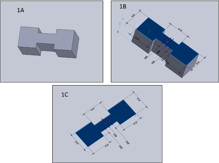
Figure 1: Computer-Aided Design (CAD) Representation of Microchannel Chamber. This image depicts the CAD drawing of the acrylic insert needed to create the microchannel chamber. 1A) Top view of CAD drawing, 1B) Side view of CAD drawing with dimensions in inches, 1C) Top view of CAD drawing with dimensions in inches Please click here to view a larger version of this figure.
- Pouring the polydimethylsiloxane (PDMS)
- In a weigh boat, mix 10 parts by weight of commercial Elastomer Base (e.g. Sylgard 184) with 1 part by weight of commercial Elastomer Curing Agent (e.g. Sylgard 194). Typically, mix 10 g of Elastomer Base to 1 g of Elastomer Curing Agent.
- Stir to combine the base and curing agent with a micropipette for 5 min.
- Set up a vacuum bell and place the weigh boat with PDMS in the vacuum for 30 min to remove trapped air bubbles.
- Turn off the vacuum and remove the weigh boat. Pour the PDMS on top of the acrylic cut-out in a small Petri dish. Make sure that the PDMS completely covers the insert.
- Allow the PDMS to cure overnight (at least 18 h) at room temperature.
- Removing the insert from PDMS
- After curing the PDMS, remove the insert by carefully cutting the PDMS around the acrylic piece with a scalpel.
2. Plating the Cells in a Microchannel Chamber
- Sterilizing the chamber for plating cells.
NOTE: While PDMS can be sterilized using ethylene oxide or other techniques, a quick UV treatment is fast, inexpensive, and sufficient for cell culture studies. The short UV treatment duration did not impair the structure or mechanical integrity of the PDMS piece.- In a standard cell culture cabinet, completely cover the chamber with 70% ethanol.
- Put the Petri dish in laminar flow hood with the lids off.
- Shut the hood and turn on the UV light. Expose to UV light for 1-2 h.
- Open the laminar flow hood and wait 15 min to reestablish flow.
- Remove the 70% ethanol.
- Wash the chambers twice with sterile phosphate buffered saline (PBS). Use 1 mL of PBS for each wash. Remove PBS and allow chambers to dry overnight with the lids off.
NOTE: Alternatively, instead of using a laminar flow hood, use a UV chamber box for sterilization if one is available. Make a UV box by placing a UV light source in the back of a cardboard box lined with aluminum foil. The aluminum foil reflects the light to allow for even illumination of the sample.
- Plating Cells in Chamber
- Count cells using Trypan blue and a hemocytometer.
- Add 100 µL of the cell suspension to 400 µL of 0.4% Trypan blue and mix gently. Apply 100 µL of this solution to the hemocytometer by gently filling both chambers under the glass coverslip.
- Use a microscope with a 10X objective to focus on the grid on the hemocytometer. Use a hand tally counter to count the live unstained cells within one set of 16 squares on the hemocytometer.
- Count cells in all 4 sets of 16 squares. Take the average cell count from the 4 sets of 16 squares and multiply by 5×10⁴. This final number is the number of cells/mL in the cell suspension.
- Add 2,500 cells to one well in each chamber. This translates to a near-confluent cell density of ~30,000/cm2.
- Allow cells to sit in the chamber for 60 min before adding media (Dulbecco's Modified Eagle's Medium, 10% fetal bovine serum, 1% penicillin-streptomycin).
- Count cells using Trypan blue and a hemocytometer.
3. Dextran Soaked Agarose Blocks
- Creating an agarose block
- Pour 3% agarose into the 3D printed mold (2 mm x 2 mm x 2 mm).
- Allow the agarose to solidify in a vacuum chamber for 30 min to remove any bubbles.
- Remove the agarose block from the mold.
NOTE: Alternatively, pour 3% agarose into small Petri dish at a thickness of 2 mm. Cut a 2 mm x 2 mm x 2 mm block using a scalpel or other cutting tool. This is not as precise as the mold system but it can be a little easier to do.
- Completely submerge the agarose block in a solution of the desired concentration of the chemotactic factor. To assess the concentration gradient formation, flush the chamber first with fluorescent dextran. The concentration and molecular weight of dextran should match that of a soluble factor or drug of interest.
- Soak the agarose block overnight.
4. Time-lapse of the Dextran Diffusion to Assess the Soluble Factor Concentration Gradient
- Place the dextran soaked agarose block in a small square well of the microchannel chamber.
- Place the microchannel chamber in a cell imaging system or use another fluorescent microscope with a time-lapse capability. Begin the time-lapse according to manufacturer's instructions.
NOTE: Results shown are taken with GFP filter, 50% light, 4/10 pH, and 4X magnification.
5. Time-lapse of Cell Growth
- Plate cells in the microchannel chamber at one end of the chamber. Here, use 3T3 fibroblasts. Add 2,500 cells to one well in the microchannel chamber. Count cells as described in 2.2.1.
- Allow cells to sit in the chamber for 60 min before adding media (Dulbecco's Modified Eagle's Medium, 10% fetal bovine serum, 1% penicillin-streptomycin). Keep the microchannel chamber with plated cells at 37 °C in 5% CO2. Cell migration through the channel can be viewed with a microscope.
- Add a marker or use a microscopic defect in the chamber as a marker in order to serve as a starting place to quantify the distance the cell front moves.
- Establish a gradient by placing an agarose gel (8 mm3 block) containing the concentrated growth factor, chemo-attractant, drug, or other factor being tested (here, 40% FBS is being used as an example) at one end of the microchannel chamber.
- Take time-lapse images of the moving cell front. Here, the results show images of the cell front that were taken every 12 h using an imaging system.
- Use pictures of the cell front to quantify cell growth rate. Calculate the growth rate by first using the MATLAB polyfit function (roipoly) to create a polygonal mask defined by the cell front, the channel walls, and the reference marker. The dimensions of this mask yield the migration distance of the front from which average cell front velocity is calculated. Other studies have utilized a similar method8.
NOTE: Compare the edge of the cell growth front on each frame to the concentration of the soluble factor at that same distance. The soluble factor concentration gradient is calculated using a 1D diffusion model approximation. - Take fluorescent images of the cells using Phalloidin and DAPI staining.
- Fix cells before staining with Phalloidin.
- Warm 4% paraformaldehyde at 37 °C.
- Add enough 4% paraformaldehyde to cover cells. Keep cells in the paraformaldehyde for 10 min.
- Rinse twice with PBS for 15 min each time. Use enough PBS to cover the cells.
- Remove PBS from cells and add enough permeabilizing solution (0.1% Triton X-100 in PBS) to cover cells. Leave solution for 10 min.
- Wash twice with PBS for 5 min each time. Use enough PBS to cover the cells.
- Remove PBS from cells and add blocking solution (1% bovine serum albumin in PBS) for 20 min.
- Remove blocking solution and wash 2 times with PBS for 5 min each time. Use enough PBS to cover the cells.
- From this point, protect cells from light with aluminum foil when not being used. Remove PBS from cells and add Phalloidin 488 diluted 1:50 in PBS for 1 h.
- Wash 2 times in PBS for 5 min for each wash. Use enough PBS to cover the cells. Then remove PBS from cells.
- Add DAPI (4',6-diamidino-2-phenylindole) diluted 1:1,000 in PBS to cells for 15 min.
- Remove DAPI and wash cells twice with PBS for 5 min for each wash.
- Remove last wash and add enough PBS to cover cells. Cells can now be imaged with a fluorescence microscope.
- Fix cells before staining with Phalloidin.
Figure 2 shows the movement of the cell front across the channel in response to a gradient of fetal bovine serum placed at the opposite end of the channel from where the cells are plated. The cell front is shown at 48 h (Figure 2A), 72 h (Figure 2B), 96 h (Figure 2C), and 120 h (Figure 2D) post plating. The movement of the cell front was tracked with these time-lapse images, and the migration distance and growth rate were calculated by the MATLAB polyfit function. From this, the average cell front velocity was found to be 13.4 µm/h (Figure 3). Figure 4 shows the fluorescent gradient established by placing a dextran soaked agarose block at one end of the channel and allowing dextran to diffuse through the channel for 12 h. A time-lapse image was taken at every hour, and the fluorescence across the distance of the channel was measured and plotted as shown in Figure 4 and compared to a 1D diffusion model.
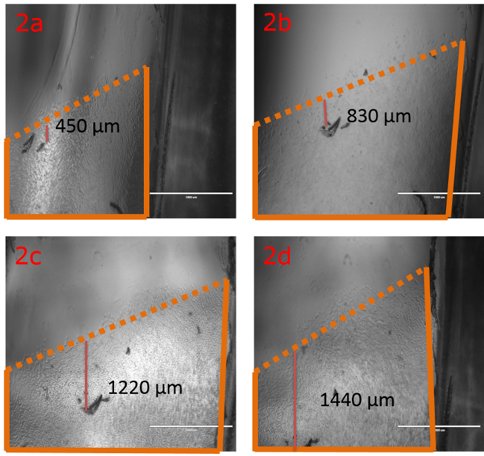
Figure 2: Cell Front Movement. Time-lapse images of the cell front in the microchannel chamber moving through the channel. The cell front marked in orange lines, and the migration distance is included. This migration distance was measured from the marking on the plate to the front of the cell front as shown. A 1 mm scale bar is shown in white. A) 48 h post plating, B) 72 h post plating, C) 96 h post plating, D) 120 h post plating. Please click here to view a larger version of this figure.
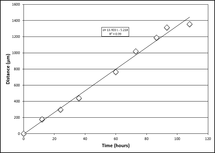
Figure 3: Growth Rate Calculation. Cell front movement tracked with time-lapse pictures. Growth rate was calculated using the MATLAB polyfit function (roipoly) to yield average cell front velocity of 13.4 µm/h. Please click here to view a larger version of this figure.
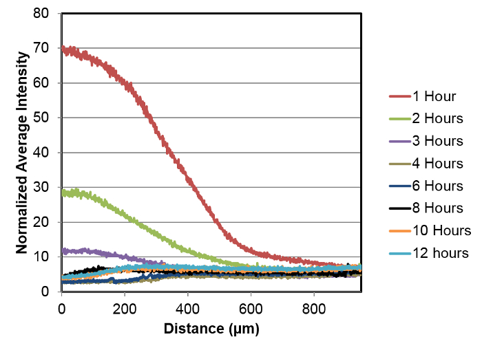
Figure 4: Fluorescent Gradient with Dextran. Fluorescent intensity measured across the distance of the channel where each line represents the gradient at a particular hour. ImageJ was used to calculate the fluorescence intensity. This can be done using the Analyze | Measure tool. First, choose Set Measurements under Analyze and select intensity. Then, choose Measure under Analyze in order to get average pixel intensity. This can be plotted versus distance in microns as shown in this figure. Please click here to view a larger version of this figure.
