Epidermal keratinocytes form a functional skin barrier and are positioned at the front line of host defense against external environmental insults. Here we describe methods for isolation and primary culture of epidermal keratinocytes from neonatal and adult mouse skin, and induction of terminal differentiation and UVB-triggered inflammatory response from keratinocytes.
The keratinocyte (KC) is the predominant cell type in the epidermis, the outermost layer of the skin. Epidermal KCs play a critical role in providing skin defense by forming an intact skin barrier against environmental insults, such as UVB irradiation or pathogens, and also by initiating an inflammatory response upon those insults. Here we describe methods to isolate KCs from neonatal mouse skin and from adult mouse tail skin. We also describe culturing conditions using defined growth supplements (dGS) in comparison to chelexed fetal bovine serum (cFBS). Functionally, we show that both neonatal and adult KCs are highly responsive to high calcium-induced terminal differentiation, tight junction formation and stratification. Additionally, cultured adult KCs are susceptible to UVB-triggered cell death and can release large amounts of TNF upon UVB irradiation. Together, the methods described here will be useful to researchers for the setup of in vitro models to study epidermal biology in the neonatal mouse and/or the adult mouse.
The skin is the largest organ in the body with the epidermis as the outer most layer. The epidermis plays a critical role in forming an intact epidermal barrier to separate the body from the environment, and thus prevents water loss and provides protection from environmental insults, such as allergens, pathogens and UVB exposure. The epidermis develops from a single layer of undifferentiated basal keratinocytes (KCs) into a multi-layered stratified epithelium consisting of a basal layer, followed by a spinous layer, granular layer, and stratum corneum. Basal KCs, consisting of both epidermal stem cells and transit-amplifying cells, are proliferative and undifferentiated. As basal KCs exit the cell cycle, the cells commit to differentiation and gradually migrate towards the surface of the epidermis, accompanied by the maturation of cell-cell junctions and formation of an epidermal permeability barrier (EPB). The KCs at the spinous layer express early differentiation markers such as Keratin 10 (K10); as the KCs migrate to the granular layer, the cells express late differentiation markers such as Filaggrin (FLG), Loricrin (LOR) and Involucrin (INV). At the stratum corneum, the KCs become terminally differentiated corneocytes, which are eventually shed off through desquamation as new cells replace them.
Calcium is considered the most physiological agent in the epidermis and triggers differentiation in vitro and in vivo in a similar manner. In normal skin epidermis, calcium ions form a characteristic "concentration gradient", increasing in concentration towards the skin surface1,2,3. The calcium concentration rises from low levels in the lowest sublayers (basal and spinous layers) to a peak in the upper granular layer and then drops to negligible levels in the most superficial layer (stratum corneum). The calcium gradient also develops coincidently with the emergence of a component permeability barrier, which supports that calcium signaling plays a critical role of KC differentiation. In vitro, low calcium (0.02-0.1 mM) maintains the proliferation of basal KCs as a monolayer, whereas high calcium (>0.1 mM) induces a rapid and irreversible commitment of the cells to terminal differentiation as demonstrated by tight-junction formation and induction of LOR and INV upon high calcium treatment to the basal KCs4,5.
In addition to barrier formation, epidermal KCs are also an important component of the skin's innate immune system. In response to pathogens or damaged-associated molecular patterns (DAMPs) released upon UVB irradiation or injury, KCs can produce large amounts of inflammatory cytokines, such as TNFα, IL6 and IFNβ, leading to immune system activation6,7,8,9. Although proper inflammatory signaling from KCs is required for pathogen clearance, uncontrolled inflammatory response may trigger the development of auto-inflammatory skin diseases, such as psoriasis and rosacea6,8.
Overall, KCs play a vital role in maintaining the intact skin barrier and initiating an immune response upon pathogen invasion or environmental insults. Therefore, primary culture of epidermal KCs is a useful technique to study the epithelial biology, KC differentiation, as well as KC-stimulated innate immune responses. The isolation and culture of primary mouse epidermal KCs can be a challenging process due to KC's susceptibility and sensitivity to various external stimulants. Here we describe a method to isolate and culture KCs from either neonatal mouse skin or adult mouse tail skin. For adult KC isolation, we do not use mouse dorsal skin because isolating sufficient quantities of viable KCs from this tissue can be difficult for the following reasons: First, the adult dorsal skin at the resting phase of the hair cycle (telogen) consists of a thin epidermis with only 1-2 layers of cells, leading to a low cell yield and inefficient separation of the epidermis from the dermis, which is the critical step for successful KC isolation. Second, the high hair follicle density that is present on adult dorsal skin further contributes to the difficulty in separating epidermis from the dermis. Instead, we routinely use tail skin as the source for adult mouse KCs as this epithelium is thicker with 3-5 layers of epidermal KCs. It also has a lower hair follicle density, which does not interfere with the epidermal separation, thus allowing KC isolation from any adult mouse tail skin regardless of the age and hair cycling stage of the mouse. The isolated neonatal KCs are seeded to gelatin-coated culture dishes, whereas collagen-coated dishes are used to seed isolated adult KCs due to the impaired ability of the adult cells to adhere compared to their neonatal counterparts. To culture mouse KCs, low calcium basal medium is supplemented with dGS, which contains epidermal growth factor (EGF), bovine transferrin, insulin-like growth factorc1 (IGF1), prostaglandin E2 (PGE2), bovine serum albumin (BSA) and hydrocortisone. Between 2-4 days after the initial plating, most of the differentiated KCs can be washed away during daily medium changes, and the remaining adherent cells show typical cobblestone morphology4, are proliferating, and do not express the early differentiation marker K10.
All animal experiments are approved by the UCSD Institutional Animal Care and Use Committee.
1. Primary Mouse KC Isolation and Culture from Neonatal Skin
2. Primary Mouse KC Isolation from Adult Tail Skin
3. In Vitro Differentiation of KCs by High Calcium
4. UVB Irradiation Mediated Cell Death and TNFα Release from KCs
High calcium induced terminal differentiation of neonatal and adult KCs. The primary mouse epidermal KCs plated and maintained at 0.06 mM CaCl2 grew as a monolayer, and individual cells had a polygonal shape with distinct intercellular space, showing a cobblestone appearance when confluent (Figure 1A and Figure 2A). Elevating the CaCl2 to 0.2 mM induced a rapid morphology change of the cells. Within 8 h after the high calcium switch, the cells became flattened and the distinct intercellular space became less apparent, and by 24 h the cell-cell adhesion with tight junctions became obvious (Figure 1A and Figure 2B). The formation of the cornified cell envelope and vertical cell stratification started around 48-72 h after the high calcium switch (Figure 2B). To analyze the actin remodeling and cell-cell tight junction formation during the high calcium switch, KCs treated with 0.2 mM CaCl2 were stained with phalloidin (Figure 1B) to measure the actin remodeling during KC differentiation13. The formation of the actin fiber-rich filopodial projections between the adjacent cells were detected as early as 3 h post calcium switch, and the actin fiber remodeling further progressed between 6-24 h, and by 48 h the cell stratification became prominent (Figure 1B).
Susceptibility of mouse KCs to UVB-triggered cell death and TNFα release. The cultured adult mouse KCs were exposed to 25 mJ/cm2 UVB irradiation, and the cell viability and TNFα released in the culture medium were analyzed. As shown by the phase contrast images (Figure 3A), as well as the cell viability assay (Figure 3B), the UVB triggered a time dependent death of the KCs. By 24 h, the majority of the cells were rounded and were detached from the dish (Figure 3A). As quantified by the cell viability assay, ~90% of cells were dead within 24 h post UVB treatment (Figure 3B; p <0.001 as calculated by one-way ANOVA multiple comparison test). Finally, we showed that TNFα, an important cytokine that is induced by UVB and drives KC apoptosis following UVB irradiation14, was abundantly secreted (~250 pg/mL within 24 h post UVB-irradiation; p <0.001) in the culture medium of UVB-treated KCs in a time dependent manner (Figure 3C).
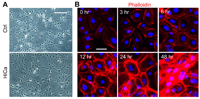
Figure 1: Primary Culture of Neonatal Mouse KCs, and High Calcium Induced Terminal Differentiation and Tight Cell-cell Junction Formation. (A). Primary neonatal mouse KCs were treated with high calcium (0.2 mM CaCl2), and phase contrast images at 4X magnification were taken at 0 hour (ctrl) and at 8 h after treatment. Scale bar = 100 µm. (B). Neonatal KCs were differentiated in the presence of 0.2 mM CaCl2, and the cells were stained with phalloidin (red) to visualize the formation of actin fiber-rich filopodial projections between the adjacent cells during differentiation. Nuclei were counterstained with DAPI, and actin fibers were stained with rhodamine phalloidin. Scale bar = 25 µm. Images were taken at 20X magnification using a fluorescence microscope set to the DAPI channel (for nuclei staining) and RFP channel (for actin staining). Please click here to view a larger version of this figure.
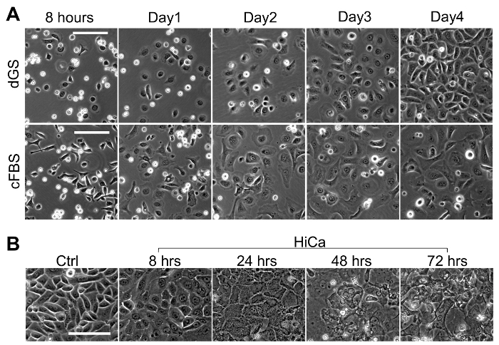
Figure 2: Primary Culture and High Calcium Induced Terminal Differentiation of Adult mouse KCs. (A). Phase contrast images at 10x magnification of primary adult mouse KCs at 8 h, 1 day, 2 days and 3 days after the initial plating. The upper panel represents cells grown in dGS, and the lower panel represents cells grown in 10% chelexed FBS (cFBS) with 8 ng/mL recombinant mouse EGF. Scale bar = 100 µm. (B). Confluent adult mouse KCs were differentiated in the presence of 0.2 mM CaCl2, and phase contrast images at 10X magnification were taken at 0 (ctrl), 8, 24, 48, and 72 h post high calcium switch. Scale bar = 100 µm. Please click here to view a larger version of this figure.
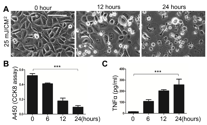
Figure 3: Primary Culture of Adult mouse KCs, and Susceptibility of Adult KCs to UVB-induced Cell Death and Release of TNFα. Primary adult mouse KCs were cultured to confluency, then exposed to 25 mJ/cm2 UVB irradiation. (A) Phase contrast images at 10X magnification taken at 12 and 24 hours post-UVB exposure showed a time dependent UVB-induced cell death. Scale bar = 100 µm. (B) The cell viability was quantified by the CCK-8 cell viability assay at 6, 8, and 24 h post-UVB treatment. (C) UVB-induced release of TNFα to the media at indicated time points was measured by ELISA. All error bars indicate mean ± SEM. The p-values were calculated by one-way ANOVA multiple comparison test (***, p <0.001) Please click here to view a larger version of this figure.
The skin epidermis functions as a critical barrier to separate and protect the body from the outside environment and damage from water loss, pathogens, heat and UV irradiation. The KCs are the predominant cell lineage of the epidermis, and primary culture of epidermal KCs is a useful tool to study and understand the biological processes of barrier formation and the response of KCs to environmental insults in vitro.
Here we describe methods to isolate and culture primary epidermal KCs from neonatal and adult mouse skin. While primary mouse KCs can be conveniently isolated from neonatal mouse skin, the isolation and successful culture of adult KCs are considered difficult due to the thinning of the epidermis and the high hair follicle density of the adult mouse dorsal skin. The method described here uses adult mouse tail skin as a convenient and abundant source of basal KCs. Because of the presence of a thick epidermis and low hair follicle density in the tail skin, the critical step of isolating KCs by separating the epidermis from the dermis becomes feasible after overnight dispase digestion of the tail skin. In contrast, our attempt to isolate KCs from adult dorsal skin using this protocol was not successful, resulting in low cell yield after isolation and low cell attachment after plating. However, successful isolation and culture of KCs from adult dorsal skin has been reported after overnight trypsin digestion of the fur-free skin15, suggesting that dispase alone may not be sufficient to extract follicular KCs from the dermis of the adult dorsal skin.
In this protocol, the primary KCs were grown in low calcium medium supplemented with dGS instead of the calcium-depleted chelexed-FBS, which was widely used in several previously published mouse KC culture protocols4,15,16,17,18. In this study, we observed that dGS is superior to chelexed FBS to culture adult mouse KCs and to maintain KC's basal cell morphology (Figure 2A). However, it is likely that the effectiveness of chelexed-FBS to maintain KC's basal morphology may depend on the FBS lot used in each study.
Using the methods presented here, we demonstrated that both the neonatal and adult mouse epidermal KCs responded to high calcium triggered terminal differentiation, tight cell junction formation and stratification (Figure 1 and Figure 2). In general, elevating the extracellular calcium level to greater than 0.1 mM triggers terminal differentiation of basal KCs4,5,17,19. It has been reported that an intermediate level of calcium (0.1- 0.16 mM) substantially induced the expression of early differentiation markers, such as K1 and K10, during the first 24 h upon calcium switch, whereas K1 and K10 was only induced in a small fraction of cells when treated with higher calcium medium (1 mM)19,20. However, the higher calcium medium ( ≥1 mM) has been widely used in many other studies to rapidly and robustly induce KC stratification and the expression of late differentiation genes, such as INV and LOR4,21,22,23. Based on our experience, an intermediate level of calcium, such as 0.2 mM CaCl2, leads to a gradual and slower onset of terminal differentiation, whereas a higher calcium levels (≥1 mM) drastically speed up the onset of KC terminal differentiation, leading to a shorter time window to study the cellular changes during differentiation. Therefore, we used 0.2 mM CaCl2 to induce KC terminal differentiation, and we have shown that this intermediate calcium level led to a gradual change of the basal cell morphology to a stratified corneocyte morphology within 72 h after the calcium switch (Figure 2B).
Our group has previously demonstrated that epidermal KCs also play an important role to initiate inflammatory responses to pathogens or DAMPs released during injury and/or UVB-irradiation6,7,8,9,10. In consistent with those in vivo observations, here we showed that cultured adult mouse KCs were highly susceptible to UVB-triggered cell death and UVB-treated KCs released a large amount of TNFα, which is a key inflammatory cytokine that activates the immune system.
In summary, the primary mouse epidermal KC cell culture provides a useful in vitro model to study the epidermal barrier formation and innate immune response of KCs, and the methods described here will be of interest to researchers pursuing studies in cutaneous biology in the neonatal mouse as well as in the adult mouse.
The authors have nothing to disclose.
This work was supported by NATIONAL INSTITUTE OF ARTHRITIS AND MUSCULOSKELETAL AND SKIN DISEASES grant (R01AR069653 to Zhang LJ), and National Institutes of Health Support (5T32AR062496-03 to CA).
C57BL/6 neonates or adult wildtype mice | Jackson Laboratory | 000664 | Wildtype mice (originally purchased from Jackson Laboratory) are breeded and maintained in animal vivarium at UCSD. |
KC basal medium (EpiLife) | Invitrogen, Carlsbad, CA | MEPICF500 | basal medium for keratinocyte culture with 0.06 mM CaCl2 |
Defined Growth Supplement (dGS) | Invitrogen, Carlsbad, CA | S0125 | defined growth supplements for culture medium |
Dispase powder | Invitrogen, Carlsbad, CA | 17105041 | enzyme to dissociate the epidermis from dermis |
Attachment Factor | Invitrogen, Carlsbad, CA | S006100 | gelatin-based coating material |
Coating Matrix | Invitrogen, Carlsbad, CA | R011K | Collagen-based coating material |
TrypLE | Invitrogen, Carlsbad, CA | 12604-013 | A gentle trypsin-like enzyme to dissociate keratinocytes from epidermal sheet |
100 μm Cell Strainer Nylon mesh | Corning | 352360 | |
CCK-8 cell viability Kit | Dojindo Molecular Technologies, Rockville, MD | CK04-11 | |
Mouse TNF (Mono/Mono) ELISA Set II | BD Biosciences, San Jose, CA | 555268 | |
Corded Hand-Held UV Lamps | Spectronics, Westbury, NY | EB-280C | |
8-watt UV tubes | Spectronics, Westbury, NY | BLE-8T312 | |
Light Inverted Microscope for cell culture | ZEISS, Jena, Germany | Axio Observer | |
Fluorescent Microscope | Olympus | BX41 |