Exploring the Potential of Mesenchymal Stem Cell Sheet on The Development of Hepatocellular Carcinoma In Vivo
Summary
Here, we present a protocol to develop an in vivo cancer model using cell sheet technology. Such a model could be very useful for the evaluation of anticancer therapeutics.
Abstract
An in vivo animal model that mimics human cancer could have various applications that deliver significant clinical information. The currently used techniques for the development of in vivo cancer models have considerable limitations. Therefore, in this study, we aim to implement cell sheet technology to develop an in vivo cancer model. Hepatocellular carcinoma (HCC) is successfully developed in nude rats using cell sheets created from HCC cell line cells. The cancer cell sheets are generated through intracellular adhesion and the formation of a stratified structure, controlled by the extracellular matrix. This allows for the HCC sheet transplantation into the liver and the creation of a tumor-bearing animal model within a month. In addition, the role of mesenchymal stem cells (MSC) in the development of this cancer model is investigated. In addition to the HCC cell line sheet, another two cell sheets are created: a sheet of HCC cells and bone marrow MSCs (BMMSCs) and a sheet of HCC cells and umbilical cord MSCs (UCMSCs). Sheets that have a combination of both HCC cells and MSCs are also capable of producing a tumor-bearing animal. However, the addition of MSCs reduces the size of the formed tumor, and this adverse effect on tumor development varies depending on the used MSCs’ source. This indicates that a cell sheet made of certain MSC subtypes could be utilized in tumor management and control.
Introduction
HCC is a primary cancer of the liver that is significantly associated with a poor prognosis. Annually, almost a half million new patients are diagnosed with HCC, representing 85% of liver cancer patients worldwide1. Hepatocarcinogenesis is not a single-form disease; rather,it is a collection of diseases that have different histopathological features and genetic and genomic variability, in addition to varied prognostic outcomes1. Therefore, the main challenges in the development of an effective therapeutic strategy for HCC are the limited knowledge of HCC biology and the lack of a suitable experimental animal model that can help understand this complex disease. An in vivo animal model that mimics human cancer is necessary for selecting candidate genes and identifying prognostic/predictive markers implicated in cancer induction, as well as for investigating different factors that may affect cancer responses to therapeutic agents.
In vitro cancer studies are still associated with major limitations. This is due to the fact that cancer cells lose many of their in vivo features when maintained in culture. The changes that occur to cells in vitro result from the absence of the whole tissue physiology in an ex vivo setting. Cancer cell-to-cell interaction (stromal, immune, vasculature, epithelial, etc.) within the tumor microenvironment reflects greatly on cancer cell characteristics2. The tumor microenvironment could alter cancer cell gene/protein expression and phenotypic characteristics, in addition to angiogenetic and metastatic potentials. The two-dimensional (2-D) in vitro culture system also lacks a suitable tissue matrix, which is necessary to regulate tumor progression. Thus, due to these limitations, in vivo models should always be utilized to support the preliminary findings of in vitro models. In this study, we use cell sheet technology to develop an in vivo animal model that elucidates the complete biological process underlying HCC.
More than a decade ago, Okano's laboratory established a novel method of tissue engineering based on cell sheet technology3. This technique utilizes a thermo-responsive culture plastic to allow reversible cell adhesion/detachment by controlling the surface hydrophobicity. This method allows a gentle harvesting of cultured cells in an intact three-dimensional (3-D) format (i.e.,cell sheet), with a well-kept extracellular matrix (ECM) and cell-to-cell interactions. The cell sheet technique requires the precoating of culture dishes with a temperature-responsive polymer poly(N-isopropylacrylamide) (PIPA Am), which is commercially available and ready for use. At a temperature below 20 °C, PIPA Am polymers become hydrated and dissolve in aqueous solutions, whereas, at a higher temperature (37 °C), the polymers become dehydrated and transform into a turbid precipitate. The polymer contains hydrophilic amide chains and hydrophobic side chains (isopropyl groups). At high temperatures, the Brownian movement of the water molecules is intensified, whereas, at a low temperature, the molecules of the water surrounding the isopropyl groups of the hydrated structure breakdown and the groups of hydrophobic isopropyl aggregate because of hydrophobic interactions. Therefore, the entire chain of the polymer aggregates and precipitates4.
In the presented study, this technique is employed to develop an HCC animal model using three different cell sheets. The first sheet employed consists of HCC cell line cells only, whereas the other two sheets consist of a combination of HCC cell line cells and MSCs from two different sources: bone marrow MSCs (BMMSCs) and umbilical cord MSCs (UCMSCs). MSCs are non-hematopoietic stromal cells that are capable of differentiating intocell derivatives of the mesenchymal lineage, including adipocytes, osteocytes, chondrocytes, and myocytes5. The reason we employ these cells when creating the cancer cell sheet is the inconsistent reporting on the effect of MSCs on cancers. It has been suggested that MSCs can have two distinct phenotypes: "MSC1", a proinflammatory phenotype, and "MSC2", an immunosuppressive phenotype6. MSCs express toll-like receptors (TLRs). TLR4 priming of MSCs increases their secretion of proinflammatory factors, whereas TLR3 priming increases their secretion of immunosuppressive factors6. An in vitro study of these two phenotypes reported that a co-culture of MSC1 with cancer cell lines attenuated cancer cell growth, while MSC2 co-culture had the opposite effect7. This implicates that MSCs can be either pro-cancer or anticancer, depending on their phenotype. Thus, in addition to developing the HCC animal model using cell sheet technology, we want to explore the effect of MSC transplants on tumor development, and whether using these cells will enhance or reduce the development of this model.
Protocol
The protocol follows the animal care guidelines of King Saud University's ethical committee. Surgical procedures, anesthetics, and other medications used on animals are approved by King Saud University's ethical committee. All experimental work is performed by appropriately trained personnel.
1. Cell Sheet Construction
- Coat the culture dishes (3.5-cm temperature-responsive culture dishes) with undiluted fetal bovine serum (FBS) and ensure to cover all the surface of the dish.
NOTE: This step is important for the detachment of the cell sheet since it supports the growth of the cells in a monolayer and prevents cell aggregation. - Incubate the dishes for 24 h at 37 °C in a humidified atmosphere containing 5% CO2 and 95% air.
- Remove the excess FBS from the coated dishes and allow the dishes to dry at room temperature (RT) for at least 1 h.
- Prepare the three cell suspensions: 1 x 10⁶ HepG2 cells, 1 x 10⁶ HepG2 cells with BMMSCs, and 1 x 10⁶ HepG2 cells with UCMSCs (in a ratio of 4:1 for both BMMSCs and UCMSCs).
- Seed the appropriate cell suspension to each labeled dish.
- Suspend all cells in DMEM culture medium supplemented with 10% FBS and 1% penicillin/streptomycin.
- Incubate the cells at 37 °C in a humidified atmosphere containing 5% CO2 and 95% air.
2. Cell Sheet Detachment
- At 100% cell confluence, take the dishes out of the 37 °C incubator.
- Incubate the HepG2 cell sheet dish for 1 h at RT, while incubating the HepG2/BMMSC and HepG2/UCMSC cell sheet dishes for 30 – 40 min at 20 °C in a humidified atmosphere containing 5% CO2 and 95% air.
NOTE: The cell sheet made from only HepG2 is fragile; reducing the temperature to 20 °C causes damages to the structure of the sheet.
3. Performance of the Surgical Procedure
Note: During the incubation time for the cell sheet detachment, start the animal procedure.
- Instructions for the preparation of the surgery area
- Conduct all surgical procedures in a sterile separate area within the laboratory, such as a hood, laminar flow cabinet, or an aseptic surgery room. Ensure that all surfaces of the surgery area are non-porous, sealed, durable, and sanitizable.
- Wear proper protective gear, including scrubs, gloves, facemasks, and head and shoe covers.
- Use surgical tools that are clean and sterilized (via autoclaving; saturated stem under high pressure) at the beginning of the surgery.
- Change gloves between rats.
- Preparation of the animal for surgery
- Use 8- to 12-week-old nude rats.
- Create injectable anesthesia for rats.
- To prepare the anesthesia solution, ketamine-xylazine, mix 2 mL of ketamine, 1 mL of xylazine (at a concentration of 20 mg/mL), and 1 mL of sterile physiologic saline (e.g., 0.9% sodium chloride) in a sterile 10-mL serum collection vial and shake well before use.
- To apply anesthesia, inject 0.2 mL of the ketamine-xylazine solution per 100 g of body weight of the rat intraperitoneally.
NOTE: The total dose of the solution is 100 mg/kg of ketamine and 10 mg/kg of xylazine.
- Remove visible dirt/debris from the surgical site.
- Check the depth of anesthesia by testing the pedal withdrawal reflex (foot pad pinch on both hind feet).
NOTE: If the foot pad pinch causes a response, repeat at a dose of 0.05 mL/100 g (approximately every 30 min), then recheck the anesthetic depth. - Disinfect the surgery area with a two-stage scrub by povidone-iodine/isopropanol.
- Cover the rat with a sterile drape to avoid contamination of the incision.
- Inject the rats subcutaneously with buprenorphine (0.01 – 0.05 mg/kg)8 prior to making the incision.
- Using a sterile scalpel, make a 7- to 10-cm cut between the skins and underlying muscles to expose the liver.
- Separate the abdominal muscle from the skin with the flat back edge of the scalpel.
- Carefully stab the linea alba using a scalpel and extend the cut with sharp scissors.
4. Cell Sheet Transplantation
Note: Cell sheet transplantation is performed on the liver .
- Collect the cell sheet from the culture dish.
- Gently tap the culture dish over the bench to detach the sheet from its surface.
- Separate the cell sheet from the dish's surface by scraping the edge of the sheet in a half circle using a sterile pipette tip.
- Gently remove the entire medium from the culture dish.
- Ensure the sheet is intact without any damage; otherwise, it cannot be used (Figure 1).
NOTE: The resulting sheets can be detected directly by the eye.
- Prepare a sterile membrane (filter paper) to harvest the cell sheet from the dish.
- First, soak the membrane with normal saline. Then, remove the excess saline with a sterile cotton pad.
- Place the wet membrane over the cell sheet, while avoiding creases and air bubbles between the membrane and the cell sheet.
- Add few drops of normal saline over the membrane and the cell sheet to collect them easier.
Note: To avoid breaking the cell sheet, do not use cold saline and lift the membrane and the cell sheet with forceps. - Leave the membrane for a few seconds to ensure that the cell sheet is properly attached to the membrane, and then collect it.
- Prepare the liver for cell sheet transplantation.
- Make sure the liver surface is dry by gently tapping it with a sterile cotton pad.
- Using sterile forceps, place the membrane with the cell sheet over a single lobe of the rat's liver.
- Add a few drops of sterile saline and apply a gentle pressure to the membrane to detach the cell sheet from it.
- Wait for 5 min before carefully removing the membrane.
NOTE: This is done to ensure the cell sheet is properly attached to the liver. - Finally, close the underlying muscles and skin incisions with 5-0 nylon sutures.
- Disinfect the surgery area with povidone-iodine.
Note: Figure 2 shows a diagram of the cell sheet transplantation procedure on the liver of a nude rat.
5. Post-surgery Care
- Immediately after the surgery, house the rat individually to avoid cannibalism or suffocation, and monitor it regularly (at least every 15 min) until it is conscious and fully ambulant.
- Then, assess the rats daily for 5 – 14 d, at least 5 d post-operatively, to ensure that there are no complications. During the daily assessment, make sure of the following.
- The wound is closed, and the sutures are intact, without being excessively tight.
- There are no signs of infection in the incision site, such as heat, excessive swelling, or purulent discharge.
- There are no signs associated with pain, such as decreased food and water consumption, weight loss, dehydration, skin tenting, or rapid and open mouth breathing. To control moderate to severe post-surgery pain, if any, inject the rats subcutaneously with buprenorphine (0.01 – 0.05 mg/kg) every 6 – 8 h9 for a minimum of 48 – 72 h postoperatively.
6. Analysis of the Transplanted Area
- One month after the transplantation, scarifice the rat and collect the transplanted area for histological and immunohistochemical analysis.
Representative Results
Tumorigenicity of Transplanted Cell Sheets in Rats:
One month after the transplantation, all transplanted cell sheets in rats' livers have developed tumors (Figure 3). The average size of the developed tumors from HepG2, HepG2/BMMSC, and HepG2/UCMSC cell sheets were 4.5 cm, 4 cm, and 2.5 cm, respectively10.
Histological Analysis of Liver Tissues:
Hematoxylin and Eosin (H&E) staining of non-transplanted rats showed livers with hepatocytes arranged in plates that anastomose with one another, and the nuclei were round, with one or two prominent nucleoli. In contrast, the tumor sections collected showed edges of viable inflamed and necrotic subcutaneous hepatocytes, with irregular nuclear contours and clear degeneration (Figure 4).
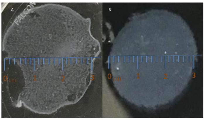
Figure 1: Fabrication of a cell sheet using a 3.5-cm temperature-responsive culture dish. (A) This panel shows a damaged cell sheet not suitable for transplantation. (B) This panel shows an intact cell sheet, ready for transplantation. Please click here to view a larger version of this figure.
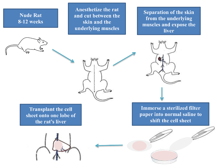
Figure 2: Cell sheet transplantation procedure in nude rats. Please click here to view a larger version of this figure.
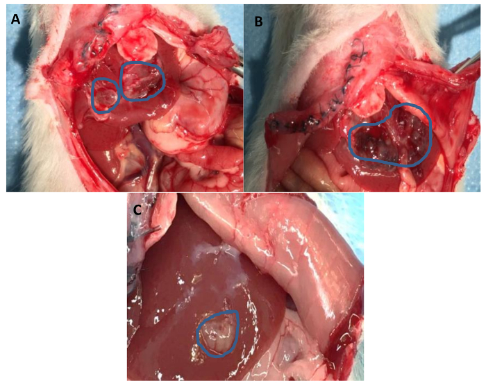
Figure 3: Tumor developed on a rat's liver following one month of cell sheet transplantation. The tumor was generated from (A) a HepG2 cell sheet (size: 4.5 cm), (B) a HepG2 and BMMSC cell sheet (size: 4 cm), and (C) a HepG2 and UCMSC cell sheet (size: 2.5 cm). The blue circles show the tumor area. This figure has been modified from previously published work10. Please click here to view a larger version of this figure.
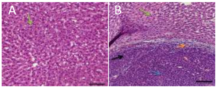
Figure 4: Histological analysis of a rat's liver. H&E staining of a rat's liver, four weeks after cell sheet transplantation. (A) This panel shows normal rat liver cells. (B) This panel shows the liver morphology after cancer cells transplantation. The black arrow indicates hepatic cancer, the green arrows indicate normal hepatocytes, the blue arrow indicates inflamed cells, and the orange arrow indicates necrotic HCC cells. The scale bars indicate 20X magnification. This figure has been modified from previously published work10. Please click here to view a larger version of this figure.
Discussion
An extensive amount of research is dedicated to developing an adequate in vivo preclinical animal model that resembles human cancers. Currently, the major approaches used to create cancer animal models involve genetic engineering and cell transplantation11. Genetically modified animal models are good tools for the identification and validation of target genes, as well as understanding the molecular mechanisms underlying drug-induced toxicity. However, this model is associated with some limitations; for instance, the modification of a given gene does not always result in the anticipated phenotype12.The cell transplantation approach is more commonly used to induce cancer in animals, and it involves the injection of cancer cell suspension. Yet, this simple and convenient approach has its own disadvantages. To generate a cell suspension, an enzymatic digestion step will be used to harvest the cancer cells. This results in the loss of some of the adhesion proteins, which will reduce the engraftment efficiency of the transplanted cells. Therefore, there is no guarantee that the transplanted cancer cells will form cancerous tissues, not to mention the difficulty of controlling the size of the induced tumor. In addition, this approach has another serious drawback, which is the rapid transition of transplanted cancer cells into the blood circulation or cell engraftment into non-targeted organs13.
The novel cell sheet technology was developed to overcome these disadvantages. Suzuki et al.14 have successfully created colon, liver, and pancreas tumor-bearing animals using the cancer cell sheet transplantation method. When compared with tumors generated via the cancer cell transplantation method, these tumors were larger and more stable. The implantation of a small tumor tissue into an animal to induce a tumor is highly dependent on the use of a transplantation method that does not require an enzymatic digestion step. Proteolytic enzymes cause damage to extracellular matrices, which affects cell-to-cell interaction. Cell sheet technology overcomes this limitation of using an invasive harvesting method, allowing the collection of an intact monolayer cell sheet, along with its associated extracellular matrix and growth factors15. Cell sheet technology also allows for long-term engraftment16,17. This is of great value in in vivo studies, where cells must be able to engraft into the host's tissue for the whole period of the tissue's life17. In addition, this technique enabled the development of a novel method of tissue engineering, without the use of biodegradable scaffolds3,15. Yet, this approach still has a few limitations. When using the cell sheet technique, the central portion of the grafted tissue may possibly become necrotic if the graft is too large. Conversely, the success chance of the transplantation may be reduced if the graft is too small. Additionally, in order to control the subsequent growth of the tumor, the size of the transplanted tissue must be uniform across all experiments. The size of the transplantable tissue is limited to 2 mm3 as a result of the induction of necrosis due to the poor oxygen environment18.
In this study, an HCC animal model was successfully created using the scaffold-free transplantation of cell sheets. Three types of cell sheets were transplanted into rats' livers: a cell sheet of HCC cell line cells, a cell sheet of HCC cell line cells and BMSCs, and a cell sheet of HCC cell line cells and UCMSCs. The transplantation of all three cell sheets was sufficient to induce tumors in recipient rats. However, it was noted that adding MSCs to the sheet reduced the size of the formed tumors. This indicates that the MSCs used, especially UCMSCs, have an adverse effect on tumor development. To conclude, cancer cell sheet transplantation offers an efficient method to create an animal model with solid tumors. Having such in vivo models could result in critical clinical information with considerable applications. In addition, as apparent by the tumor size reduction, UCMSCs limit tumor development and propagation. This indicates that some MSC subtypes could be used in a cell-based approach for the treatment of cancer.
Divulgazioni
The authors have nothing to disclose.
Acknowledgements
The authors would like to thank the staff of the Experimental Surgery and Animal Laboratory at the College of Medicine, King Saud University, for their cooperation and support-especially Hussain Almukhayzim and Hisham Aloudah. The authors would also like to acknowledge the media team at King Saud bin Abdul-Aziz University for preparing the visual material-especially Muath bin Ghannam and Abdulwahab Alsulami.
Materials
| Reagents | |||
| FBS | Gibco/Invitrogen | 10270106 | |
| DMEM high glucose | Sigma | D5671-500ML | |
| Penicillin/streptomycin | Life Technology | 15070063 | |
| Sterile physiologic saline | Sigma | S0817-1GA | |
| Human HepG2 cell line | ATCC, USA | HB-8065 | |
| Human bone marrow MSCs cell line | PromoCell, USA | C-12974 | |
| human umbilical cord tissue MSCs | PromoCell, USA | C-12971 | |
| Ketamine 50% | Rompun, Bayer | ||
| Xylazine 2% | Rompun | 23076-35-9 | |
| Alphadine® solution. | Riyadh Pharma | LBL0816 | |
| Disposables: | |||
| 15mL Polypropylene High Clarity PP Centrifuge Tube | Falcon | 352097 | |
| 3.5 cm sterile UpCell culture dishes with the filter paper (membrane) | Sigma | 174904-1CS | |
| 100-1000 µl Pipette Tips | Sigma | CLS4868-1000EA | |
| Basic Procedure Drape | Thermofisher | PMD5293.0 | |
| Equipment | |||
| Plus pipette, variable volume | Eppendorf® Research® | Z683779-1EA | |
| Tissue culture incubator 37 °C, 5% CO2 | Any brand | ||
| Biological safety cabinet | Any brand | ||
| Tissue culture incubator 20 °C, 5% CO2 | Any brand | ||
| Sterile surgical tools and nude rats: | |||
| Forceps | |||
| Scissors | |||
| scalpel | |||
| Nylon Suture 5-0 | Accutome | AB-3854S | Monofilament, Lancet |
| 1 ml Tuberculin Syringes | Fisher Scientific | 14-826-88 | |
| Nude rats | Charles river | ||
Riferimenti
- Marra, M., et al. Molecular targets and oxidative stress biomarkers in hepatocellular carcinoma: an overview. Journal of Translational Medicine. 9, 171 (2011).
- Yamada, K. M., Cukierman, E. Modeling tissue morphogenesis and cancer in 3D. Cell. 130 (4), 601-610 (2007).
- Kushida, A., et al. Decrease in culture temperature releases monolayer endothelial cell sheets together with deposited fibronectin matrix from temperature-responsive culture surfaces. Journal of Biomedical Materials Research. 45 (4), 355-362 (1999).
- Okano, T., Yamada, N., Sakai, H., Sakurai, Y. A novel recovery system for cultured cells using plasma-treated polystyrene dishes grafted with poly (N-isopropylacrylamide). Journal of Biomedical Materials Research Part A. 27 (10), 1243-1251 (1993).
- Chamberlain, G., Fox, J., Ashton, B., Middleton, J. Concise review: mesenchymal stem cells: their phenotype, differentiation capacity, immunological features, and potential for homing. Stem Cells. 25 (11), 2739-2749 (2007).
- Waterman, R. S., Tomchuck, S. L., Henkle, S. L., Betancourt, A. M. A new mesenchymal stem cell (MSC) paradigm: polarization into a pro-inflammatory MSC1 or an Immunosuppressive MSC2 phenotype. PLoS One. 5 (4), e10088 (2010).
- Waterman, R. S., Henkle, S. L., Betancourt, A. M. Mesenchymal stem cell 1 (MSC1)-based therapy attenuates tumor growth whereas MSC2-treatment promotes tumor growth and metastasis. PLoS One. 7 (9), e45590 (2012).
- Curtin, L. I., et al. Evaluation of buprenorphine in a postoperative pain model in rats. Comparative Medicine. 59 (1), 60-71 (2009).
- . Guidelines on Anesthesia and Analgesia in Rats – ULAM Guidelines and SOPs – Michigan Medicine Confluence Available from: https://wiki.med.umich.edu/display/ULAMGSOP/Guidelines+on+Anesthesia+and+Analgesia+in+Rats (2017)
- Alshareeda, A. T., Sakaguchi, K., Abumaree, M., Mohd Zin, N. K., Shimizu, T. The potential of cell sheet technique on the development of hepatocellular carcinoma in rat models. PLoS One. 12 (8), e0184004 (2017).
- Russo, J., Russo, I. H. Atlas and histologic classification of tumors of the rat mammary gland. Journal of Mammary Gland Biology and Neoplasia. 5 (2), 187-200 (2000).
- Lin, J. H. Applications and limitations of genetically modified mouse models in drug discovery and development. Current Drug Metabolism. 9 (5), 419-438 (2008).
- Driscoll, J. S. The preclinical new drug research program of the National Cancer Institute. Cancer Treatment Reports. 68 (1), 63-76 (1984).
- Suzuki, R., Aruga, A., Kobayashi, H., Yamato, M., Yamamoto, M. Development of a novel in vivo cancer model using cell sheet engineering. Anticancer Research. 34 (9), 4747-4754 (2014).
- Chen, G., et al. Application of the cell sheet technique in tissue engineering. Biomedical Reports. 3 (6), 749-757 (2015).
- Matsuura, K., Haraguchi, Y., Shimizu, T., Okano, T. Cell sheet transplantation for heart tissue repair. Journal of Controlled Release. 169 (3), 336-340 (2013).
- Matsuura, K., Shimizu, T., Okano, T. Toward the development of bioengineered human three-dimensional vascularized cardiac tissue using cell sheet technology. International Heart Journal. 55 (1), 1-7 (2014).
- Folkman, J. What is the evidence that tumors are angiogenesis dependent?. Journal of the National Cancer Institute. 82 (1), 4-6 (1990).
