In this article, we provide a detailed protocol for the expression of fluorescent fusion proteins in murine bone marrow derived dendritic cells and macrophages. The method is based on the transduction of bone marrow progenitors with retroviral constructs followed by differentiation into macrophages and dendritic cells in vitro.
Dendritic cells and macrophages are crucial cells that form the first line of defense against pathogens. They also play important roles in the initiation of an adaptive immune response. Experimental work with these cells is rather challenging. Their abundance in organs and tissues is relatively low. As a result, they cannot be isolated in large numbers. They are also difficult to transfect with cDNA constructs. In the murine model, these problems can be partially overcome by in vitro differentiation from bone marrow progenitors in the presence of M-CSF for macrophages or GM-CSF for dendritic cells. In this way, it is possible to obtain large amounts of these cells from very few animals. Moreover, bone marrow progenitors can be transduced with retroviral vectors carrying cDNA constructs during early stages of cultivation prior to their differentiation into bone marrow derived dendritic cells and macrophages. Thus, retroviral transduction followed by differentiation in vitro can be used to express various cDNA constructs in these cells. The ability to express ectopic proteins substantially extends the range of experiments that can be performed on these cells, including live cell imaging of fluorescent proteins, tandem purifications for interactome analyses, structure-function analyses, monitoring of cellular functions with biosensors and many others. In this article, we describe a detailed protocol for retroviral transduction of murine bone marrow derived dendritic cells and macrophages with vectors coding for fluorescently-tagged proteins. On the example of two adaptor proteins, OPAL1 and PSTPIP2, we demonstrate its practical application in flow cytometry and microscopy. We also discuss the advantages and limitations of this approach.
Myeloid cells represent an indispensable part of our defense mechanisms against pathogens. They are able to rapidly eliminate microbes, as well as dying cells. In addition, they are also involved in regulating tissue development and repair and in maintaining homeostasis1,2,3. All myeloid cells differentiate from common myeloid progenitors in the bone marrow. Their differentiation into many functionally and morphologically distinct subsets is to a large extent controlled by cytokines and their various combinations4. The most intensively studied myeloid cell subsets include neutrophilic granulocytes, macrophages and dendritic cells. Defects in any of these populations lead to potentially life-threatening consequences and cause severe dysfunctions of the immune system in humans and mice1,2,3,5,6.
Unlike neutrophilic granulocytes, dendritic cells and macrophages are tissue resident cells and their abundance in immune organs is relatively low. As a result, the isolation and purification of primary dendritic cells and macrophages for experiments requiring a large number of these cells is expensive and often impossible. To solve this problem, protocols have been developed to obtain large amounts of homogenous macrophages or dendritic cells in vitro. These approaches are based on the differentiation of murine bone marrow cells in the presence of cytokines: macrophage colony-stimulating factor (M-CSF) for macrophages and granulocyte-macrophage colony-stimulating factor (GM-CSF) or Flt3 ligand for dendritic cells7,8,9,10,11,12. Cells generated by this method are commonly described in the literature as bone marrow derived macrophages (BMDMs) and bone marrow derived dendritic cells (BMDCs). They have more physiological properties in common with primary macrophages or dendritic cells than with corresponding cell lines. Another major advantage is the possibility of obtaining these cells form genetically modified mice13. Comparative studies between wild-type cells and cells derived from genetically modified mice are often critical for uncovering novel functions of genes or proteins of interest.
Analysis of subcellular localization of proteins in living cells requires the coupling of a fluorescent label to the protein of interest in vivo. This is most commonly achieved by expressing genetically encoded fusion construct composed of an analyzed protein coupled (often via a short linker) to a fluorescent protein (e.g., green fluorescent protein (GFP))14,15,16. The expression of fluorescently tagged proteins in dendritic cells or macrophages is challenging. These cells are generally difficult to transfect by standard transfection procedures and the efficiencies tend to be very low. Moreover, the transfection is transient, it generates cellular stress and achieved intensity of fluorescence might not be sufficient for microscopy17. In order to obtain a reasonable fraction of these cells with a sufficient level of transgene expression, the infection of bone marrow progenitor cells with retroviral vectors and their subsequent differentiation into BMDMs or BMDCs has become a very efficient approach. It has allowed for the analysis of the proteins of myeloid origin in their native cellular environment, both in a steady state or during processes that are critical for immune response such as phagocytosis, immunological synapse formation or migration. Here, we describe a protocol that allows stable expression of fluorescently tagged proteins of interest in murine bone marrow derived macrophages and dendritic cells.
All methods described here have been approved by the Expert Committee on the Welfare of Experimental Animals of the Institute of Molecular Genetics and by the Academy of Sciences of the Czech Republic.
1. Reagent Preparation
2. Production of Retrovirus
CAUTION: Although retroviral vectors are relatively safe when compared to other types of viral vectors, they still pose a potential safety hazard. Therefore, it is crucial to work with the utmost care and appropriate protective equipment, and to adhere to all safety regulations and legal requirements for working with viral particles.
3. Murine Bone Marrow Cell Isolation
4. Bone Marrow Cell Differentiation into Bone Marrow Derived Macrophages
5. Bone Marrow Cell Differentiation into Bone Marrow Derived Dendritic Cells
6. Production of BMDMs and BMDCs Expressing EGFP-tagged Protein of Interest
Signaling adaptor proteins are usually small proteins without any enzymatic activity. They possess various interaction domains or motifs, which mediate binding to other proteins involved in signal transduction, including tyrosine kinases, phosphatases, ubiquitin ligases and others21. For the demonstration of the functionality of this protocol myeloid cell adaptors PSTPIP2 and OPAL1 were selected. PSTPIP2 is a well characterized protein involved in the regulation of inflammatory response22. It is a cytoplasmic protein which can also be recruited to cellular membranes via its F-bar domain. Second protein is a transmembrane adaptor OPAL1, expected to be associated with cellular membranes. Its physiological function is still unknown. However, in acute lymphoblastic leukemia, expression of OPAL1 is associated with better prognosis23.
cDNA constructs coding for PSTPIP2 or OPAL1 fused via a short linker (GSGGGS or Myc-tag, respectively) to EGFP at the C-terminus were cloned into the pMSCV retroviral vector using standard methods of cDNA cloning. This construct was then transfected into Plat-E cells together with the packaging vector pCL-Eco. The resulting supernatants containing retroviruses were used for the transduction of bone marrow cells, followed by the differentiation into BMDMs and BMDCs. The efficacy of Plat-E transfection was evaluated by flow cytometry after the collection of the second virus-containing supernatant. Mean transfection efficiency was 62% for PSTPIP2-EGFP and 53% for OPAL1 and the results were highly reproducible (Figure 1A, B). OPAL1 construct seemed to be more toxic for Plat-E cells (assessed by the appearance of floating/dying cells in culture), resulting in a reduction in the percentages of transfected cells.
Differentiation status of the bone marrow derived macrophages and dendritic cells (transduced with PSTPIP2-EGFP and OPAL1-EGFP retroviral constructs) was assessed by flow cytometry. Mature macrophage population is defined by CD11b and F4/80 expression, while dendritic cells express the CD11c lineage marker. More than 90% of cells in both types of culture were positive for their respective markers (Figure 2A, B). Finally, we determined the expression level of PSTPIP2-EGFP and OPAL1-EGFP constructs in BMDMs and BMDCs by a simple flow cytometry measurement of EGFP fluorescence. The mean percentage of EGFP-positive macrophages was 71% for PSTPIP2-EGFP and 62% for OPAL1-EGFP (Figure 3A). In case of dendritic cells, the efficiency was lower, 32% for PSTPIP2 and 9% for OPAL1 (Figure 3B). The results of multiple experiments demonstrate the reproducibility of this method (Figure 3C).
We typically do not determine the virus concentration in the supernatants that we use in infections. We prefer to use the virus supernatants fresh, immediately after collection, while the virus titer determination requires three additional days. As a result, the information on virus titer can only be obtained ex post. However, it can still be useful when addressing technical issues and problems. To assess the virus concentration in supernatants from Plat E cells transfected with PSTPIP2-EGFP and OPAL1-EGFP constructs, we incubated NIH-3T3 cells with serially diluted virus-containing supernatants collected from these transfected Plat-E cells and determined virus titer exactly as described by Zjablovskaja et al in previously published JoVE article24. In three independent experiments, the virus titer ranged from 1.1 x 10⁶ to 4.4 x 10⁶ TU/mL. We did not observe any substantial differences between PSTPIP1-EGFP and OPAL1-EGFP constructs and between supernatants from day 1 and day 2. When these supernatants were used for bone marrow cell infections according to the protocol we are describing in this article, the multiplicity of infection (MOI) ranged from 1.1 to 4.4. Interestingly, within this range, we did not observe any correlation between MOI and infection efficiency.
In Figure 4, PSTPIP2-EGFP and OPAL1-EGFP expressed in BMDMs and BMDCs were visualized by confocal microscopy. Fully differentiated macrophages and dendritic cells have a characteristic shape. The change in morphology from small rounded progenitor cells to the large cells of irregular shapes confirms successful differentiation. In macrophages, PSTPIP2 was cytoplasmic with partial localization at the plasma membrane. OPAL1 appeared to be also partially targeted to the plasma membrane. The rest was likely associated with intracellular membranes, such as the endoplasmic reticulum and Golgi complex. However, to confirm this localization, specific organelle markers would have to be used. In dendritic cells, the membrane localization was less apparent.
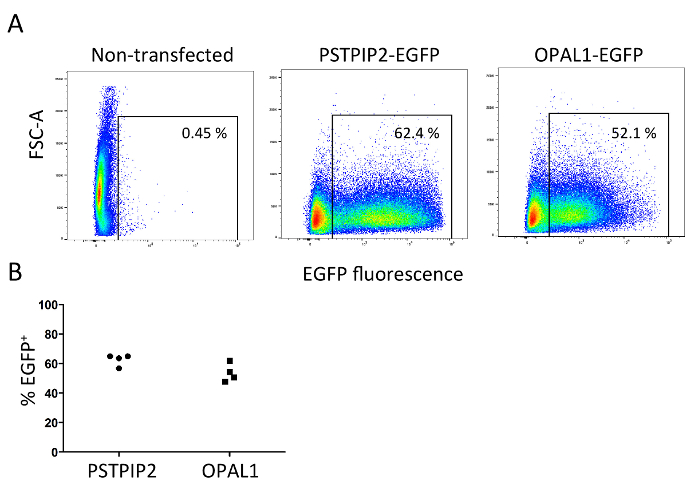
Figure 1: Efficiency of Plat-E cell transfection. For transfection of Plat-E cells, two constructs encoding adaptor proteins PSTPIP2 and OPAL1 fused with EGFP (PSTPIP2-EGFP and OPAL1-EGFP) were cloned into the pMSCV vector. Standard PEI transfection was performed. The efficacy of transfection was evaluated by flow cytometry of the Plat-E cells after the collection of the second viral supernatant. (A). Representative flow cytometry plot. (B). Graph showing results of four independent experiments. Please click here to view a larger version of this figure.
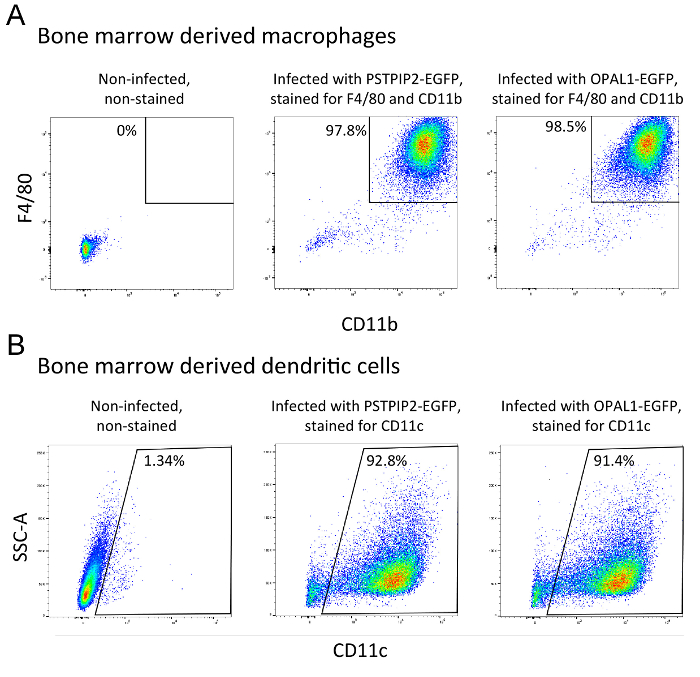
Figure 2: Assessment of the differentiation status of BMDMs (A) and BMDCs (B). Surface expression of specific macrophage and dendritic cell lineage markers was measured by flow cytometry at day 8 of cultivation. Dead cells were gated out based on their side and forward scatter properties and staining with Hoechst 33258. The results are representative of at least 3 independent experiments. Please click here to view a larger version of this figure.
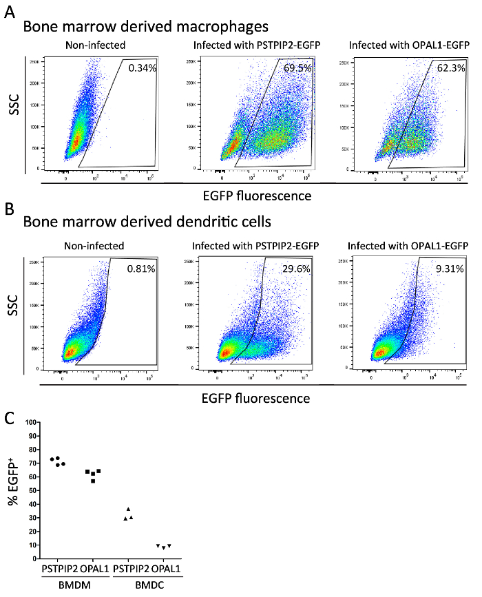
Figure 3: Assessment of the expression of PSTPIP2-EGFP and OPAL1-EGFP. EGFP fluorescence in BMDMs (A) and BMDCs (B) retrovirally transduced with PSTPIP2-EGFP and OPAL1-EGFP constructs was measured by flow cytometry at day 8 of cultivation. (C). Graph showing the results of multiple independent experiments. BMDMs and BMDCs were gated as in Figure 2. Please click here to view a larger version of this figure.
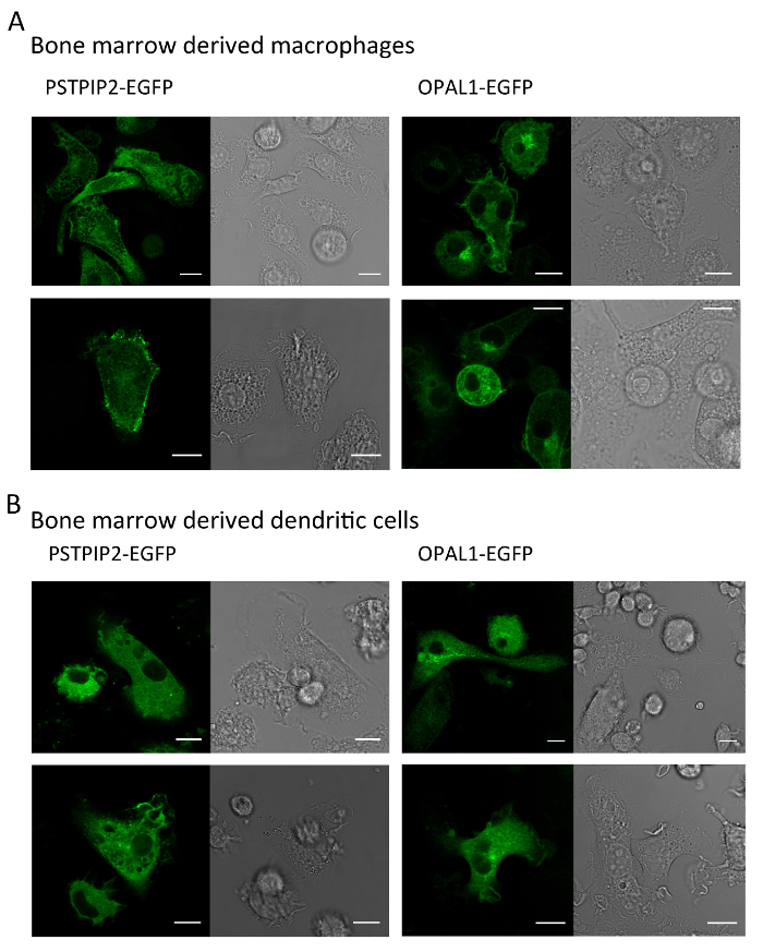
Figure 4: Representative images of macrophages and dendritic cells expressing PSTPIP2 or OPAL1. BMDMs (A) and BMDCs (B) expressing PSTPIP2 and OPAL1 were visualized by live imaging confocal microscopy. EGFP fluorescence in green is shown on the left side of each panel, bright field image on the right. Scale bar = 10 µm. The results are representative of at least three independent experiments. Please click here to view a larger version of this figure.
The expression of protein of interest in target cells is a key step in many types of biological studies. Differentiated macrophages and dendritic cells are difficult to transfect by standard transfection and retroviral transduction techniques. Bypassing the transfection of these differentiated cells with retroviral transduction of bone marrow progenitors, followed by differentiation when they already carry the desired construct, is a critical step allowing the expression of ectopic cDNAs in these cell types. An example of successful use of this method can be found in our recent publication25. Here, we provide a cost-effective protocol for achieving stable expression of the construct of choice in bone marrow-derived dendritic cells and macrophages using this approach. The procedure we present is relatively inexpensive and simple, yet delivering very good results. Reagents used in this protocol allow for its routine use even under a relatively restrictive budget. The protocol for Plat-E transfection employs PEI as a transfection reagent. Compared to other chemical transfection agents, PEI is of a very low cost, while its efficiency is similar to the majority of other widely used compounds. However, PEI can be replaced with many different commercially available transfection reagents in this step without any loss of efficiency. As a guiding principle, transfection protocols known to work with commonly used HEK293 cells typically perform well with Plat-E cells, too. Another cost-effective measure is the utilization of cytokine-containing supernatants instead of purified recombinant cytokines. The use of these supernatants requires some optimization. However, when the standard protocol for their preparation is established and followed, the variability between individual batches of these supernatants becomes very low, usually requiring no changes in working concentrations between individual lots. The efficiencies of BMDM and BMDC differentiation with these supernatants are, in our experience, identical to purified cytokines.
In addition to retroviral transduction, other well-established methods of mammalian cell transfection exist, including chemical transfection (typically using cationic lipids or cationic polymers forming complexes with DNA), electroporation and the use of other types of viral vectors, mainly adenoviral and lentiviral systems26,27,28,29,30. Although very high titers and efficiencies can be achieved with adenovirus-based gene delivery, the preparation of corresponding plasmids and viral particles are more difficult and time-consuming than in the case of retroviral systems29. In addition, adenovirus elicits inflammatory response in dendritic cells and macrophages29,31,32 and for optimum performance in murine hematopoietic cells mouse strain carrying transgenic adenovirus receptor is required33. On the other hand, the generation of lentiviral particles carrying the gene of interest is a relatively simple process, virtually identical to the one utilized for retroviruses. In contrast to the ecotropic retroviral vectors used in this protocol, lentiviruses are capable of infecting non-proliferating cells of multiple species, including humans34. This may be an important advantage under specific experimental conditions. However, this feature also greatly compromises the safety of these vectors. In our opinion, for gene delivery to BMDMs and BMDCs, retroviruses provide the best balance of efficiency, safety and ease of use. Retroviral DNA constructs can be easily prepared using simple standard molecular cloning techniques. Virus is produced by packaging the cell lines directly to the culture supernatant and further virus purification is usually not necessary. Ecotropic retroviruses also do not readily infect human cells, which makes their use relatively safe. However, there also are some general disadvantages associated with the use of retroviral vectors. The major limiting factor is that these viruses infect only proliferating cells35. This feature does not significantly affect the protocol described here, but it limits the range of applications where retroviral vectors can be used. The size of the gene of interest that can be cloned into these vectors is also limited and the viral particle titer decreases with increasing insert size. With pMSCV vectors, we usually start seeing effects of insert size at around 3 kbp. With further increases in insert size, the infection efficiency gradually declines.
The chemical transfection and electroporation are easier to use, safer and less time-consuming than any of the virus-based procedures26,27,28,30. However, in BMDMs and BMDCs, they can stimulate responses to foreign nucleic acids31 and they generate more cellular stress. In addition, some chemical transfection reagents increase cell auto-fluorescence that may interfere with flow cytometry or microscopy analyzes. Due to the transient nature of expression, they can only be used with mature differentiated BMDMs or BMDCs. In contrast, the sequences introduced with retroviral vectors are permanently integrated into the genome of the target cells and allow for a stable long-lasting expression compatible with the time scale of the differentiation protocols34,35. However, this feature also increases the risk of insertional mutagenesis. Due to the relatively random nature of the vector integration, its effects on large populations of cells are limited. However, they may be visible at the level of individual cells. Additional problems may arise when a construct expressed from the retroviral vector affects dendritic cell or macrophage differentiation, resulting in failure to generate differentiated BMDMs or BMDCs from the infected progenitors. To some extent, this may be overcome by adjusting the infection conditions to achieve low expression levels (e.g., by reducing the virus titer) or through the use of an inducible expression system. The use of EGFP fused to the protein of interest or as a reporter also allows for sorting of cells with expression level corresponding to experiment goals and limitations. Finally, we should also mention problems common to all transfection/transduction procedures. These include mainly overexpression artefacts, such as protein misfolding, mislocalization and toxicity36. Protein toxicity could be the reason why OPAL1 was relatively difficult to express. Its example clearly illustrates that the nature of the expressed protein can substantially affect the effectiveness of this method. However, despite this, we were able to obtain sufficient quantities of OPAL1-EGFP expressing cells for microscopy analysis with this method, demonstrating its usefulness even when dealing with difficult targets. In addition, it would be possible to increase the percentages of transduced cells by FACS sorting if required by a particular application. The low infection efficiency can also be partially overcome by increasing viral particle concentration using various methods or ready-to-use kits. In our hands, ultrafiltration of the viral supernatant on centrifugal filters with a molecular weight cut off of 100 kDa has proven to provide the best balance between efficiency and required effort.
Bone marrow derived macrophages and dendritic cells are widely used tools in phagocyte immunology. They are more physiologically relevant than available cell lines. They can be generated in relatively high numbers and, at the same time, lack the genetic heterogeneity and instability characteristic of cell lines. Another advantage is that they can be generated from genetically modified mice to study the effects of genetic modification on a relatively abundant and homogenous cell population. This is particularly useful in biochemical studies, where relatively large amounts of cells are typically required. The ability to transduce these cells with cDNA constructs opens up additional possibilities of research based on the reconstitution or complementation of genetic defects in these cells and structure-function analysis.
The authors have nothing to disclose.
This work was supported by Czech Science Foundation (GACR) (project number 16-07425S), by Charles University Grant Agency (GAUK) (project number 923116) and by institutional funding from the Institute of Molecular Genetics, Academy of Sciences of the Czech Republic (RVO 68378050).
DMEM | Thermo Fisher Scientific, Waltham, MA, USA | 15028 | |
Fetal bovine serum (FBS) | Thermo Fisher Scientific, Waltham, MA, USA | 10270 | For media suplementation |
KHCO3 | Lachema, Brno, Czech Republic | N/A | |
NH4Cl | Sigma-Aldrich (Merck, Kenilworth, NJ, USA) | A9434 | |
Penicillin | BB Pharma AS, Prague, Czech Republic | N/A | PENICILIN G 1,0 DRASELNÁ SOL' BIOTIKA |
Streptomycin | Sigma-Aldrich (Merck, Kenilworth, NJ, USA) | S9137 | Streptomycin sulfate salt powder |
Gentamicin | Dr. Kulich Pharma, Hradec Králové, Czech Republic | N/A | |
Polyethylenimine, linear, MW 25,000 | Polyscience, Warrington, PA, USA | 23966 | |
Polybrene | Sigma-Aldrich (Merck, Kenilworth, NJ, USA) | H9268 | |
EDTA | Sigma-Aldrich (Merck, Kenilworth, NJ, USA) | E5134 | |
PBS | Prepared in-house by media facility of IMG ASCR, Prague, Czech Republic | N/A | |
APC anti-mouse/human CD11b Antibody, clone M1/70 | BioLegend (San Diego, CA, USA) | 101212 | flow cytometry analysis |
PE anti-mouse F4/80 Antibody, clone BM8 | BioLegend (San Diego, CA, USA) | 123110 | flow cytometry analysis |
APC anti-mouse CD11c Antibody, clone N418 | BioLegend (San Diego, CA, USA) | 117310 | flow cytometry analysis |
M-CSF | PeproTech (Rocky Hill, NJ, USA) | 315-02 | |
GM-CSF | PeproTech (Rocky Hill, NJ, USA) | 315-03 | |
Hoechst 33258 | Thermo Fisher Scientific, Waltham, MA, USA | H1398 | flow cytometry analysis use at 1-2 µg/ml |