1. Reagent Preparation
- 0.05 M HCl
- Prepare 0.05 M HCl by adding 208 µL of 37% HCl to 50 mL of distilled water.
- High-performance liquid chromatography eluent
- Prepare high-performance liquid chromatography (HPLC) eluent by dissolving 6.9 g of sodium dihydrogen phosphate monohydrate, 7.1 g of disodium hydrogen phosphate, 8.7 g of sodium chloride, and 0.7 g of sodium azide in 1 L of water. Mix well and check the pH. Pass the eluent through a 0.1-µm cutoff sterile filter and degas before use. Acceptance range: pH 6.2 – 7.0 (if not, adjust with NaOH [1 M] or HCl [5 M]).
2. Synthesis of Citrate-coated Iron Oxide Nanoparticles
- Dissolve 75 mg of FeCl3·6H2O and 80 mg of citric acid trisodium salt dihydrate in 9 mL of water.
NOTE: These quantities provide 12 mL of final purified nanoparticles ([Fe] ~1.4 mg·mL-1). Quantities can be scaled down to obtain a final volume of 2.5 mL. - Put the mixture in the microwave-adapted flask.
- Load a dynamic protocol in the microwave. Set the temperature to 120 °C, the time to 10 min, the pressure to 250 psi, and the power to 240 W.
- Add 1 mL of hydrazine hydrate to the reaction.
NOTE: Hydrazine hydrate starts iron reduction. Therefore, a change in the appearance of the solution, from light yellow to brown, is observed. - Start the microwave protocol.
- Meanwhile, rinse a gel filtration desalting column with 20 mL of distilled water.
- Once the protocol has finished, allow the flask to cool at room temperature.
- Pipette 2.5 mL of the final mixture onto the column and discard the flow-through.
NOTE: The microwave stops the protocol at 60 °C; the nanoparticles can be added directly to the gel filtration column at 60 °C. - Add 3 mL of distilled water to the column and collect the nanoparticles in a glass vial.
NOTE: Nanoparticles can be stored at room temperature for 1 week. After this time, nanoparticle aggregation appears, increasing their hydrodynamic size.
3. Synthesis of 68Ga Core-doped Iron Oxide Nanoparticles (68Ga-C-IONP)
- Put 75 mg of FeCl3·6H2O and 80 mg of citric acid trisodium salt dihydrate into the microwave-adapted flask.
- Elute the 68Ge/68Ga generator using the recommended volume and concentration of HCl, according to the vendor (in our case, 4 mL of 0.05 M HCl). After the injection of that volume in the self-shielded generator, (4 mL of) 68GaCl3 is obtained, ready to use without further processing.
NOTE: Follow the corresponding radioactivity safety measures for steps 3.2 – 3.12. 68Ga is a positron and gamma emitter isotope. The use of the appropriate safety measures to avoid exposure to radiation by the operator is crucial. Researchers must follow an ALARA (as low as reasonably achievable) protocol using typical shielding and radionuclide-handling procedures. Moreover, the use of a ring, body badges, and a contamination detector is mandatory. - Add 4 mL of 68GaCl3 to the microwave-adapted flask. This volume can be smaller, depending on the generator activity and desired activity of final nanoparticles.
- Pipette 5 mL of distilled water into the flask and mix well.
- Load a dynamic protocol in the microwave. Set the temperature to 120 °C, the time to 10 min, the pressure to 250 psi, and the power to 240 W.
- Add 1 mL of hydrazine hydrate to the reaction.
NOTE: Hydrazine hydrate starts iron reduction. Therefore, a change in the appearance of the solution, from light yellow to brown, is observed. - Start the microwave protocol.
- Meanwhile, rinse a gel filtration desalting column with 20 mL of distilled water.
- Once the protocol has finished, allow the flask to cool at room temperature.
- Pipette 2.5 mL of the final mixture onto the column and discard the flow-through.
NOTE: The microwave stops the protocol at 60 °C; the nanoparticles can be directly added to the gel filtration column at 60 °C. - Add 3 mL of distilled water to the column and collect the nanoparticles in a glass vial.
- Calculate radiolabeling efficiency using a NaI well-type detector. This parameter typically measures the activity of the 68Ga incorporated in the reaction. After synthetic and purification processes, the activity of the purified sample is measured. Because of the short half-life of 68Ga, the initial activity has to be corrected at time (t). Normalization with time follows the standard equation:
NT = N0 · e-λt
Here,
NT: Counts at time (t)
N0: Counts at time (t) = 0
λ: Decay constant
t: Elapsed time

NOTE: Radiolabeling efficiency should be between 90% – 95%.
4. Analysis of 68Ga Core-doped Iron Oxide Nanoparticles (68Ga-C-IONP)
- Dynamic light scattering
- Use dynamic light scattering (DLS) to measure the hydrodynamic size of 68Ga-C-IONP. Pipette 60 µL of the sample into a cuvette and perform three size measurements per sample. To ensure reproducibility, this should be repeated with several nanoparticle batches.
- Colloidal stability
- Assess the colloidal stability of 68Ga-C-IONP by measuring the hydrodynamic size of the sample after incubation in different buffers (PBS, saline, and mouse serum) for different times, ranging from 0 to 24 h. Incubate 500 µL of the sample in each buffer at 37 °C. At the selected times, take 60-µL aliquots and pipette them into DLS cuvettes to measure their hydrodynamic size.
- Electron microscopy
- Analyze the core size of 68Ga-C-IONP using transmission electron microscopy (TEM) and annular dark-field imaging (STEM-HAADF) (ref TEM protocol: NIST – NCL Joint Assay Protocol, PCC-X, Measuring the Size of Nanoparticles Using Transmission Electron Microscopy).
- Gel filtration radio-chromatogram
- Fractionate the elution into 500-µL aliquots during the gel-filtration purification step and measure the radioactivity present in each one using an activimeter; thus, rendering a gel-filtration chromatogram.
- Radiochemical stability of 68Ga-C-IONP
- Incubate 68Ga-C-IONP in mouse serum for 30 min at 37 °C (repeated 3x). After that time, purify the nanoparticles by ultrafiltration and measure the radioactivity present in the nanoparticles and filtrate. No activity should be detected in the different filtrates.
- Relaxometry
- Measure longitudinal (T1) and transverse (T2) relaxation times in a relaxometer at 1.5 T and 37 °C. Four different concentrations of 68Ga-C-IONP (2 mM, 1 mM, 0.5 mM, and 0.25 mM) should be measured. Plot relaxation rates (r1=1/T1, r2=1/T2) against iron concentration. The slope of the curve obtained renders r1 and r2 values.
- MR and PET phantom images
- Acquire in situ MR (T1-weighted sequence) and PET phantom images for a series of dilutions of 68Ga-C-IONP (0 mM, 1 mM, 6.5 mM, and 9.0 mM) to observe the increasing signal in correlation with the PET activity and MRI.
68Ga-C-IONP were synthesized by combining FeCl3, 68GaCl3, citric acid, water, and hydrazine hydrate. This mixture was introduced into the microwave for 10 min at 120 °C and 240 W under controlled pressure. Once the sample had cooled down to room temperature, the nanoparticles were purified by gel filtration to eliminate unreacted species (FeCl3, citrate, hydrazine hydrate) and free 68Ga (Figure 1).
The hydrodynamic size of 68Ga-C-IONP was measured using dynamic light scattering (DLS). This revealed a narrow size distribution (PDI 0.2) and mean hydrodynamic size of 7.9 nm. Measurements of five different syntheses proved method reproducibility (Figure 2a). The zeta potential of several 68Ga-C-IONP syntheses was measured to analyze nanoparticle surface charge; the mean value obtained was -36.5 mV. 68Ga-C-IONP was incubated in different media at 37 °C during different times to ensure nanoparticle stability in biological solutions. The hydrodynamic size was measured at different times, revealing 68Ga-C-IONP hydrodynamic size suffers no significant changes, meaning the sample is stable in different buffers and serums (Figure 2b). Because of the fast heating achieved using microwave technology, nanoparticles present ultra-small core sizes of about 4 nm. Electron microscopy images revealed homogeneous core sizes and the absence of aggregation (Figure 2c). A gel filtration chromatogram of 68Ga-C-IONP shows a main radioactivity peak corresponding to the nanoparticles, followed by a reduced peak that corresponds to free 68Ga (Figure 2d). The radiolabeling yield calculated after sample purification was 92%. This excellent radiolabeling yield was translated into a specific activity relative to an iron amount of 7.1 GBq/mmol Fe. The potential of 68Ga-C-IONP as a contrast agent for MRI was checked by measuring longitudinal (r1) and transversal (r2) relaxation times. These were measured for five different 68Ga-C-IONP syntheses at 37 °C and 1.5 T. An excellent mean r1 value of 11.9 mM-1·s-1 and a modest r2 value of 22.9 mM-1·s-1 were obtained, yielding an average r2/r1 ratio of 1.9, meaning 68Ga-C-IONP is ideal for T1-weighted MRI (Figure 2e). To confirm this hypothesis, the capability of 68Ga-C-IONP to produce T1 contrast in an MRI and PET signal was checked with the acquisition of PET and MR phantom images at different 68Ga-C-IONP concentrations. As the iron concentration increases, so does the positive contrast in MR phantom. An increasing iron concentration implies an increasing 68Ga concentration as well; hence, the PET signal is increasingly intense (Figure 2f).
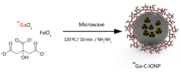
Figure 1: Synthetic steps followed in the protocol. Precursors are added in a microwave flask and introduced into the microwave upon hydrazine hydrate addition at 120 °C for 10 minutes, after which nanoparticles are obtained. Please click here to view a larger version of this figure.
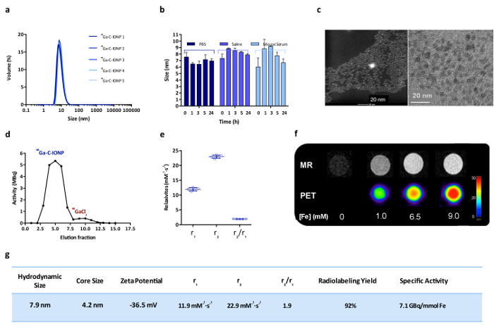
Figure 2: 68Ga-C-IONP characterization. (a) This panel shows the hydrodynamic size distribution (volume weighted) of five different syntheses of 68Ga-C-IONP. (b) This panel shows the hydrodynamic size (maximum peak in volume, mean ± SD) of 68Ga-C-IONP in PBS, saline, and mouse serum (from t = 0 h to t = 24 h). (c) These are STEM-HAADF (left) and TEM (right) images of 68Ga-C-IONP. The scale bars are 20 nm. (d) This panel shows a gel filtration radio-chromatogram. (e) This panel shows the longitudinal (r1) and transversal (r2) relaxivity values, and the r2/r1 ratio for five 68Ga-C-IONP syntheses (mean ± SD). (f) These are MR and PET phantom images of different 68Ga-C-IONP concentrations. (g) This is a table summarizing the main 68Ga-C-IONP characteristics. Please click here to view a larger version of this figure.
