We describe a method of using polyethyleneimine (PEI)-coated superparamagnetic iron oxide nanoparticles for transfecting macrophages with siRNA. These nanoparticles can efficiently deliver siRNA to macrophages in vitro and in vivo and silence target gene expression.
Because of their critical role in regulating immune responses, macrophages have continuously been the subject of intensive research and represent a promising therapeutic target in many disorders, such as autoimmune diseases, atherosclerosis, and cancer. RNAi-mediated gene silencing is a valuable approach of choice to probe and manipulate macrophage function; however, the transfection of macrophages with siRNA is often considered to be technically challenging, and, at present, few methodologies dedicated to the siRNA transfer to macrophages are available. Here, we present a protocol of using polyethyleneimine-coated superparamagnetic iron oxide nanoparticles (PEI-SPIONs) as a vehicle for the targeted delivery of siRNA to macrophages. PEI-SPIONs are capable of binding and completely condensing siRNA when the Fe:siRNA weight ratio reaches 4 and above. In vitro, these nanoparticles can efficiently deliver siRNA into primary macrophages, as well as into the macrophage-like RAW 264.7 cell line, without compromising cell viability at the optimal dose for transfection, and, ultimately, they induce siRNA-mediated target gene silencing. Apart from being used for in vitro siRNA transfection, PEI-SPIONs are also a promising tool for delivering siRNA to macrophages in vivo. In view of its combined features of magnetic property and gene-silencing ability, systemically administered PEI-SPION/siRNA particles are expected not only to modulate macrophage function but also to enable macrophages to be imaged and tracked. In essence, PEI-SPIONs represent a simple, safe, and effective nonviral platform for siRNA delivery to macrophages both in vitro and in vivo.
Macrophages are a type of innate immune cells distributed in all body tissues, albeit in different amounts. By producing a variety of cytokines and other mediators, they play critical roles in the host defense against invading microbial pathogens, in tissue repair following injury, and in maintaining tissue homeostasis1. Due to their importance, macrophages have continuously been the subject of intensive research. However, despite its prevalence in gene regulation and function studies, siRNA-mediated gene silencing is less likely to succeed in macrophages because these cells—particularly, primary macrophages—are often difficult to transfect. This can be ascribed to a relatively high degree of toxicity associated with most well-established transfection approaches in which the cell membrane is chemically (e.g., with polymers and lipids) or physically (e.g., by electroporation and gene guns) disrupted to let siRNA molecules cross the membrane, thereby drastically reducing macrophages' viability2,3. Furthermore, macrophages are dedicated phagocytes rich in degradative enzymes. These enzymes can damage the integrity of siRNA, weakening its silencing efficiency even if gene-specific siRNA has been delivered into the cell3,4. Therefore, an effective macrophage-targeted siRNA delivery system needs to protect the integrity and stability of siRNA during delivery4.
It is increasingly evident that dysfunctional macrophages are implicated in the initiation and progression of certain common clinical disorders like autoimmune diseases, atherosclerosis, and cancer. For this reason, modulating macrophage function with, for instance, siRNA, has been emerging as an attractive methodology for treating these disorders5,6,7. Although much progress has been made, a major challenge of siRNA-based treatment strategy is the poor cell specificity of systemically administered siRNA and the insufficient siRNA uptake by macrophages, which consequently lead to undesired side effects. Compared with free nucleic acid therapeutics that usually lack optimal cell selectivity and often lead to off-target adverse effects, drug-loaded nanoparticles (NPs), owing to their spontaneous propensity of being captured by the reticuloendothelial system, can be engineered for passive targeting to macrophages in vivo, allowing for improved therapeutic efficacy with minimal side effects8. Current NPs explored for the delivery of RNA molecules include inorganic nanocarriers, various liposomes, and polymers9. Among them, polyethyleneimine (PEI), a type of cationic polymers capable of binding and condensing nucleic acids into stabilized NPs, shows the highest RNA delivering capacity9,10. PEI protects nucleic acids from enzymatic and nonenzymatic degradation, mediates their transfer across the cell membrane, and promotes their intracellular release. Although initially introduced as a DNA delivery reagent, PEI was subsequently demonstrated to be an attractive platform for in vivo siRNA delivery, either locally or systemically9,10.
Superparamagnetic iron oxide nanoparticles (SPIONs) have shown great promise in biomedicine, owing to their magnetic properties, biocompatibility, comparable size to biologically important objects, high surface-area-to-volume ratio, and easily adaptable surface for bioagent attachment11. For instance, because of their potential utility as a contrast agent and rapid uptake by macrophages, SPIONs have emerged as a favorite clinical tool to image tissue macrophages12. While SPIONs have also been extensively studied as nucleic acid delivery vehicles11,13,14,15, to our knowledge, the literature contains few reports of SPIONs as a carrier for macrophage-targeted siRNA delivery. For gene delivery by SPIONs, their surface is usually coated with a layer of hydrophilic cationic polymers onto which negatively charged nucleic acids can be electrostatically attracted and tethered. Here, we present a method for synthesizing SPIONs whose surface is modified with low-molecular-weight (10 kDa), branched PEI (PEI-SPIONs). These magnetic nanoplatforms are then employed to condense siRNA, forming PEI-SPION/siRNA complexes that enable siRNA transport into the cell. We reason that spontaneous phagocytosis of SPIONs by cells of the reticuloendothelial system16, coupled with the strong ability of binding and condensing nucleic acids by PEI, renders PEI-SPIONs suitable for the efficient transport of siRNA into macrophages. The data presented here support the feasibility of PEI-SPION/siRNA-mediated gene silencing in macrophages in culture as well as in vivo.
All methods involving live animals were performed in accordance with the animal care and use guidelines of Southeast University, China.
1. Preparation of PEI-SPIONs
2. Preparation and Agarose Gel Electrophoresis of PEI-SPION/siRNA NPs
3. Transfection of RAW264.7 Macrophages In Vitro
4. Systemic Delivery of siRNA to Macrophages in Rats with Experimental Arthritis
The size and zeta potential of PEI-SPIONs prepared with this protocol were in the range of 29 – 48 nm (polydispersity index: 0.12 – 0.23) and 30 – 48 mV, respectively. They were stable in water at 4 °C for over 12 months without obvious aggregation. To evaluate their siRNA binding ability, PEI-SPIONs were mixed with siRNA at various Fe:siRNA weight ratios. Figure 1 shows that when the Fe:siRNA weight ratio reaches 4 and above, the band of free siRNA was completely missing, implying successful siRNA binding to PEI-SPIONs. A major concern of PEI in biomedical application is its toxicity, which is a result of the strong positive charge, particularly at high molecular weights and high doses. As shown in Figure 2A, PEI-SPIONs with a zeta potential of 30.5 and 37 mV did not exhibit apparent cytotoxicity at concentrations up to 30 μg Fe/mL, which is about twofold higher than the concentration (15 μg Fe/mL) normally used for cell transfection. However, PEI-SPIONs with a zeta potential of 48 mV were toxic even at the lowest dose examined (10 μg Fe/mL). Therefore, PEI-SPIONs possess a charge-dependent toxicity. Since cationic charge is not important for the NP uptake by macrophages19, we suggest that PEI-SPIONs with an average zeta potential not higher than +37 mV are used for the siRNA transfer, although siRNA binding would decrease the charge to some extent and alleviate cytotoxicity18.
To test the potential application of PEI-SPIONs for siRNA delivery to macrophages, the in vitro transfection was performed with the murine macrophage cell line RAW 264.7. As analyzed by flow cytometry, more than 90% of the cells were transfected with fluorescently labeled PEI-SPION/siRNA complexes at 15 µg Fe/mL (Figure 2B). With respect to transfection efficiency, there was actually no difference between PEI-SPION/siRNA NPs formed at Fe:siRNA weight ratios of 4 and 8, although, under the latter condition, the NPs that were formed were smaller in size and weaker in positive charge because a lesser amount of siRNA was loaded per particle. We also assessed the effect of PEI-SPION/siRNA concentration on cellular internalization by Prussian blue staining. As shown in Figure 2C, the blue spots within the transfected cells were minimally detectable at 7.5 µg Fe/mL, but clearly visible at 15 µg Fe/mL. Interestingly, increasing the PEI-SPION/siRNA concentration to 32 µg Fe/mL did not increase the staining intensities, probably because the PEI-SPION/siRNA uptake was saturated at concentrations around 15 µg Fe/mL. In addition, the ability of PEI-SPIONs for mediating the siRNA transfer was corroborated in primary macrophages18, and the method presented here had a high siRNA transfection efficiency in rat peritoneal macrophages, equivalent to that in RAW264.7 cells. The peritoneal macrophages transfected with PEI-SPIONs harboring specific siRNA showed a significant decrease in the target mRNA level as compared with nonspecific siRNA (Figure 2D), implying that siRNA could escape from endocytosis vesicles into the cytoplasm and reach the RNAi machinery.
We previously also investigated the in vivo cellular uptake of systemically administered PEI-SPION/siRNA complexes in rats with adjuvant arthritis18. We analyzed PEI-SPION/siRNA transfection efficiency in phagocytic macrophages and nonphagocytic T lymphocytes. As shown in Figure 3, CD11b+ cells took up PEI-SPION/siRNA complexes more efficiently than CD3+ cells at any time point in all of the organs examined, indicating that PEI-SPION/siRNA NPs preferentially target macrophages18. Notably, a high-level accumulation of the NPs was observed in inflamed joints18, suggesting that the PEI-SPION can be an attractive platform for the systemic delivery of siRNA therapeutics in rheumatoid arthritis whose pathogenesis is linked to macrophage dysfunction and for which local siRNA administration is not a favorite choice due to the involvement of multiple organs of the disease.
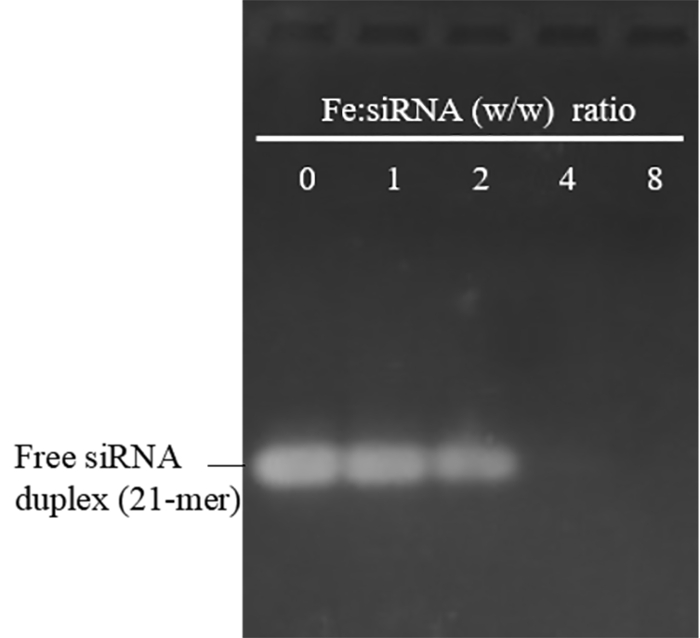
Figure 1: Agarose gel electrophoresis of PEI-SPION/siRNA complexes formed at various Fe:siRNA (w/w) ratios. A Fe:siRNA ratio of 0 represents free siRNA duplexes without PEI-SPIONS. The average size and zeta potential of the free PEI-SPIONs used here were 30 nm and 45 mV. siRNA could completely bind to PEI-SPIONs when the Fe:siRNA ratio reaches 4 and above, consistent with the previous results using PEI-SPIONs with an average size of 48 nm and a zeta potential of 30.5 mV18. The absence of retarded bands (PEI-SPION/siRNA complexes) may reflect inaccessibility of siRNA to EB during staining, an indication of strong siRNA binding, and/or the condensing ability of PEI-SPIONs. Please click here to view a larger version of this figure.
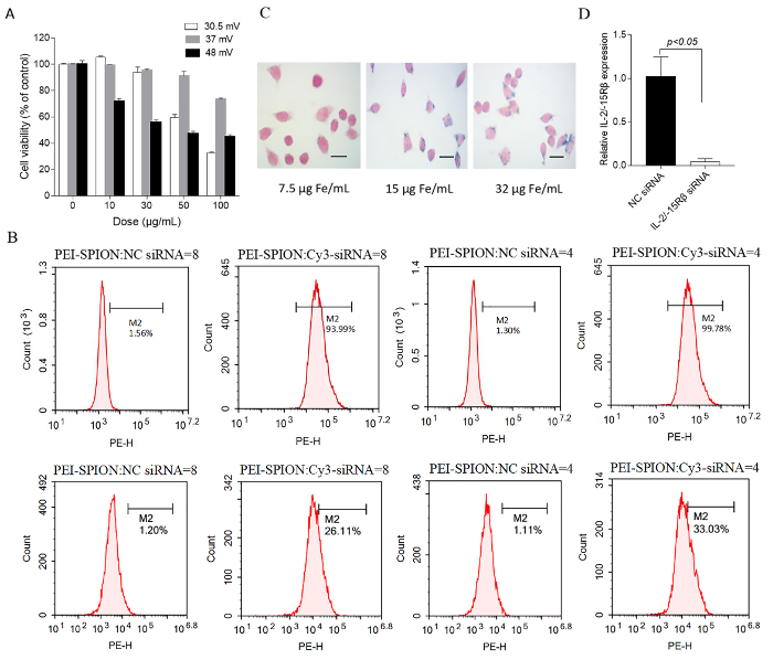
Figure 2: Biological characterization of PEI-SPION and PEI-SPION/siRNA NPs. (A) This panel shows a cell viability assay. RAW 264.7 cells were treated for 16 h with the indicated doses of PEI-SPIONs bearing different zeta potential and, then, an MTS assay was performed. The cell viability was normalized against the control (no particle exposure). The data are the mean ± SD of duplicate wells. (B) This panel shows a flow cytometric analysis of the PEI-SPION/Cy3-siRNA uptake by RAW 264.7 cells. The cells were incubated for 24 h with 15 µg Fe/mL (upper panel) or 5 μg Fe/mL (lower panel) of PEI-SPIONs complexed with Cy3-labeled siRNA at a Fe:siRNA (w/w) ratio of 4 and 8, respectively. Nonspecific (NC) siRNA represents non–fluorescent siRNA. M2: gated region; Pe-H: Cy3 fluorescence intensity. The siRNA transfection efficiency of PEI-SPIONs used here (37.8 nm, 48 mV) was similar to a previous study using PEI-SPIONs with an average size of 48 nm and a zeta potential of 30.5 mV18. (C) This panel shows an analysis of PEI-SPION/siRNA uptake by visualizing cellular iron deposits. RAW 264.7 cells were incubated with 7.5, 15, and 32 μg Fe/mL PEI-SPIONs (48 nm, 30.5 mV) complexed with siRNA (Fe:siRNA = 8) and stained by Prussian blue. The scale bars are 20 μm. (D) This panel shows an in vitro validation of the silencing efficiency of siRNA delivered by PEI-SPIONs. A specific siRNA-targeting rat IL-2/-15 receptor β chain was loaded onto PEI-SPIONs (48 nm, 30.5 mV) at Fe:siRNA = 8 and, then, transfected into rat peritoneal macrophages. An NC siRNA was used as control. The gene silencing effect was assessed by quantitative PCR. The cells were incubated with the complexes at 15 μg Fe/mL. The data are the mean ± the SD of triplicate wells. Panel D has been modified from Duan et al.18 with permission from the publisher. Please click here to view a larger version of this figure.
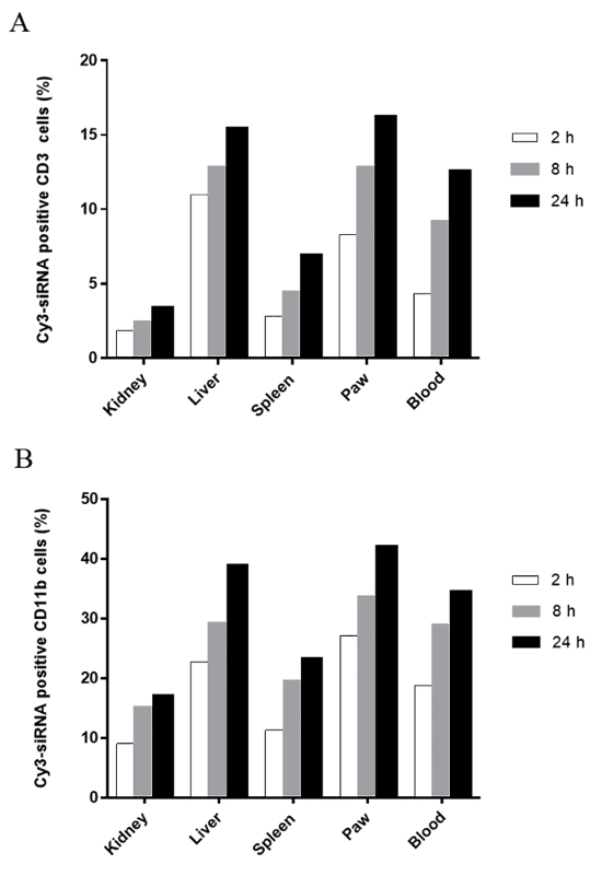
Figure 3: In vivo cellular uptake of PEI-SPION/siRNA NPs. Three arthritic rats were injected intravenously with a single dose of 0.3 mg/kg Cy3-siRNA formulated with PEI-SPION (48 nm, 30.5 mV). A rat injected with PBS was used as a control. Blood, spleen, liver, kidney, and inflamed joints were collected at 2, 8, and 24 h after the injection. The cellular uptake of PEI-SPION/Cy3-siRNA NPs was assessed by flow cytometry using (A) anti-CD3 and (B) anti-CD11b monoclonal antibodies. The percentages are of Cy3-siRNA uptake within the gated CD3+ or CD11b+ cells. The results shown here are representative of two independent experiments. This figure has been modified from Duan et al.18 with permission from the publisher. Please click here to view a larger version of this figure.
Macrophages are refractory to transfect by commonly used nonviral approaches, such as electroporation, cationic liposomes, and lipid species. Here we described a reliable and efficient method to transfect macrophages with siRNA. Using the present protocol, over 90% of macrophage-like RAW 264.7 cells (Figure 2B) and rat peritoneal macrophages18 can be transfected with siRNA without significant impairment of the cell viability. This method depends on the delivery platform PEI-SPION, which is a nanocarrier composed of a core of iron oxide and a shell of PEI. So, the first key step of the protocol is the synthesis of PEI-SPIONs suitable for siRNA delivery. Usually, PEI-coated SPIONs are prepared from oleic acid-coated SPIONs by a ligand-exchange method in which oleic acid is directly exchanged from the surface of SPIONs by PEI or its derivatives15, generating hydrophilic NPs with a positively charged surface. In the case presented here, the oleic acid capped on the surface of SPIONs was replaced by water-soluble dimercaptosuccinic acid, and then PEI was loaded onto SPION surfaces through electrostatic interactions. This method is gentle and easy to prepare in large quantities, and the synthesized NPs have excellent stability in water20. It is well-known that PEI is cytotoxic, and the toxicity correlates strongly with its molecular weight21. To ensure safety, an important consideration when synthesizing PEI-SPIONs is that these particles are coated with low-molecular-weight PEI, which is 10 kDa in this protocol. The undesired toxic effect of PEI is mainly mediated by its positive charge; therefore, the measurement of the zeta potential of PEI-SPIONs is essential, and the value should be not higher than 37 mV. A decrease in positive charge can be achieved simply by reducing the PEI content. Another critical step for the successful application of this siRNA delivery system is the optimization of the Fe:siRNA ratio by gel retardation. It seems reasonable to make PEI-SPION/siRNA complexes at low Fe:siRNA ratios under which siRNA molecules are still capable of binding to PEI-SPIONs. In this circumstance, small amounts of PEI-SPIONs can be used, thus minimizing its potential cytotoxicity.
In case a desired silencing efficiency or therapeutic effect is not produced, check the transfection efficiency by flow cytometry or fluorescence microscopy using the carrier loaded with a fluorescently labeled siRNA. Alternatively, the PEI-SPION/siRNA uptake can be examined by conventional Prussian blue staining, which is sensitive enough to detect single granules of iron in cells. If the transfection efficiency is indeed low, it may be required to optimize transfection conditions such as cell density, transfection time, and the dose of PEI-SPION/siRNA particles. The cell passage number can also affect the efficiency of transfection22. In most cases, an insufficient silencing effect is not caused by an insufficient PEI-SPION/siRNA uptake, as the present PEI-SPION system has been demonstrated to facilitate an effective siRNA transfer to macrophages. Sometimes, combining several siRNAs targeting the same gene can be a good strategy to enhance knockdown efficiency. It is noteworthy that, although RNAi generally occurs within 24 h of transfection, the onset and duration of gene silencing depend on the turnover rate of the target, the rate of dilution and longevity of siRNA, and even the concentration of serum in the medium. Thus, time course experiments may be needed to accurately determine the time point of maximal effect2,22. For in vivo application, the therapeutic efficacy also depends on whether, and to what extent, the siRNA target contributes to disease phenotypes; thus, the choice of an appropriate siRNA target is critical for expected results.
There are several advantages of this protocol for delivering siRNA to macrophages. (1) The method is easy to perform and is a cheap way to produce PEI-SPIONs in large quantities, and the NPs produced are stable in water for over 12 months if they are kept at 4 °C. (2) Spontaneous phagocytosis of SPIONs by macrophages facilitates an effective PEI-SPION-mediated siRNA transfer, resulting in high transfection efficiency. It is expected that besides RAW 264.7 cells and rat peritoneal macrophages, this approach is applicable to other macrophage cell lines and primary macrophages, as long as the dose for transfection is optimized. (3) siRNA transfection is fast and easy to conduct compared with other macrophage transfection methods such as nucleofection, which is time-consuming and requires a Nucleofector device2. (4) PEI-SPION can be an ideal vehicle for macrophage-targeted systemic siRNA delivery in certain disease models. Macrophages play critical roles in the development and progression of various chronic inflammatory disorders, as well as tumors; and one conspicuous histological feature of these diseases is the abnormal blood vessels with leaky endothelium. Hence, due to the enhanced permeability and retention effect, systemically administered drug-loaded NPs tend to accumulate in diseased tissues and are readily captured by local macrophages, leading to enhanced specificity, reduced side effects, and improved therapeutic efficacy. In a rat model of adjuvant arthritis, intravenously injected PEI-SPION/siRNA complexes were taken up by ~40% of the CD11b+ cells during the first 24 h after the injection18. In contrast, when cationic liposome was used as a carrier for systemic siRNA delivery in mice, less than 5% of the CD11b+ cells in the arthritic joints entrapped the siRNA lipoplexes23. Moreover, owing to their magnetic properties, the application of an external magnetic field may further facilitate the accumulation of PEI-SPION/siRNA complexes in the target tissues and increase their cellular uptake. Also noteworthy is that such siRNA-loaded NPs can be used not only for modulating macrophage function but also for imaging macrophage to provide diagnostic information, monitor treatment efficacy, and predict patients' clinical outcomes12.
However, limitations associated with this protocol do exist. The PEI-SPION system exhibits a narrow range of dosage for siRNA delivery. The maximum uptake occurred when RAW264.7 cells were exposed to PEI- SPION/siRNAs at a concentration of 15 µg Fe/mL (Figure 2B and 2C). Increasing the PEI-SPION/siRNA concentration to 32 µg Fe/mL did not result in an increase in cellular uptake (Figure 2C) but, on the contrary, might increase the risk of inducing cell death due to the intrinsic toxicity of PEI. On the other hand, decreasing PEI-SPION/siRNA to 5 or 7.5 µg Fe/mL obviously reduced its uptake by RAW264.7 cells (Figure 2B and 2C). Thus, we propose that the optimal PEI-SPION/siRNA concentration for in vitro macrophage transfection is ~15 µg Fe/mL (the final concentration in a well). Another limitation that needs to be taken into consideration is the possible effect of PEI-SPIONs on macrophage activity. Nanoparticles may induce the immune response24, depending on their surface modification, surface charge, size, shape, and even on the methodology used to synthesize them. Mulens-Arias et al. recently reported that PEI-coated SPIONs trigger macrophage activation25. The method of PEI-SPION synthesis presented here differs significantly from that of Mulens-Arias et al., and therefore, whether PEI-SPIONs prepared based on the present protocol trigger macrophage activation awaits further investigation. However, to unambiguously address this concern, we suggest that, in addition to the PEI-SPION complexed with scramble siRNA, the vehicle itself (PEI-SPION only) can serve as another control when using the present protocol. Finally, this protocol is not suitable for the delivery of DNA due to its relatively large size.
In summary, we presented here a method of using PEI-coated SPIONs as a vehicle for siRNA transfection in macrophages. These NPs can efficiently deliver siRNA into immortalized macrophage cell lines, as well as into primary macrophages in vitro, and functionally induce gene silencing without affecting cell viability at the optimal dose for transfection. Moreover, PEI-SPIONs may be used for in vivo siRNA delivery to macrophages, making it possible to image, as well as modulate, macrophages whose dysfunction contributes to the development and progression of many chronic inflammatory disorders and cancers.
The authors have nothing to disclose.
This work was supported by the National Natural Science Foundation of China (81772308) and the National Key Research and Development Program of China (No. 2017YFA0205502).
DMEM | Gibco | C11995500BT | Warm in 37°C water bath before use |
Fetal bovine serum | Gibco | A31608-02 | |
Penicillin/streptomycin (1.5 ml) | Gibco | 15140122 | |
Tetrazolium-based MTS assay kit | Promega | G3582 | For cytotoxicity analysis |
RAW 264.7 cell line | Cell Bank of Chinese Academy of Sciences, Shanghai, China | TCM13 | |
Tissue culture plates (6-well) | Corning | 3516 | |
Tissue culture dishes (10 cm) | Corning | 430167 | |
RNase-free tubes (1.5 ml) | AXYGEN | MCT-150-C | |
Centrifuge tubes (15 ml) | Corning | 430791 | |
Trypsin | Gibco | 25200-056 | |
Wistar rats | Shanghai Experimental Animal Center of Chinese Academy of Sciences |
||
Bacillus Calmette–Guérin freeze-dried powder | National Institutes for Food and Drug Control, China |
for inducing adjuvant arthritis in rats | |
siRNA | GenePharma (Shanghai, China) | ||
Cy3-siRNA | RiboBio (Guangzhou, China) | ||
Polyethyleneimine (10 kDa) | Aladdin Chemical Reagent Co., Ltd. | E107079 | |
Ammonia water | Aladdin Chemical Reagent Co., Ltd. | A112077 | |
Oleic acid | Aladdin Chemical Reagent Co., Ltd. | O108484 | |
Dimethylsulfoxide | Aladdin Chemical Reagent Co., Ltd. | D103272 | |
FeSO4•7H2O | Sinopharm Chemical Reagent Co., Ltd | 10012118 | |
FeCl3•6H2O | Sinopharm Chemical Reagent Co., Ltd | 10011918 | |
Dimercaptosuccinic acid | Aladdin Chemical Reagent Co., Ltd. | D107254 | |
ultrafiltration tube | Millipore | UFC910096 | |
Tetramethylammonium hydroxide solution | Aladdin Chemical Reagent Co., Ltd. | T100882 | |
Particle size and zeta potential analyzer | Malvern, England | Nano ZS90 |