Here, we present a protocol for culturing the gut microbiota of the colon in vitro, using a series of bioreactors that simulate the physiological conditions of the gastro intestinal tract.
The human gut microbiota plays a vital role in both human health and disease. Studying the gut microbiota using an in vivo model, is difficult due to its complex nature, and its diverse association with mammalian components. The goal of this protocol is to culture the gut microbiota in vitro, which allows for the study of the gut microbiota dynamics, without having to consider the contribution of the mammalian milieu. Using in vitro culturing technology, the physiological conditions of the gastro intestinal tract are simulated, including parameters such as pH, temperature, anaerobiosis, and transit time. The intestinal surface of the colon is simulated by adding mucin-coated carriers, creating a mucosal phase, and adding further dimension. The gut microbiota is introduced by inoculating with the human fecal material. Upon inoculation with this complex mixture of bacteria, specific microbes are enriched in the different longitudinal (ascending, transverse and descending colons) and transversal (luminal and mucosal) environments of the in vitro model. It is crucial to allow the system to reach a steady state, in which the community and the metabolites produced remain stable. The experimental results in this manuscript demonstrate how the inoculated gut microbiota community develops into a stable community over time. Once steady state is achieved, the system can be used to analyze bacterial interactions and community functions or to test the effects of any additives on the gut microbiota, such as food, food components, or pharmaceuticals.
The gut microbiota is a community of micro-organisms that reside in the human gastrointestinal tract (GIT). This community reaches maximum concentration in the colon, which is estimated to hold 1013-1014 bacteria, from 500-1,000 species, that live in symbiosis with the colon milieu1,2. The composition and functionality of the gut microbiota change spatially along the GIT, forming region specific communities, with the most diversity found distally2,3,4,5. For each anatomical region, separate microbial communities reside in the lumen and on the mucosal lining6. The lumen community has more direct access to nutrients as substrates move through the luminal compartment7. Despite this, some bacteria reside preferentially in the mucus layer, utilizing mucin produced by the colon cells as an energy source1,5,8. The difference in microenvironments between the luminal and mucosal phases results in divergence and the development of phase specific communities. Together, these communities provide metabolic functions, such as nutrient metabolism and the production of vitamins, and immunological functions, such as preventing the colonization of human pathogens1,3,9. The gut microbiota also works functionally in conjugation with the human colon cells3.
As an important part of the human GIT, it is not surprising that the gut microbiota is known to contribute to both host health and disease status3,9,10,11,12. A shift in the gut microbial population has been associated with multiple human diseases, including GIT disorders like intestinal bowel disease (IBD) and intestinal bowel syndrome (IBS), but also other diseases, such as obesity, circulatory disease, and autism3,9,10,11,12. Metabolites produced from the gut microbiota have a global effect, reaching locations far from the gut12,13. For example, the gut-brain axis is associated with mental disorders like anxiety and depression14. Therefore, studying the gut microbiota is important to multiple fields of research, and is applicable to many diseases, even those not often associated with the GIT.
While it is widely acknowledged that studying the gut microbiota is important, it is a complicated endeavor. Multiple animal models are available, from small animals like zebrafish, rats, and mice, to larger ones like monkeys and pigs15-19. However, the application of these animals in terms of the human gut microbiota is not straightforward, since these animals have a unique bacterial community that has evolved based on environment and diet, and they are anatomically distinct from humans20,21. The use of human subjects removes the question of relevance yet introduces another set of challenges. Human studies are expensive, time consuming, and are ethically constrained11. Moreover, confounding factors influence the gut microbiota in human studies, including age or developmental stage, environment, diet, medication, and genetic factors2,4,22. There are also restrictions on what can be tested in humans, and which type of samples can be harvested at what times4.
One critical disadvantage of using an in vivo system to study the gut microbiota is the presence of mammalian components. The gut microbiota and human cells interact with one another, and in an in vivo setting, it is impossible to distinguish the two. The metabolites produced by the gut microbiota are taken in by the colon cells, so measurements cannot be calculated with precision. Therefore, any mechanistic study must be limited to end-point measurements11. Another major disadvantage for in vivo studies is the inability to harvest samples from the different regions of the GIT longitudinally23. This does not allow for the assessment of changes that may occur in the microenvironments of the colon over time12. Many in vivo studies, including human studies, rely on analysis of fecal samples to detect changes to the gut microbiota12. While this is informative, it does not provide data on the gut microbiota across the GIT and does not differentiate between the luminal and mucosal communities5,6,7,8.
For the gut microbiota, the application of an in vitro method is required to study the dynamics of the bacterial community, without interference from the mammalian components. Using an in vitro method allows for the tight control of environmental conditions10, testing of multiple parameters simultaneously, and the ability to sample longitudinally, and in large volumes11. Since an in vitro method utilizes a mechanical device and not a host, no considerations are needed for age, environment, diet, or genetic background. These systems can be used to test either the entire gut microbiota community, only selected organisms, or even single strains. Importantly, in vitro results are reproducible, yet retain a level of diversity comparable to in vivo studies11,22.
Depending on the hypothesis in question and the desired results, in vitro studies can be performed in numerous ways. They can utilize single-vessel systems and simple methods, such as incubating samples with fecal homogenate24 or performing single batch cultures over the course of 24-48 h25. They can also be accomplished using single-vessel systems and more complex methods, such as using a chemostat system to produce a stable gut microbial community11. However, the use of a single reactor can over-simplify the microbiota12 since it only represents one section of the colon, even though the colon is composed of the ascending, transverse, and descending regions.
In order to study the gut microbiota community that develops in the different regions of the colon (the ascending, transverse, and descending regions), a complex, multi-stage system can be employed. In these systems, multiple vessels are set up to mimic the different regions of the colon, so the gut microbiota of the ascending, transverse, and descending regions are cultivated independently. These vessels are connected, using pumps to move substrates in sequence, from the ascending to the transverse to the descending colon regions, mimicking the flow of nutrients through the GIT.
The objective of this study was to demonstrate how a 5-stage in vitro culture system (see Table of Materials) can be used to cultivate the gut microbiota community, and to demonstrate community dynamics in terms of stability and composition. In this system, one vessel represents the stomach and one represent the small intestine. The colon is divided into three regions (ascending, transverse, descending), with one vessel representing each region26. In this experimental setup, two complete systems were run in parallel, with Unit 1 containing mucin carriers to represents the mucosal surface and Unit 2 containing no mucin carriers. The communities that developed in the luminal and mucosal phases of each region were compared to each other, and to the fecal inoculum over time using 16S rRNA gene sequencing and SCFA analysis. The results presented demonstrate the type of community, both in terms of composition and functionality, which can be produced from this type of in vitro system.
1. Materials and Preparations
NOTE: The defined medium is purchased as a powder (see Table of Materials). The composition of the defined medium in g/L is the following: Arabinogalactan (1.2), Pectin (2.0), Xylan (0.5), Glucose (0.4), Yeast extract (3.0), Special peptone (1.0), Mucin (2.0), L-cysteine-HCl (0.2).
2. Set up, Inoculation, and Running of the System
3. Harvesting Samples from the System
NOTE: During the experiment, samples can be harvested from the luminal or mucosal phase of any region at any time, following the below guidelines.
4. DNA Extraction, Sequencing, and Analysis
NOTE: DNA is extracted using the CTAB DNA extraction method with physical homogenization in a fume hood29. Following extraction, a spectrophotometer is used to quantify the amount of DNA in each sample.
5. Short Chain Fatty Acid (SCFA) Detection and Analysis
The above protocol describes set up, inoculation, and running of a 5-stage in vitro system to study the gut microbiota of the colon. To generate the data presented below, following DNA extraction, 16S rRNA marker gene DNA sequencing of the V1V2 region was performed using the high throughput sequencing (e.g., MiSeq Illumina platform) by the Microbiome Center at the Children’s Hospital of Philadelphia27. QIIME (Quantitative Insight into Microbial Ecology) version 1.928 was used to process the sequencing data and statistical analysis was performed on the R environment for statistical computing29. Taxonomic assignments were generated using the Greengenes 16S reference database30,31. OTU relative abundance values were calculated by dividing the OTU read count by the total number of reads in the sample. SCFA levels were quantified using the protocol described.
The in vitro communities achieve a steady state equilibrium
The ability to study the gut microbiota and short chain fatty acid (SCFA) production in vitro with a multi-stage system requires that the microbial communities reach a steady state. This occurs after inoculation when the taxa have become established in their niches, and the composition of the community and their metabolites are no longer fluctuating. In a steady state, the community and its products remain the same over time. For in vitro gut microbiota studies, stability is a key requirement; without stability, it is impossible to determine whether or not observed changes are occurring due to the experimental conditions, or if they are due to variation. According to what has been proposed in the literature, a community can be considered stable when there is 80% similarity between time points32.
Using the results of 16S rRNA marker gene sequencing, Principal Coordinates Analysis (PCoA) plots based on unweighted and weighted UniFrac distances were generated for the communities that developed in each region of the system over time (Figure 2). An unweighted analysis compares the communities in terms of the presence/absence of species. The weighted analysis compares them based not only on the presence/absence of species but also considers their abundance. For Unit 1, this included both the luminal and mucosal phases, and for Unit 2 only the luminal phase. Based on this analysis, it was observed that the community changed appreciably between days 1 and 7. However, from day 11 post inoculation until the end of the experiment, the samples occupied a small region of the PCoA space. This was true for sample-sample distance scores based on OTU presence-absence (Figure 2A) or on OTU abundance (Figure 2B). Thus, it was observed that, after 11 days, the abundance and the types of bacteria were stable from one sample point to the next. This pattern was observed for both the luminal and mucosal phases of all three colon regions of Unit 1 and the luminal phase of Unit 2. These results illustrated that both the luminal and mucosal phases reach stability and that this occurred at the same time.
System stability was also probed by analyzing the production of short chain fatty acids (SCFAs) over time. The three most prominent SCFAs (propionic acid, butanoic acid, and acetic acid)38 were measured in each sample over the course of the experiment using gas chromatography GC/MS. These measurements revealed that propionic acid, butanoic acid, and acetic acid fluctuated from the start of the experiment until day 15 post inoculation (Figure 3A-C). After day 15, the amounts of these SCFAs produced in each colon region remained constant, with only minimal changes occurring until the end of the experiment (Figure 3A-C). The difference between time points was an average of 6.8% for propionic acid, 7.2% for acetic acid, and 8.02% for butanoic acid. This suggests that similar to the community composition, the metabolic properties of the community entered a steady state, as indicated by the production of stable amounts of SCFAs over time. Both Unit 1 and Unit 2 produced similar amounts of SCFAs, with no significant differences between the two (p > 0.05). This indicated that the production of SCFAs was not affected by the presence or absence of the mucosal community. It should be noted that the point of stability determined in this experiment is similar to that reported previously, in which it was stated that community stabilization in a 5-stage in vitro system occurred approximately 2 weeks post inoculation33,34,35.
The communities developed in the in vitro system are similar to the inoculum
The second required element for an in vitro gut microbiota experiment is that the community developed in the model preserves the microbial diversity of the fecal inoculum. Since the system used in this experiment provides for three distinct colon regions it cannot be expected that any one region be exactly the same as the fecal inoculum. However, it is expected that the members of each community are derived from the fecal inoculum, and all together maintain a similar level of diversity as the inoculum.
Based on the results of 16S rRNA marker gene sequencing, the average, stable community for the luminal and mucosal phase of each colon region was determined (Days 15-28 post inoculation) and compared to the community of the fecal inoculum (Figure 4A). These results demonstrate that the communities which developed in the individual colon regions of the in vitro system were similar to the fecal inoculum in composition. The two most prominent orders in the reactor, Clostridiales and Bacteroidales, matched those observed in the inoculum. However, several low-abundance bacterial orders in the colon regions were different from the inoculum, with the most prominent differences between orders Burkholderiales and Synergistales (Figure 4A).
The alpha diversity for in vitro system was compared to the inoculum, as another measure of community structure after stabilization. The Shannon index, calculated for each region over time, reached a similar level of diversity as the inoculum for all regions, except for luminal samples from the ascending region (Figure. 4B). Taken together, these results demonstrate that the in vitro culture system was able to produce a community comparable to the fecal inoculum, both in terms of composition and diversity (Figure 4A-B).
Using an in vitro system allows for the development of the region and the phase specific communities
The three colon regions represented in this system are maintained at different pH values and receive different nutrient supplies. (This was mentioned in the protocol section). Based on this, it is expected that the communities in these regions will differentiate 33. The stable (Days 15-28) luminal communities in both Unit 1 and Unit 2 were determined at the family level and plotted according to relative abundance (Figure 5). The divergence between the three colon regions in terms of the abundance of specific taxa, demonstrates that each region develops a unique community. This is also supported by the results of Figure 2 and Figure 4A. In Figure 2, the mature communities that developed in each region cluster at different locations in the PCoA chart. In Figure 4A, the communities in each colon region differed in the percentages of the dominant order members.
For this experiment, Unit 1 was provided with mucosal carriers, while Unit 2 had no mucin carriers. Based on this experimental design, the contribution of the mucosal surface could be examined. For comparison purposes, the stable (Days 15-28) luminal and mucosal communities for Unit 1, at the family level, were plotted together according to relative abundance (Figure 6). It is clear that the composition of the luminal and mucosal communities for all three colons differ in the abundance of some taxa, the most prominent being Lachnospiraceae and Bacteroidaceae. These results also demonstrate that the mucosal communities in the three regions are different from each other. For example, Clostridiaceae is enriched in the mucosal communities of the descending and transverse regions, and Veillonellaceae is higher in the mucosal phase of the ascending colon, but not in the transverse or descending colon regions. Taken together, the results presented in these figures show that there is a clear difference between the communities in each phase and each region for some taxa, illustrating that the composition between the regions is similar and that there is a difference in abundance.
Although analysis of DNA sequencing reveals the development of a region-specific community, there is no apparent difference in the production of SCFAs between regions. The average ratio of Acetic Acid: Propionic Acid: Butanoic Acid for the stable community (Day 15-28) was calculated for each region (Figure 7A). While the ratios of the different SCFA remained fairly similar, there is an increase in the total amounts of SCFAs produced between the ascending, transverse and descending regions, with the highest levels found in the descending region (Figure 7B). This was true for both Unit 1 and Unit 2.
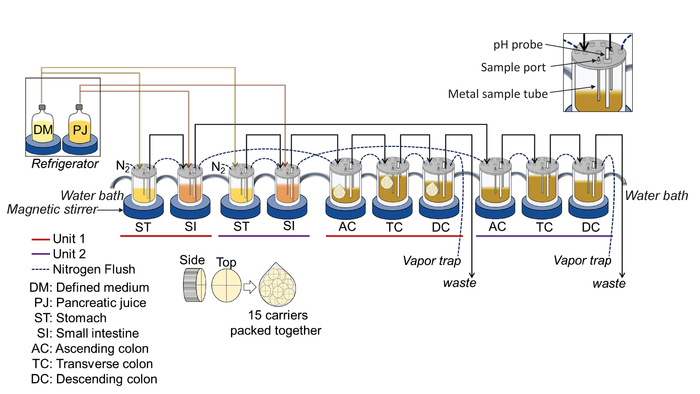
Figure 1: Illustration of the 5-stage, in vitro experimental design. The complete system consists of the following components: A circulating water bath, nitrogen flow, a set of glass bioreactors, a set of magnetic stirrer bars and magnetic stirrers, pH probes, a computer-controlled console containing 40 peristaltic pumps, a computer monitor, and a refrigerator. The main system is composed of a set of bioreactors mimicking the stomach, small intestine, and the ascending, transverse, and descending colon regions. Two complete units are set up to run in parallel, providing for an experimental and a control group. This means that 10 bioreactors, 10 pH probes, and 10 magnetic stirrers are required for this experiment. Please click here to view a larger version of this figure.
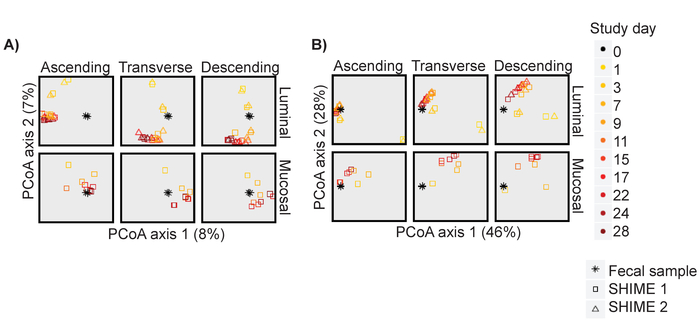
Figure 2: The in vitro community stabilizes by day 11 post inoculation. PCoA analysis based on (A) Unweighted and (B) Weighted Unifrac distances for the luminal and mucosal phase of each region over time for Unit 1 and for the luminal phase of Unit 2. The plot is faceted into the components after calculating the PCoA axes. Please click here to view a larger version of this figure.
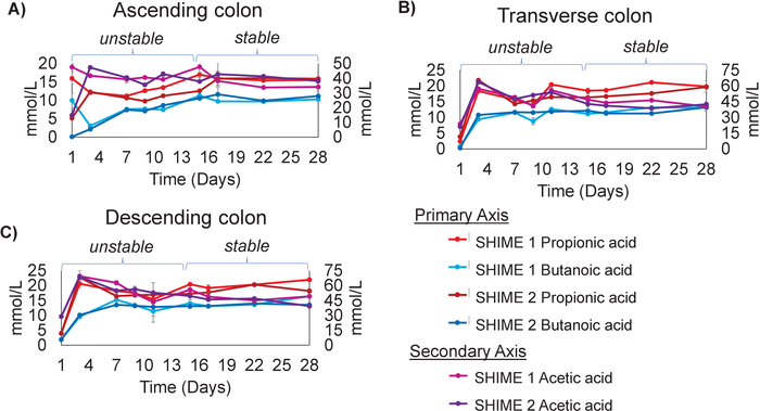
Figure 3: The production of SCFAs by the in vitro system stabilize by day 15 post inoculation. Measurements of Propionic Acid, Butanoic Acid, and Acetic Acid over time for the (A) Ascending colon (B) Transverse colon and (C) Descending colon. The experiment was performed in triplicate. The results represent an average of three independent measurements, with error bars depicting the standard deviation. Please click here to view a larger version of this figure.
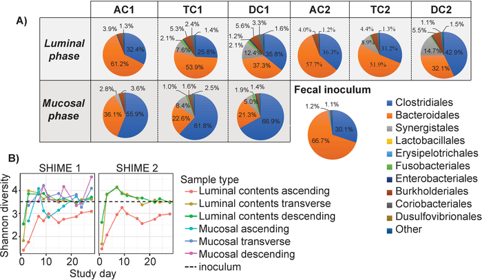
Figure 4: Comparison of the stable communities from the in vitro system to the fecal inoculum. (A) The stable communities (D15-28), at the order level were averaged and formatted in pie charts for both the mucosal and luminal phase of each colon region, and for the fecal inoculum. (B) The Shannon diversity for the mucosal and luminal phase of each colon region compared to the fecal inoculum (black dotted line). Please click here to view a larger version of this figure.
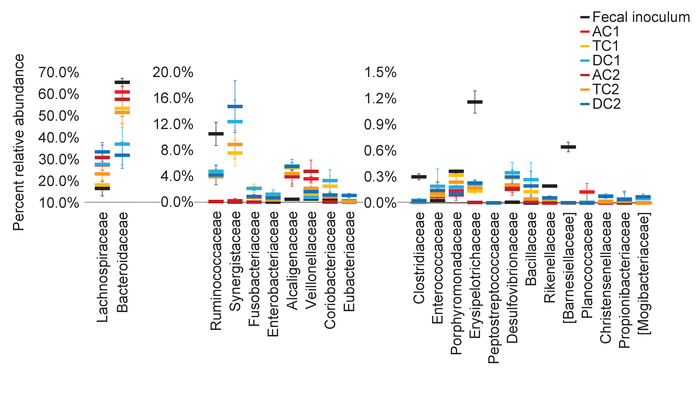
Figure 5: The luminal and mucosal phase in each colon region promote the growth of distinct communities. The average relative abundance, at the family level, for the stable communities (D15-28) for the luminal and mucosal phase, and the inoculum, were calculated and plotted together for each colon region. Error bars represent the standard deviation between timepoints. AC1 = Ascending colon Unit 1; TC1 = Transverse colon Unit 1; DC1 = Descending colon Unit 1; AC2 = Ascending colon Unit 2; TC2 = Transverse colon Unit 2; DC2 = Descending colon Unit 2. Please click here to view a larger version of this figure.
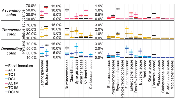
Figure 6: Each colon region of the in vitro system develops a unique community. The average relative abundance, at the family level, for the stable communities (D15-28) in each colon region, and the fecal inoculum, were calculated and plotted together. The experiment was performed in triplicate. The results represent an average of three independent measurements, with error bars depicting the standard deviation. Please click here to view a larger version of this figure.
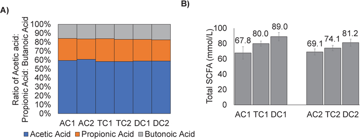
Figure 7: The ratio of Acetic Acid: Propionic Acid: Butanoic Acid is similar for each colon region. (A) The amounts of acetic acid, propionic acid, and butanoic acid for the stable communities (D15-28) of each colon region were calculated and converted to a ratio. (B) The ratios were plotted as percentages for each colon region. Error bars represent the standard deviation between timepoints. AC1= Ascending colon Unit 1; TC1 = Transverse colon Unit 1; DC1 = Descending colon Unit 1; AC2 = Ascending colon Unit 2; TC2 = Transverse colon Unit 2; DC2 = Descending colon Unit 2. Please click here to view a larger version of this figure.
In vitro culturing systems have been developed to study the gut microbiota of the large intestine. They use apparatuses designed to simulate the physiological conditions of the gastro intestinal tract, promoting the growth of a mature gut microbial community for each region of the colon33. While the concept is logical and comprehensible, the actual running of in vitro culturing systems to study the gut microbiota requires precision and an understanding of what is required and expected to produce reliable results.
The required elements for an in vitro gut microbiota experiment are that the community must reach stability after inoculation and that it must maintain the microbial diversity of the fecal inoculum. If the experiment is run properly, these two required elements are achieved readily and predictably. Critical steps in the protocol that will affect system stability and diversity are the following: The preparation of defined medium and pancreatic juice must be accurate, and delivery of these substrates must be consistent. Anaerobic conditions must be maintained during the experiment. The pH of each intestinal region must be accurately maintained over time. Changes in these three parameters during the experiment may result in population fluctuations and variable metabolite production.
Using this type of multi-stage in vitro system has several advantages. Notably, they simulate the different regions of the colon. They can also be designed to simulate in vitro digestion of food in the upper GIT, including the enzymatic components from saliva, and the addition of pancreatin and bile. A mucosal phase can be incorporated into each colon reactor, adding further complexity8. This results in the development of the region and the phase specific microbial communities that can be individually analyzed and compared. These types of systems can be readily manipulated, depending on experimental design through physical reconfiguration, alterations of the physiological parameters such as pH, temperature, or transit time, or inoculation with specific fecal samples26. Importantly, research has illustrated that the communities developed in these in vitro systems represent the gut microbial community11,26,34.
While the use of multi-stage in vitro systems can be used to mechanistically study the gut microbiota, they do have limitations. First, while the physiological conditions are programmable, they are fixed using specific average values, thus representing the average person. This is a simplification since there is considerable inter-individual variatiability12,23. Second, although the lack of mammalian components is one reason to use an in vitro system, it also must be considered a limitation. The lack of certain mammalian components, like the immune system, can result in differences between the in vitro model and the corresponding in vivo microenvironment12,23. However, these limitations do not detract from the value and insight that can be gained from using in vitro systems to mechanistically study the gut microbiota. Finally, the cultures are grown in glass bioreactors with no absorption of water or metabolites. This is an important difference, and the effect can be seen in the measurement of SCFAs where the total amounts of SCFAs are increasing from the ascending to transverse to descending colon regions (Figure 7). This is opposite of what is observed in vivo, where the highest concentrations of SCFAs are found proximally, in the ascending region, and the lowest concentrations found distally36. However, when the net production of SCFAs is considered for each region, then it can be determined that the most SCFA production is occurring in the ascending colon. It should also be mentioned here that bacterial load was not measured in this experiment. Differences in bacterial load between the regions in this system may have an effect on the total amounts of SCFAs produced.
In conclusion, it is well known that the gut microbiota plays a role in both human health and disease, and it is considered to function as a mediator between diet and metabolic health37,38. Here, the application of an in vitro system to study the gut microbiota was discussed, and the results from an experiment demonstrating the development of a stable community were presented. An in vitro system has many advantages and promotes the development of a complex and dynamic community, which is illustrated in the described experiment. This type of system is best used to study the interactions and changes within the gut microbiota community in response to external factors, such as food components and medicines. The results of these in vitro studies can then be supplemented with in vivo studies, to gain a deep understanding of the function and contribution of the gut microbiota to both health and disease.
The authors have nothing to disclose.
Ms. Audrey Thomas-Gahring is acknowledged for her GC/MS work. We would also like to thank Massimo Marzorati for editing the manuscript.
TWINSHIME | Prodigest | NA | |
defined medium (Adult M-SHIME growth medium with starch) | Prodigest | NA | |
Masterflex tubing | cole Parmer | NA | |
Urine Drainage bag | Bard | NA | |
Labsorb | Sigma-Aldrich | NA | |
Fecal sample | Openbiome | NA | |
Syringes | Becton Dickson | NA | |
Defined medium | Prodigest | NA | |
Oxgall Bile | Becton Dickson | NA | |
Pancreatin | Sigma-Aldrich | NA | |
Glass ware | Ace Glass | NA | |
Porcine mucin | Sigma-Aldrich | NA | |
Bacteriological agar | Sigma-Aldrich | NA | |
Sterilization pouches | VWR | NA | |
BeadBug | Benchmark Scientific | NA | |
Triple-Pure High Impact Zirconium 0.1mm Bead beater tube | Benchmark Scientific | NA | |
RNAse free, DNAse free, sterile water | Roche | NA | |
Shimadzu QP2010 Ultra GC/MS | Shimadzu | NA | |
Stabilwax-DA column, 30m, 0.25mm ID, 0.25µm | Restek | NA | |
plastic mucin carriers | Prodigest | NA |