In this study, a feeding system for nymphal and larval stages of hard tick was developed using a capsule attached to laboratory mouse. The feeding capsule is made from flexible materials and remains firmly attached to the mouse for at least one week and allows comfortable monitoring of tick feeding.
Ticks are obligatory blood feeding parasites at all stages of development (except eggs) and are recognized as vectors of various pathogens. The use of mouse models in tick research is critical for understanding their biology and tick-host-pathogen interactions. Here we demonstrate a non-laborious technique for the feeding of immature stages of hard ticks on laboratory mice. The benefit of the method is its simplicity, short duration, and the ability to monitor or collect ticks at different time points of an experiment. In addition, the technique allows attachment of two individual capsules on the same mouse, which is beneficial for a variety of experiments where two different groups of ticks are required to feed on the same animal. The non-irritating and flexible capsule is made from easily accessible materials and minimizes the discomfort of the experimental animals. Furthermore, euthanasia is not necessary, mice recover completely after the experiment and are available for re-use.
Ticks are important vectors of several pathogens and represent a serious risk to animal and human health1. Setting up an effective feeding system is crucial when studying their biology, tick-host-pathogen interactions, or establishing effective control measures. Currently, several artificial feeding systems, which avoid the use of live animals are available for ticks2,3,4 and these should be utilized whenever experimental conditions allow. However, in various experimental settings these systems fail to appropriately mimic the specific physiologic features and the use of live animals is necessary to achieve relevant results.
Laboratory mice are commonly used for the study of many biological systems and are routinely utilized as hosts for feeding ticks5,6,7,8,9. The two most common methods of feeding immature ticks on mice include free infestations and the use of confinement chambers attached to the mouse. Free infestations are primarily used for larval stages and engorged ticks can drop to an area where they can be recovered. Confinement chambers are usually composed of acrylic or polypropylene caps which are glued to the mouse’s back. The first technique is an effective natural system for tick feeding but does not allow close monitoring during the experiment because the individual ticks are dispersed in different parts of the host body. Additionally, engorged ticks that drop to a recovery area can become contaminated with feces and urine10,11,12,13,14 that may severely affect the tick fitness or they can be damaged or eaten by the mouse if there is no separation between the animal and the recovery area15. Chamber-based systems allow the confinement of ticks to a defined area, however, the gluing process is laborious and the caps are often weakly adherent to the glue and thus they often detach during the experiment16,17,18,19. The caps are also stiff, uncomfortable, and lead to skin reactions, which prevent the re-use of the mice and necessitates their euthanasia after the experiment.
In our previous study, we successfully developed an effective system using chambers made of ethylene-vinyl acetate (EVA) foam for feeding ticks on laboratory rabbits20. Herein, we adapted this system to a mouse model and propose a simple and clean method to feed immature hard tick stages in closed capsules made from EVA-foam. Specifically, our system uses elastic EVA-foam capsules glued to the shaved mice back with fast drying (3 min), non-irritating latex glue. This technique allows firm and long-lasting attachment of capsules to the experimental mouse, as well as effective tick infestation/collection during the entire course of the experiment. The flat capsule is made from flexible materials and does not impede manipulation of the mouse for blood collection or other purposes. The system is suitable mainly for the nymphal tick stages, but with slight modification it can be used for feeding larvae as well. The method can be completed by one single experienced person and extensive training is not required.
Please note that this protocol can be only applied when all welfare and safety measures are met in the laboratory. This protocol received permission to use mice for tick feeding by the Ethics Committee for Animal ExperimentsComEth Anses/ENVA/UPEC, Permit Numbers E 94 046 08. For the endpoint, the animals were exposed to CO2 for 9 min in two phases of 4 and 5 min each one.
1. Preparation of the capsule
2. Preparation of the mice before tick infestation
NOTE: In this study, 10 – 12 weeks old female experimental mice (strain C57BL/6 and BALB/cByJ) were maintained in standard cages with food and water offered ad libitum (Green line ventilated racks at -20 Pa) at the French Agency for Food, Environmental and Occupational Health & Safety (ANSES) accredited animal facilities in Maisons-Alfort, France. Animals were monitored twice daily by experienced technicians for any abnormal skin reactions, health problems or complications.
3. Tick Infestation
4. Collection of Ticks
5. Recovery of the mice
We propose the detailed step-by step method for feeding immature hard tick stages in EVA-foam capsules applied to a mouse’s back (Figure 2). This non-laborious protocol is suitable for various types of experiments when precise tick monitoring and collection is required. The main advantages of this method are its simplicity, easily accessible cost-effective materials, and short duration. In addition, we succeeded in attaching two capsules to one mouse individual (Figure 2K) allowing to us to feed two different groups of ticks on the same animal. The use of the highly effective, fast-drying, and non-irritating latex glue ensures that the capsule is firmly attached within 3 min. Also, the capsule remained attached for at least one week (Figure 2J) which was enough time for engorgement of most of the immature hard tick species21,22,23,24. Due to the capsule elasticity, further manipulation of the mouse for blood collection or other purposes was very convenient. This procedure also allows complete recovery of the mice after the experiments (Figure 3D) giving the opportunity to reuse the animals and avoid euthanasia. Our system has been successfully used to feed Ixodes ricinus nymphs (Figure 4). A moderate to high engorgement success rate was achieved in C57BL/6 and BALB/cByJ mouse strains, respectively. In both cases all nymphs finished the feeding within 4 – 5 days, while the majority (~75%) dropped off on the fourth day.
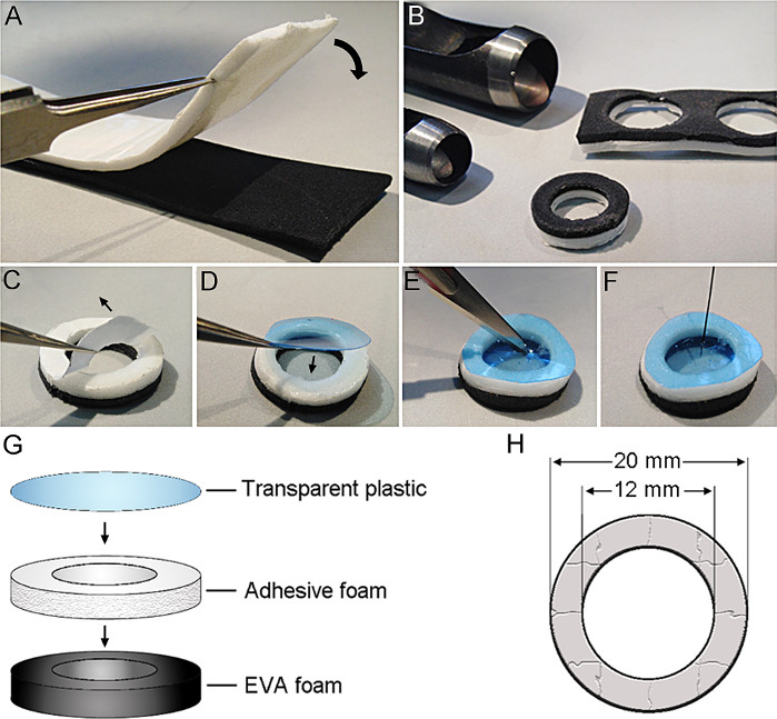
Figure 1: EVA-foam capsule preparation. (A) Attachment of EVA-foam (black) and adhesive double sticky foam (white). (B) Cutting 20 mm diameter outer and 12 mm inner circle using leather hole punches. (C) Removal of the paper protection tape from the adhesive double sticky foam. (D) Attachment of the transparent plastic to the capsule. (E) Cutting the slit in the transparent plastic with a scalpel. (F) Creation of holes using an entomological pin in the plastic. (G-H) Schematic drawing of the different parts of the capsule and dimensions. Please click here to view a larger version of this figure.
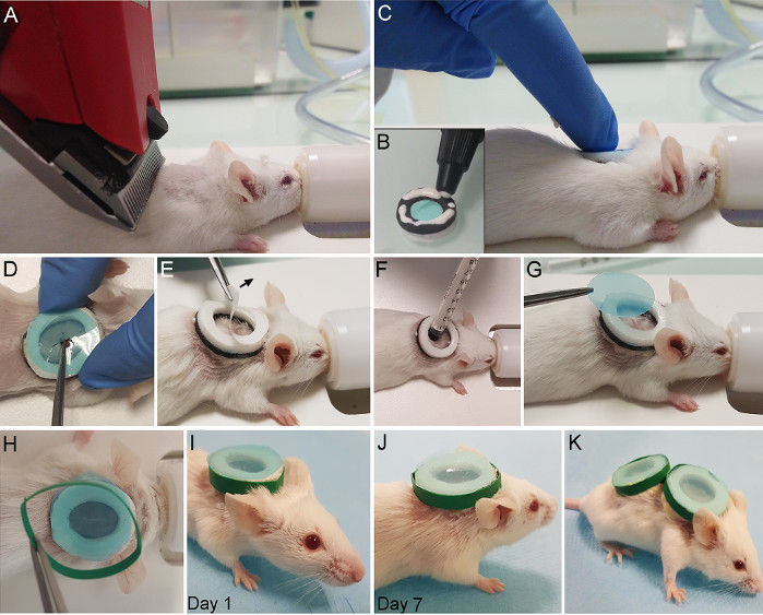
Figure 2: Gluing the capsule to the mice and tick infestation. (A) Shaving mouse’s back anterior part. (B) Application of the latex glue to the EVA-foam side of the capsule. (C) Attachment of capsule to the mouse. (D) Placing the nymph in the capsule via the cut in the transparent plastic. (E) Peeling the paper protection tape from the adhesive double sticky foam before larvae infestation. (F) Injections of larvae inside the capsule using a cut syringe. (G) Closing the capsule with the transparent plastic. (H) Placing a protective plastic band around the capsule. (I) Mouse with the attached capsule – 1st day. (J) Mouse with the attached capsule – 7th day. (K) Mouse with two capsules attached. Please click here to view a larger version of this figure.
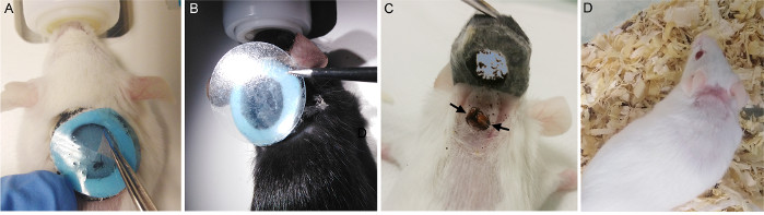
Figure 3: Tick collection and mouse recovery. (A) Cutting cross-shape opening for tick collection. (B) Resealing the capsule with adhesive plastic patch. (C) Capsule removal from a euthanized mouse. Arrows show the attached ticks. (D) Recovered mouse after dropped off capsule. Please click here to view a larger version of this figure.
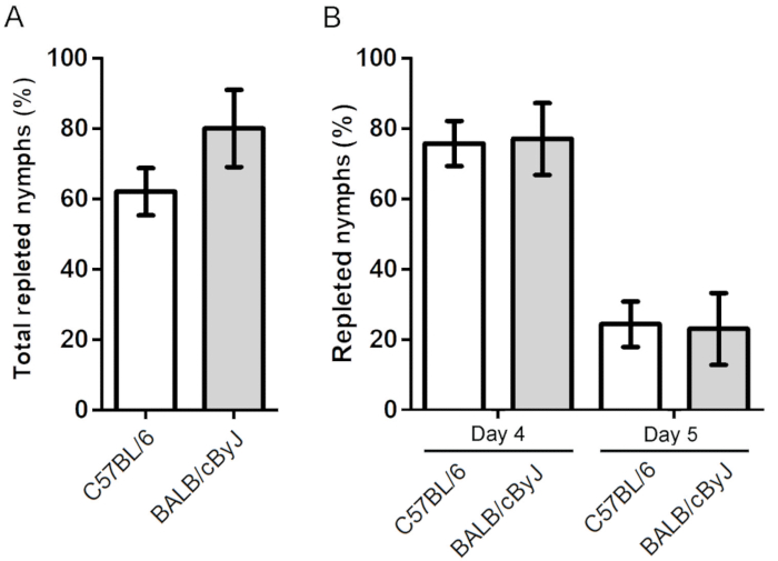
Figure 4: Engorgement success and feeding duration of Ixodes ricinus nymphs feeding on mice. (A) Total percentage of engorged nymphs in C57BL/6 and BALB/cByJ mice. (B) Duration of nymph engorgement in C57BL/6 and BALB/cByJ mice. The (n) numbers for infested nymphs are 130 and 25 for 15 individual C57BL/6 and 5 individual BALB/cByJ mice, respectively. Please click here to view a larger version of this figure.
The most critical step in the protocol is firm gluing of the capsule to the mouse skin. Therefore, the latex glue should be homogenously applied to the entire EVA-foam surface of the capsule and constant pressure for 3 minutes should be applied, especially to the left and right side of the capsule. We also recommend placement of the capsule as far forward on the back as possible to avoid its removal by the mouse using its rear paws. In our experiments, only the adhesion of the EVA-foam and latex glue to the mouse skin has been validated and we cannot guarantee the achievement of same results using different materials.
During our experiments, detachment of the capsule from the skin within first seven days was not observed. We strongly recommend protecting the outer surface of the capsule using the plastic band (Figure 2H). If the protective band is damaged over the course of tick feeding, it can be replaced with a new one. The diameter of the capsule can be modified for different mouse strain sizes. We suggest monitoring the feeding ticks at least twice daily and to collect engorged ticks immediately after detachment to avoid their desiccation.
The number of infested ticks is limited by the capsule diameter, as well as the host size. In our experiments we used maximum of 20 nymphs or 100 larvae of I. ricinus for one mouse. For the larger size ticks such Amblyomma or Hyalomma sp., etc the number of infested ticks should be reduced to avoid harm to the host from blood loss19,26,27. Therefore, this technique is not suitable for the maintenance of tick rearing colonies, where large numbers of ticks are required to feed. For this purpose, larger hosts like rabbits or sheep are recommended20,27 to reduce overall animal requirement.
Our technique is suitable for various types of experiments where a mouse model is required, and it is necessary to keep ticks in enclosed area for easy collection and/or monitoring of their biological parameters. Compared to other techniques10,11,12,13,14,15,16,17,18, this simple protocol greatly reduces the overall anesthesia time (approximately 5 minutes) per mouse and the fast drying, non-irritating latex glue does not cause harm to the animal. The highly adhesive EVA-foam capsule protects the tick feeding area and minimizes the risk of lost, damaged, or eaten ticks as reported in free infestation systems10,11,12,13,15. The great advantage of the proposed technique is the flat-shape capsule and its firm long-lasting attachment to the skin allowing easy manipulation with the mouse if required. Special attention has been paid on usage of elastic and non-irritating materials to reduce the discomfort to the experimental animals allowing complete recovery of the mouse host after experiment (Figure 3D).
The method is expected to be used for a variety of the experiments when studying tick-host-pathogen interactions, tick manipulation of host immune systems, evaluating different tick control measures or tick biology.
The authors have nothing to disclose.
We acknowledge the technical assistance of Alain Bernier French National Institute of Agricultural Research (INRAE), and Océane Le Bidel (ANSES). The study was supported by the DIM One Health – Région Île-de-France (Acronym of the project: NeuroPaTick). The mice were purchased by ANSES. Dr. Jeffrey L. Blair is acknowledged for reviewing the earlier version of the manuscript.
EVA-foam 2 mm thick, (low density) | Cosplay Shop | EVA-45kg (950/450/2 mm) | It can be ordered also via Amazon (ref. no. B07BLMJDXD) |
Heat Shrink Tubing Electric Wire Wrap Sleeve | Amazon | B014GMT1AM | Different diameters of Heat Shrink Tubing are available via Amazon. |
Mice BALB/cByJ | Charles River | Strain code 627 | |
Mice C57BL/6 | Charles River | Strain code 664 | |
No-toxic Latex Glue | Tear mender | Fabric & Leather Adhesive | Also available also via Amazon (ref. no. B001RQCTUU) |
Punch Tool Hand Art Tool | Amazon | B07QPWNGBF | Saled by amazon as Leather Working Tools 1-25mm Round Steel Leather Craft Cutter Working for Belt Strap |
PVC Binding Covers Transparent | Amazon | B078BNLSNP | Any transparent PVC sheet of ticknes between 0.150 mm to 0.180 mm is suitable |
Self Adhesive Pad Sponge Double Coated Foam Tape | Amazon | B07RHDZ35J | Saled by amazon as 2 Rolls Double Sided Foam Tape, Super Strong White Mounting Tape Foam |
Transparent seal stickers (20 mm diameter circles) | Amazon | B01DAA6X66 |