The Doppler Blood Flow Imaging technique was used to evaluate the effectiveness of our model. In short, we compared the data from the control group with the data from the RVA group to determine the success of our creation. Figure 4A depicts a typical flux associated with the control animal, while Figure 4B depicts the results obtained from an RVA. The numeric 1-8 represents the various events associated with I/R phases. In brief, numeric 1-3 correspond to the phase of normoxia, while a sharp decline in flux at points 3 and 4 represents the events associated with the decrease in blood flow in the RVA. Once an 80% or greater drop-down had been achieved, the RVA was left raised for the next 5 min. This was the phase of ischemia (numeric 4-5). Following a 5 min lifting of the RVA, the RVA was released, which is represented by numeric 6 and 7. The flux from point 7 onward represents the reperfusion phase, which occurs after the RVA has achieved normal blood flow levels, which was the phase of reperfusion. This particular experiment demonstrated the effectiveness of I/R modeling in the 3-day developed chick embryo.
To verify the utility of our model, we have studied the expression patterns of proteins, RNA, and DNA through ELISA, western blotting, qRT-PCR, and gel electrophoresis analysis. In brief, we divided the 3-day developed eggs into three experimental groups: control, I/R, and Treatments + I/R. A significant difference in expression of proteins, genes, and DNA integrity was observed between their respective I/R and control groups. As discussed below, drug treatment to the I/R group effectively improved the outcome observed in this group compared to the I/R treated group alone; this is consistent with our prior publication on which this protocol is based1. Standard laboratory procedures were followed for ELISA18, western blotting19, qRT-PCR20, and gel electrophoresis21, which are not covered in this paper (Figure 5, Figure 6, Figure 7, Figure 8).
Ischemia-reperfusion stimulates several processes, including the formation of reactive oxygen species (ROS) that are detrimental to tissue. The detrimental effects caused by the intrinsic antioxidant body defense systems' response to ROS is an important feature of I/R injury. We examined the activities of oxygen regulatory proteins 150 (ORP150), cytoplasmic superoxide dismutase 1 (SOD1), and catalase in ischemic vessels. When compared to the control group, I/R elevated the activity of ORP150, cytoplasmic SOD1, and catalase (Figure 5A–C). However, supplementation with N-Acetyl-L-Cysteine (NAC), a ROS quencher, mitigated the I/R group's oxidative stress.
To assess our model's inflammatory potential, we used ELISA to examine IL-1β and TNF-α expression (Figure 6). Both interleukins were overexpressed in the I/R group as compared to the control group, indicating that our model has the potential to be used in inflammatory investigations. Next, we tested the expression of the NOD-like receptor pyrin domain-containing protein 3 (NLRP3) inflammasome pathway22,23, and pro-inflammatory molecule, NF-kβ24,25, which are involved in amplifying inflammation, to confirm the usefulness of this model for inflammatory studies. In response to I/R generated in the RVA, this investigation found evidence of activation of the NLRP3 inflammasome (Figure 7A), as well as NF-kβ (Figure 7B). However, treatment with Naringenin, a well-known anti-inflammatory drug, ameliorated the inflammatory effects, as observed in the drug-treated groups.
Ischemia-reperfusion activates programed cell death pathways26,27,28. We studied the effects of I/R on apoptosis and autophagy pathways in the chick embryo. Figure 8A depicts the effect of I/R on caspase-3 and zVAD-fmk. Figure 8B–D demonstrate that the I/R group had higher expression of LC3II as well as LC3II/I ratio (autophagosomes marker), Beclin1 (a significant regulator of autophagy in mammalian cells), and Atg7 (needed for basal autophagy) than the control group, respectively, at protein levels. In contrast, Figure 8E shows the effect of the I/R on mRNA levels. The addition of 3-MA, an autophagy inhibitor, however, reversed the results. These findings suggest that the model might be used to investigate different cell death processes (e.g., necroptosis). Figure 9 shows how efficiently the changes at the DNA level can be studied using our Hook-I/R model.
Finally, we compare the results from the Hook-I/R model and the middle cerebral artery occlusion (MCAO) model to see how effective our model is in comparison to other models. To summarize, I/R-treated RVAs had higher levels of the apoptotic protein cleaved caspase-3, the autophagy proteins Beclin1 and Atg7, and the inflammatory interleukins TNF-α and IFN-Ƴ than control RVAs. Interestingly, whether analyzing inflammatory stress or cellular death pathways, the Hook-I/R model produced results that were extremely similar to those obtained by the MCAO model (Figure 10).
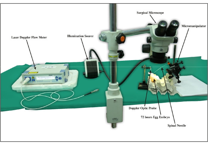
Figure 1: A schematic diagram of a typical set-up of the Hook-I/R chick embryo ischemia-reperfusion model. The picture shows the Hook-I/R chick embryo ischemia-reperfusion experimentation requirements, such as the stereo zoom surgical microscope, laser Doppler blood flowmeter, micromanipulator, spinal needle, 72 h developed chick embryos, and an illumination source.”Please click here to view a larger version of this figure.
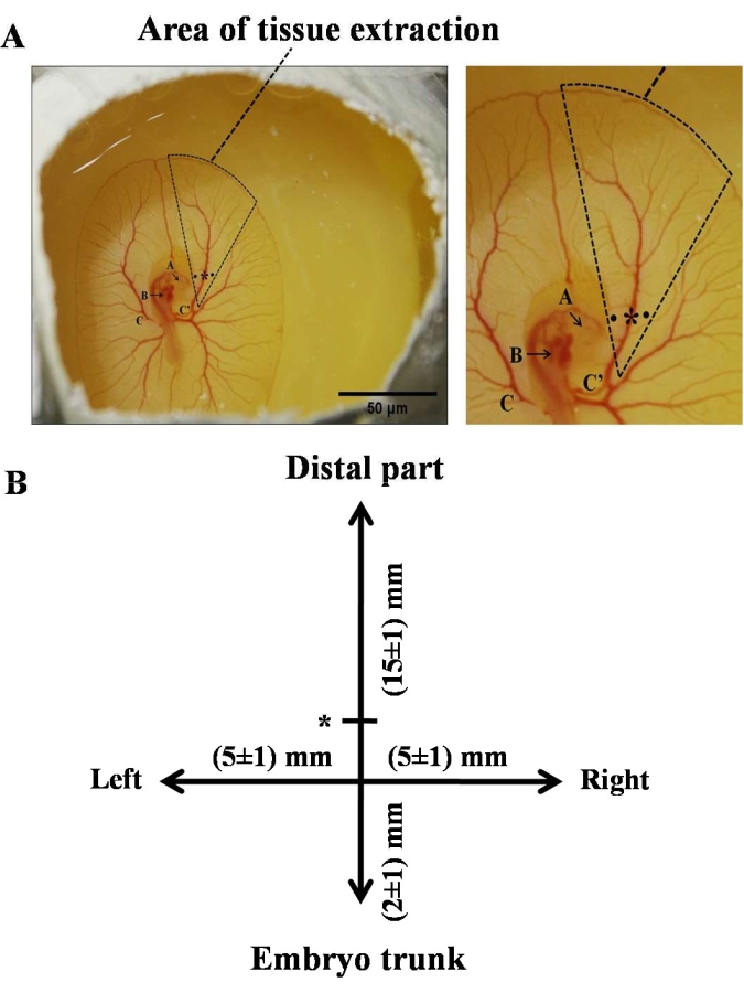
Figure 2: Representative image of a 3-day chick embryo-exhibiting site of occlusion and area of tissue excision. (A) The triangle shows the location of excision of the tissue for western blotting, RNA, and DNA isolation. The star and dots represent the site of the occlusion and the holes created on the left and right side of the RVA to insert the needle beneath the artery to lift it, respectively. A magnified image of the area of tissue extraction is also provided along with it. The notation A represents the eye, B represents the heart, C represents the left of the vitelline artery, and C' represents the right of the vitelline artery. (B) A schematic representation of the right side of the chick embryo demonstrates the area of tissue excision in the vicinity of RVA. The straight line represents the RVA emerging from the embryo trunk. The star on the vertical line symbolizes the position of the Laser Doppler blood flow probe. The intersection denotes the site of occlusion. Excision of the arteries was done up to 15 ± 1 mm (distal from the trunk), 5 ± 1 mm each on the left and right side of the artery, and 2 ± 1 mm toward the trunk. All the measurements show distances from the site of occlusion. Please click here to view a larger version of this figure.
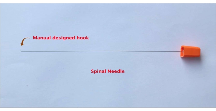
Figure 3: Representative picture of a spinal needle with a custom-made hook. Please click here to view a larger version of this figure.
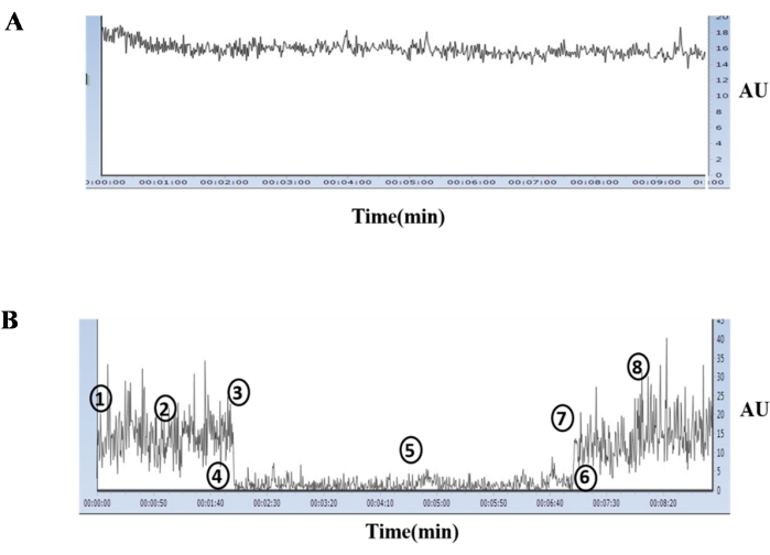
Figure 4: Representation of a typical Laser Doppler blood flowmeter signal. A typical Laser Doppler blood flowmeter signal was measured on a 3-day chick embryo artery from a control group egg (A). Events at baseline (1), during normoxia (2), immediate pre-lifting of RVA (3), immediate post-lifting of RVA (4), during ischemia (5), immediate pre-releasing of RVA (6), immediate post-reperfusion (7), during reperfusion (8) in the ischemia treated group as recorded by the Laser Doppler blood flowmeter (B). This figure was adopted from Fauzia et al., 2018 (Frontiers in Pharmacology; under a CC-BY license)1. Please click here to view a larger version of this figure.
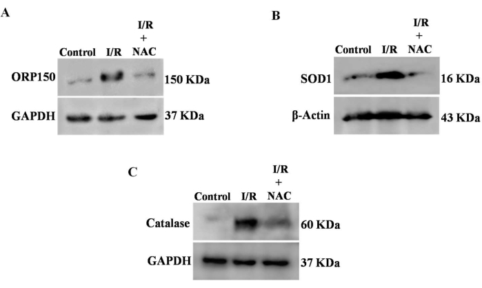
Figure 5: Effect of ischemia-reperfusion on the expression of oxidative stress marker proteins. Western blotting was used to determine the ORP150, SOD1, and catalase expression levels. (A) ORP150 expression was increased after I/R damage. NAC, a pan-ROS inhibitor, lowered ORP150 to a level comparable to that of control. (B) Following I/R, SOD1 expression was elevated. SOD1 expression was likewise decreased following NAC treatment. (C) The I/R event induced the expression of catalase. RVA treated with I/R and exposed to NAC showed a decrease in catalase levels. GAPDH (A,C) and β-Actin (B) were used as internal controls. Please click here to view a larger version of this figure.
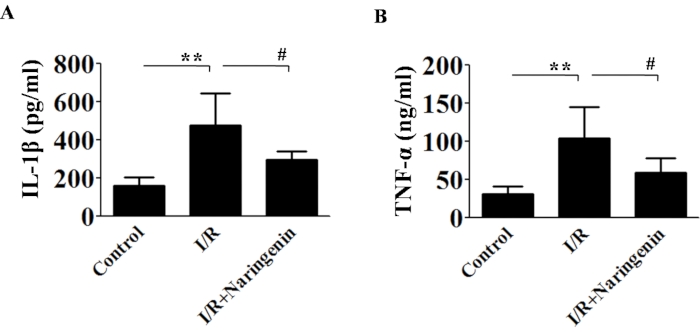
Figure 6: Estimation of IL-1β and TNF-α using ELISA. ELISA was used to quantify IL-1β and TNF-α, and the results revealed that there was an increase in IL-1β and TNF-α expression, which was mitigated by the addition of Naringenin. For this study, the ANOVA followed by the Newman-Keuls test was applied. Error bars represent mean ± SD, n = 3. Please click here to view a larger version of this figure.
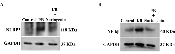
Figure 7: Effect of ischemia-reperfusion on the expression of inflammation marker proteins. (A) I/R treatment resulted in increased expression of NLRP3. Incubation of I/R treated RVA with Naringenin decreased the expression of NLRP3. (B) Similar to NLRP3, the expression of NF-kβ was also increased after I/R treatment. The treatment of Naringenin reduced the expression of NF-kβ. GAPDH was used as an internal control. Please click here to view a larger version of this figure.
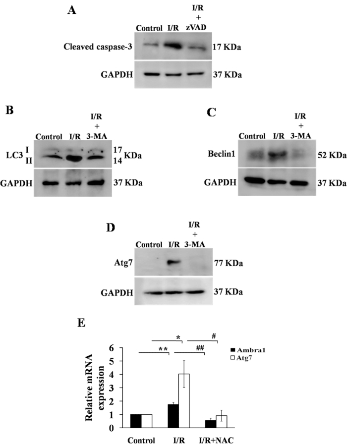
Figure 8: Effect of ischemia-reperfusion on the apoptotic and autophagic status of RVA cells. (A) To explore the induction of apoptosis by the I/R process, the expression of cleaved caspase 3 was evaluated using western blotting. Following I/R damage, the level of cleaved caspase 3 was increased. However, treatment with zVAD-fmk, an apoptosis inhibitor, decreased cleaved caspase 3 expression. (B–D) To evaluate the induction of autophagy following I/R, the expression of LC3II/I, Beclin1, and Atg7 was determined. Following I/R, the expression of all autophagic markers increased, while the exposure of I/R-treated RVA to the autophagy inhibitor 3-MA decreased the expression of all proteins to control levels. As an internal control, GAPDH was used. (E) qRT-PCR was used to confirm the induction of autophagy and determine the Ambra1 and Atg7 mRNA expression levels. Both genes showed a considerable increase in expression in the I/R group compared to control, which was restored to near-normal levels following NAC therapy. GAPDH was used as an internal control for qRT-PCR experiments. For experiment (E), the ANOVA, followed by the Newman-Keuls test, was applied. Error bars represent mean ± SD, n = 3. Please click here to view a larger version of this figure.
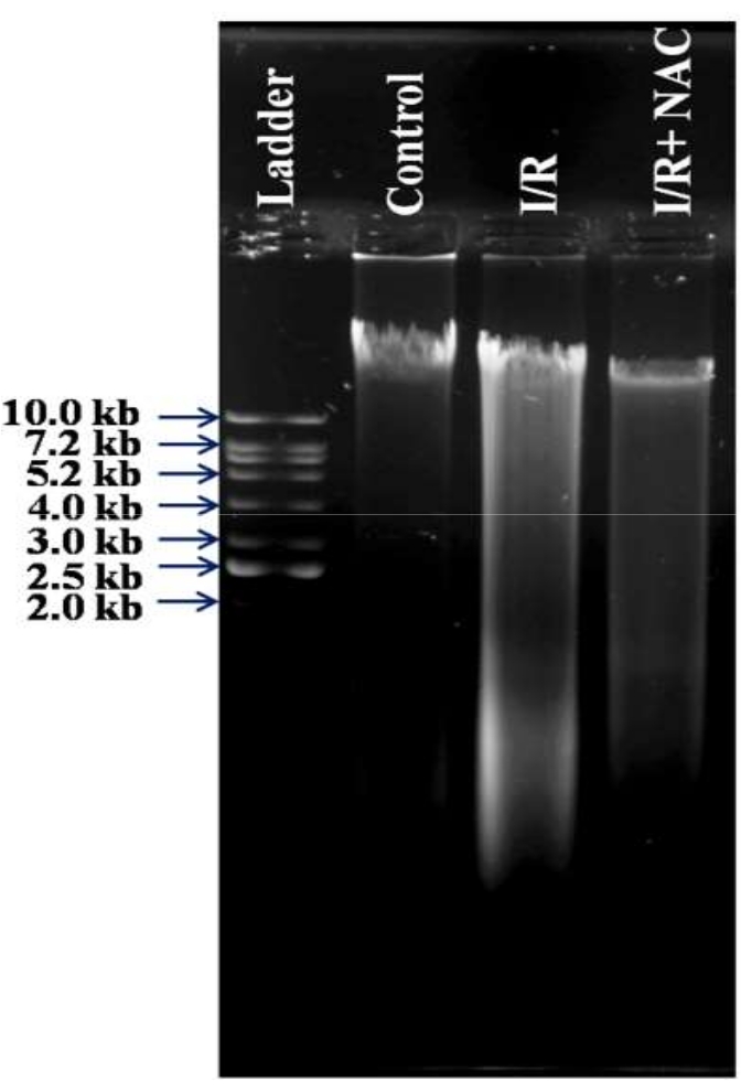
Figure 9: Effect of ischemia-reperfusion on DNA damage. To determine if I/R induces DNA nicks, we examined DNA damage using gel electrophoresis. I/R resulted in genomic DNA degradation, as shown by smearing in the I/R sample. NAC therapy of I/R-damaged RVA reduced DNA damage. A 1 kb DNA ladder was employed. Please click here to view a larger version of this figure.
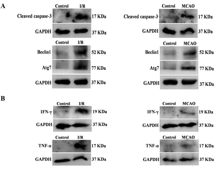
Figure 10: Comparison of the Hook-I/R model vs. the MCAO model. A comparison was made between the data generated by the Hook-I/R model and the data generated by the MCAO model. (A) The expression of the apoptotic protein caspase-3 and the autophagy proteins Beclin1 and Atg7 was higher in I/R treated RVAs when compared to the expression of the same proteins in control RVAs, which was in agreement with the findings obtained in the MCAO experiments. (B) TNF-α and IFN-Ƴ levels were found to be elevated in both the RVA and MCAO groups when compared to their respective controls. Please click here to view a larger version of this figure.
| Name of Material/Equipment | Concentration | Autoclave | Storage | ||
| Reagents for Ringer’s solution | |||||
| Sodium chloride | 123 mM | ||||
| Potassium chloride | 4.96 mM | ||||
| Calcium chloride | 1.53 mM | ||||
| Sterile distilled water | 100 mL | ||||
| pH | 7.4 | 15 min at 121 °C | R.T. | ||
| Reagents for 0.9% normal saline | |||||
| Sodium Chloride | 154mM | ||||
| Sterile distilled water | 100 mL | ||||
| pH | 7.4 | 15 min at 121 °C | R.T. | ||
| Reagents for 70% ethanol | |||||
| 70% ethanol | 70 mL | ||||
| Sterile distilled water | 30 mL | Not required | R.T. | ||
| Reagents for 1x Phosphate Buffer Saline | |||||
| Sodium chloride | 136.8 mM | ||||
| Potassium chloride | 26.8 mM | ||||
| Potassium phosphate monobasic anhydrous | 14.6 mM | ||||
| di-Sodium hydrogen phosphate heptahydrate | 5.37 mM | ||||
| Sterile distilled water | 100 mL | 15 min at 121 °C | R.T. | ||
Table 1: Recipe of solutions used in this study.
